Heart Institute of Hospital das Clínicas of FMUSP and School of Public Health of USP Mailing address: William A. Chalela – InCor Av. Dr. Enéas C. Aguiar, 44 -05403-000 - São Paulo, SP – E-mail: [email protected]
W illiam A. Chalela, Antonio P. Mansur, José M. Aldrighi
São Paulo, SP - Brazil
Noninvasive Diagnostic Evaluation for Chest
Pain in Women
Women are eight times less likely to develop myocar-dial infarction than men are; nonetheless, when myocarmyocar-dial ischemic disease is present, it has a worse evolution 1,2. No
explanations for this fact are available, although specula-tions exist about the inadequate manner of diagnosis.
It is worth emphasizing that in studies about coronary disease, women are either a minority or excluded from the study protocols. Therefore, most of the information regar-ding coronary disease in women comes from studies con-ducted in men. In fact, invasive cardiovascular procedures - either diagnostic or therapeutic - are frequently less indi-cated for women who bear confirmed coronary disease. However, it is not yet clearly established whether this diffe-rence is due to a larger use of such procedures in men or to a smaller indication in women.
Typical angina is more prevalent in women, although angiographic studies have revealed that all forms of angina, including the typical type, are associated on a smaller scale with coronary disease in women when compared with that men. The CASS 3 study reveals that 62% of women with
de-fined angina had ischemic disease compared with 40% of those with probable angina and 4% with nonischemic pain. We propose to critically evaluate the main noninvasive diagnostic methods for chronic coronary disease in women. Electrocardiogram - The association between elec-trocardiographic abnormalities at rest and a higher inciden-ce of cardiovascular diseases is well documented in men but not in women. When ranking different levels of rest electro-cardiographic abnormalities in men and women aged 40 to 64 years and with higher risk of cardiovascular disease-in-duced death, De Bacquer et al 4 observed higher relative
risk for men. High-degree abnormalities were directly associated with mortality in both sexes, but low-degree ab-normalities were not. The abab-normalities considered as high
degree were the presence of depression of the ST segment, T-wave inversion, complete or 2nd degree atrioventricular
block, branch (left or right) block, frequent extrasystoles and atrial fibrillation or flutter; low-degree abnormalities were a deviated QRS axis complex with high or low voltage and other alterations in ventricular repolarization. De Bacquer et al 4 demonstrated that depression of the ST
seg-ment in the rest electrocardiogram was associated with hi-gher cardiovascular mortality, and was equally important in both sexes. The T-wave abnormalities were also more pre-dictive for men, whereas arrythmias were more prepre-dictive for women. It is worth emphasizing that low-degree abnor-malities did not have predictive values for either sex.
Stress test - Several studies 3-7 have revealed a
redu-ced amount of noninvasive diagnostic interventions for co-ronary disease in women. This is so because of the lower prevalence of ischemic disease in women, in addition to a hi-gher number of false-positives 8, ie, the specificity of the
conventional effort test is lower in women, compared with that observed in men. It is important to consider the proba-bility that coronary disease in women, based on age and symptoms, frequently is between low and medium, espe-cially if it is considered before menopause. Low probability induces patients with an estimated <20% to have the disea-se, and is limited to women who do not have any major risk factor and who do not have more than a medium risk factor or two minor risk factors for coronary disease, for instance women of child-bearing age with atypical angina. Patients with high probability, with an estimated >80% chance of having the disease, have two or more major risk factors or a major one associated with a medium or minor risk factor. Mo-derate probability is defined as risk between 20 and 80% 9.
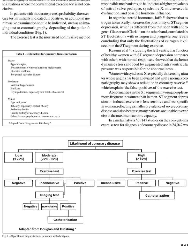
The classification of risk factors for coronary disease in women is presented in Table I.
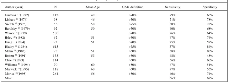
In patients with low pretest probability, no indication exists for diagnostic testing, because the posttest probabi-lity, in case of a positive result, will not be high enough to in-dicate an invasive examination, as seen by Bayes theorem 8
that observed frequent false-positive results.
On the other hand, the exercise test should be
ted in women with a high probability of coronary disease. In this situation, the imaging tests add little information, be-cause false-positive results are not frequent, in addition to the low probability of false-negatives in females.
The use of imaging tests, such as nuclear imaging and echocardiography associated with the exercise test is restricted to situations where the conventional exercise test is not con-clusive.
In patients with moderate pretest probability, the exer-cise test is initially indicated; if positive, an additional no-ninvasive examination should be indicated, such as an ima-ging test or coronariography, depending of the patient’s individual conditions (Fig. 1).
The exercise test is the most used noninvasive method
for the evaluation of ischemic heart disease. However, it is less accurate and has a greater potential for false-positives in women, which is at least partially explained by the Bayesi-an principles, based on the fact that, in most female patients with pretest probability for coronary disease, the perfor-mance on the exercise test is relatively low. Among the likely responsible mechanisms, to be indicate a higher prevalence of mitral valve prolapse, syndrome X, microvascular dysfunction, and possible hormone influence.
In regard to steroid hormones, Jaffe 10 showed that
es-trogen taken orally increases the possibility of ST segment depression, which is different from that seen with andro-gens; Glasser and Clark 11, on the other hand, correlated the
ST fluctuations with estrogen and progesterone levels, concluding that only the fluctuations of estrogen levels occur on the ST segment during exercise.
Kusumi et al 12, studying the left ventricular function
of healthy women with ST segment depression compared with others with normal responses, showed that the hemo-dynamic stress induced by augmented intraventricular pressure was responsible for the abnormal tests.
Women with syndrome X, especially those using nitra-tes whose angina has been alleviated and with a normal coro-nariography may show a reduction in coronary reserve 13,
which explains the false-positives of the exercise test. Abnormalities in the ST segment in young people are more frequent in women than in men. ST segment depres-sion on induced exercise is less sensitive and less specific in women, reflecting a smaller prevalence of severe coronary disease and also because many patients are unable to exer-cise at the maximum aerobic capacity.
In a metaanalysis 8 of 147 studies on the conventional
exercise test for diagnosis of coronary disease in 24,047
wo-Table I - Risk factors for coronary disease in women
Major Typical angina
Postmenopause without hormone replacement Diabetes mellitus
Peripheral vascular disease
Moderate
Arterial hypertension Smoking
Dyslipidemias, especially low HDL-cholesterol
Minor Age >65 years
Obesity, especially central obesity Sedentary habits
Family history of coronary disease Other factors (psychosocial, hemostatic, etc.)
Adapted from Douglas and Ginsburg 9.
Likelihood of coronary disease
Low (< 20%)
Moderate (20% - 80%)
Exercise test
Negative Inconclusive Positive Inconclusive Positive Negative
Imaging test
Negative Inconclusive Positive
Catheterization
Catheterization Exercise test
High (> 80%)
Adapted from Douglas and Ginsburg 9
men, 68% sensitivity and 77% specificity were detected. Table II summarizes some studies that show the accuracy of the ST segment for diagnosis of coronary disease in wo-men, with mean sensitivities of 66% and specificities of 67%. Variations among the studies are due to the different criteria used to define coronary disease, to the selection of patients (inclusion of previous heart attack, multi-vessel di-seases), and to the different characteristics of the test, in-cluding the ST segment-based positivity and the type of exercise accomplished 8.
At the Heart Institute 14 of the HC-FMUSP, the tests
considered positive or to indicate ischemia had ST segment depression of horizontal or downsloping morphology or both ≥1mm for men and 2mm for women, when the upslo-ping with a 2mm for men and 3mm for women situated at 80ms of the J point. The ST segment elevation 1mm is also considered a positive response. Using these criteria, up to a 40% reduction in false-positive results has been noticed, confirmed by the normal results of the myocardial perfu-sion scintigraphy.
The accuracy of the exercise test in women may be higher if the levels of ST segment depression are analyzed with other parameters.
The exercise test provides other parameters of major importance for the diagnosis and prognosis of coronary di-sease, including physical capacity, hemodynamic res-ponse, and the presence or absence of cardiac symptoms on exercise, which must be analyzed together with electrocar-diographic alterations.
Another strategy to differentiate true from false results is the retest with a beta-blocker. Marcomichelakis et al 29
stu-died 100 patients with positive effort tests, 50 of which did not have obstructive coronary disease. The retest with a beta-blocker normalized the ST segment modifications in all patients with false-positive results.
Because it is difficult to foresee the probability of coro-nary disease in women, some authors have preferred the asso-ciation of exercise testing with imaging as an initial alternative.
The need for an additional test must always be based on the analysis of clinical parameters and results of the initial exercise test. Although a large number of false-positive re-sults occur due to the physical stress, not enough data are available to justify the incorporation of imaging tests with the initial routine for the diagnosis of coronary disease. Women with moderate risk and negative exercise tests have a low probability of coronary disease; therefore an additio-nal test is not necessary. If, however, the test is positive, another test is required, due to the elevated incidence of he-art attack or hehe-art disease-induced death. If the result is in-conclusive for any reason, another method must be indica-ted, for instance, pharmacological stress for patients who cannot adequately accomplish the physical stress test.
Myocardial perfusion scintigraphy - Myocardial perfu-sion scintigraphy with tomography slices (SPECT, Single Pho-ton Emission Computed Tomography) is more accurate for diagnosis of coronary disease than the conventional exercise test. Few studies have been conducted concerning the use of myocardial perfusion scintigraphy as a noninvasive method for diagnosis of coronary artery disease in women. For SPECT30
with201 TI, the sensitivity varies from 71% to 86% and the
specificity from 81% to 91%. Just as with men, several factors may affect the accuracy of myocardial perfusion scintigraphy in women: choice of patients, percentage of maximum heart rate reached during the exercise, type of image acquisition (the plain, not currently in use or in tomography slices), criteria of interpretation, and level of tissue attenuation.
Artifacts suggestive of irreversible defects may arise as a consequence of tissue attenuation; thus, in women, the left breast may attenuate the antero-lateral wall on the plain study, and the apical-lateral wall on the tomography. Fat de-posits on the thoracic left lateral wall may lessen the inferior left ventricle area, especially in obese patients. When the persistent defect of the antero-lateral region was considered an artifact due to attenuation of mammary tissue, Friedman and co-workers 31 observed an increase in specificity from
88% to 97%, and, similarly, in Hung et al 32, the specificity
Table II - Sensitivity and specificity of the exercise test in women
Author (year) N Mean Age CAD definition Sensitivity Specificity
Guiteras 15 (1972) 112 49 >70% 79% 66%
Linhart 16 (1974) 98 46 >50% 71% 78%
Sketch 17 (1975) 56 50 >75% 50% 78%
Barolsky 18 (1979) 92 50 >50% 60% 68%
Weiner 19 (1979) 580 >70% 76% 64%
Ilsley 20 (1982) 62 51 >50% 67% 74%
Hung 21 (1984) 92 51 >70% 75% 59%
Hlatky 22 (1984) 613 >75% 57% 86%
Melin 23 (1985) 93 51 >50% 58% 80%
Robert 24 (1991) 135 53 >50% 68% 48%
Chae 25 (1993) 114 >50% 66% 60%
Williams 26 (1994) 70 60 >50% 67% 51%
Marwick 27(1995) 118 60 >50% 77% 56%
Morise 28(1995) 264 56 >50% 46% 74%
Mean 66% 67%
varied from 81% to 91%. Different strategies have been pro-posed to improve the specificity of images with 201Tl in
wo-men; however, the breast size, position, and density still limit test performance.
Myocardial perfusion scintigraphy with 99m
Tc-SESTA-MIBI may diminish the tissue attenuation-induced artifact; in fact, the high-energy 99mTc compared with 201Tl causes
the former to reduce these artifact effects in approximately 15% of patients. SPECT specificity with 99mTc-SESTAMIBI
in women is significantly better than that obtained with
201Tl. In Taillefer and colleagues’ study 30, where 115 women
were evaluated, the specificities were 84.4% for 99m
Tc-SESTAMIBI and 62.7% for 201Tl; however, the sensitivities
for detection of stenosis in coronary arteries 70% were simi-lar, ie, 84.3% and 80.4%, respectively. In this same study, when Gated-SPECT was analyzed, the specificity changed from 84.4% to 92.2%. Gated-SPECT is a technique that joins two methods; thus, with a single approach to the patient, it evaluates simultaneously the myocardial perfusion and the qualitative and quantitative parameters of ventricular function, improving therefore the test’s accuracy. This tech-nique may be performed by both perfusion markers; however, with 201Tl, acquisition time must be bigger in order
to obtain better image quality.
Echocardiogram - In addition to allowing the diag-nosis of coronary disease, the stress (physical or pharma-cological) echocardiogram helps to recognize artifacts, specifically those caused by the mammary tissue. No-netheless, the echocardiographic imaging in obese patients, those with thoracic deformities, or lung diseases is technically more difficult to interpret and of poorer quality.
For the diagnosis of coronary disease, both echocar-diography and myocardial perfusion scintigraphy may be performed in association with the exercise test or to pharma-cological stress testing.
The sensitivity of the echocardiogram associated with physical stress is comparable between both sexes, but is in-fluenced by the modality of exercise used. For instance, with the treadmill, the echocardiographic imaging is acqui-red before and immediately after the exercise, which is dif-ferent from the exercise test with the ergometric bicycle; thus, Sawada et al 33 verified that in the exercise
echocardio-gram, the sensitivity was 80% with the treadmill and 100% with the bicycle, and the specificity was 94% and 73%, res-pectively. The accuracy, however, was similar, 87% for the treadmill and 84% for the bicycle.
The advantage of pharmacological stress over exer-cise associated with echocardiography includes obtaining higher quality images, due to the absence of movement and respiratory interference, and the easiness to control the
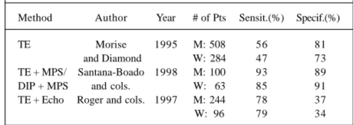
Table III - Noninvasive tests for the diagnosis of coronary disease in
men and women 35
Method Author Year # of Pts Sensit.(%) Specif.(%)
TE Morise 1995 M: 508 56 81
and Diamond W: 284 47 73 TE + MPS/ Santana-Boado 1998 M: 100 93 89 DIP + MPS and cols. W: 63 85 91 TE + Echo Roger and cols. 1997 M: 244 78 37
W: 96 79 34
Pts- patients; Sensit.- sensitivity; Specif.- specificity; TE- exercise test; M-men; W- woM-men; MPS- myocardial perfusion scintigraphy with tomography slices; Dip- dipiridamol; Echo- echocardiogram.
stress reached. Recent studies show a sensitivity of phar-macological stress between 76 and 93%, the specificity of 43 to 94%, and accuracy of 58 to 95% 34.
Some studies suggest that the echocardiography and nuclear studies have similar sensitivity and specificity, but have greater sensitivity and specificity compared with the exercise test (Table III).
Another study 36, a metaanalysis to detect coronary
disease in women, shows the preeminence of exercise echo-cardiography: 19 studies about the conventional exercise test for diagnosis of coronary disease in 3,721 women, re-ported 61% sensitivity and 70% specificity; in five studies on myocardial perfusion scintigraphy in 842 women, the sensitivity was 78% and the specificity 64%; in three stu-dies about the exercise echocardiogram in 290 women, sen-sitivity was 86% and specificity was 79%.
Conclusion
The present review critically analyzes the main comple-mentary examinations used to diagnose coronary disease in women. The stress electrocardiogram and myocardial perfu-sion scintigraphy are the most commonly used noninvasive methods for detecting coronary artery disease.
Stress echocardiography appears promising, requi-ring, however, more consistent studies.
Myocardial perfusion scintigraphy with tomography slices is still the method of choice for detection of coronary disease and, more recently, the Gated SPECT has gained fa-vor, because in effort-induced ischemia, this method allows identification of the main events of the ischemic cascade, ie, the heterogeneity of coronary flow.
1. Hendel RC. Myocardial infarction in women. Cardiology 1990; 77: 41-57. 2. Greenland P, Reicher-Reiss H, Goldbourt U, Behar S, and the Israeli SPRINT
In-vestigators. In-hospital and 1-year mortality in 1524 women after myocardial in-farction. Circulation 1991; 83: 484-91.
3. Weiner DA, Ryan TJ, McCabe CH, et al. Exercise stress testing: correlations among history of angina, ST-segment response and prevalence of coronary artery disease in the Coronary Artery Surgery Study (CASS). N Engl J Med 1979; 301: 230-5.
4. De Bacquer D, Pereira LSM, De Backer G, Henauw S, Kornit M. The predictive value of electrocardiographic abnormalities for total and cardiovascular disease mortality in men and women. Eur Heart J 1994; 15: 1604-10.
5. Ayanian JZ, Epstein AM. Differences in the use of procedures between women and men hospitalized for coronary heart disease. N Engl J Med 1991; 325: 221-5. 6. Steingart RM, Packer M, Hamm P, et al. Sex differences in the management of
coro-nary artery disease. N Engl J Med 1991; 325: 226-30.
7. Bickell NA, Pieper KS, Lee KL, et al. Referral patterns for coronary artery disease treatment. Gender bias or good clinical judment? Ann Intern Med 1992; 116: 791-7.
8. Gibbons RJ, Balady GJ, Beasley JW, et al. ACC/AHA guidelines for exercise tes-ting. A report of the American College of Cardiology/ American Heart Associa-tion. Task Force on Practice Guidelines (Committee on Exercise Testing). J Am Coll Cardiol 1997; 30: 260-315.
9. Douglas PS, Ginsburg GS. The evaluation of chest pain in women. N Engl J Med 1996; 334: 1311-5.
10. Jaffe MD. Effect of testosterone cypionate on post exercise ST segment depression. Br Heart J 1977; 39: 1217-22.
11. Glasser SP, Clark PI. Interpretation of exercise test results in women. Pract Car-diol 1988; 14: 85-90.
12. Kusumi F, Bruce RA, Ross MA, Trimble S, Voigt AE. Elevated arterial pressure and post-exertional ST segment depression in middle aged women. Am Heart J 1976; 92: 576-83.
13. Marcus ML. Coronary circulation in health and disease. McGraw-Hill, New York, 1983.
14. Alfieri RG, Moffa PJ, Lima EV, Chalela WA, Moraes AP, Pereyra PA. Aspectos do teste de esforço na cardiopatia isquêmica. Rev Bras Med (Cardiol) 1986; 5: 254-65. 15. Guiteras VP, Chaitman BR, Waters DD, et al. Diagnostic accuracy of exercise ECG
lead systems in clinical subsets of women. Circulation 1972; 65: 1465-73. 16. Linhart JW, Laws JG, Satinsky JD. Maximum treadmill exercise
electrocardiogra-phy in females patients. Circulation 1974; 50: 1173-8.
17. Sketch MH, Mohiuddin SM, Lynch JD, Zencka AE, Runco V. Significant Sex dif-ferences in the correlations of electrocardiographic exercise testing and coronary arteriograms. Am J Cardiol 1975; 36: 169-73.
18. Barolsky SM, Gilbert CA, Faruqui A, Nutter DO, Schlant RC. Differences in Electrocardiographic response to exercise of women and men: a non-Bayesian factor. Circulation 1979; 60: 1021-7.
19. Weiner DA, Ryan TJ, McCabe CH, et al. Exercise stress testing: correlations among history of angina, ST-segment response and prevalence of coronary-artery disease in the Coronary Artery Surgery Study (CASS ). N Engl J Med 1979; 301: 230-5.
20. Ilsley C, Canepa-Anson R, Westgate C, Webb S, Rickards A, Poole-Wilson P.
In-fluence of R wave analysis upon diagnostic accuracy of exercise testing in women. Br Heart J 1982; 48: 161-8.
21. Hung J, Chaitman BR, Lam J, et al. Noninvasive diagnostic test choices for the evaluation of coronary artery diasease in women: a multivariate comparison of cardiac fluoroscopy, exercise electrocardiography and exercise thallium myocar-dial perfusion scintigraphy. J Am Coll Cardiol 1984; 4: 8-16.
22. Hlatky MA, Pryor DB, Harrell FE Jr, Califf RM, Mark DB, Rosati RA. Factors af-fecting sensitivity and specificity of exercise electrocardiographiy: multiva-riable analysis. Am J Med 1984; 77: 64-71.
23. Melin JA, Wijns W, Vanbutsele RJ, et al. Alternative diagnostic strategies for co-ronary artery disease in women: demonstration of the usefulness and efficiency of probability analisis. Circulation 1985; 71: 535-42.
24. Robert AR, Melin JA, Detry JM. Logistic discriminant analysis improves diag-nostic accuracy of exercise testing for coronary artery disease in women. Circula-tion 1991; 83: 1202-9.
25. Chae SC, Heo J, Iskandrian AS, Wasserleben V. identification of extensive corona-ry artecorona-ry disease in women by exercise single-photon emision computed tomo-graphic (SPECT) thallium imaging. J Am Coll Cardiol 1993; 21: 1305-11. 26. Williams MJ, Marwick TH, O’Gorman D, Foale RA. Comparison of exercise
echocardiography with na exercise score to diagnose coronary artery disease in women. Am J Cardiol 1994; 74: 435-8.
27. Marwick TH, Anderson T, Williams MJ, et al. Exercise echocardiography is na ac-curate and cost-efficient technique for detection of coronary artery disease in wo-men. J Am Coll Cardiol 1995; 26: 335-41.
28. Morise AP, Diamond GA, Detrano R, Bobbio M. Incremental value of exercise electrocardiography and thallium-201 testing in men and women for the presence and extent of coronary artery disease. Am Heart J 1995; 130: 267-76. 29. Marcomichelakis J, Donaldson R, Green J, et al. Exercise testing after
beta-blo-ckade: improved specificity and predictive value in detecting coronary heart di-sease. Br Heart J 1980; 43: 252-61.
30. Taillefer R, DePuey G, Udelson JE, Beller GA, Latour Y, Reeves F. Comparative diagnostic accuracy of Tl-201 and Tc-99m Sestamibi SPECT imaging (perfusion and ECG-gated SPECT) in detecting coronary artery disease in women. J Am Coll Cardiol 1997; 29: 69-77.
31. Friedman TD, Greene AC, Iskandrian AS, Hakki AH, Kane AS, Segal BL. Exercise thallium 201 myocardial scintigraphy in women: correlation with co-ronary angiography. Am J Cardiol 1982; 49: 1632-7.
32. Hung J, Chaitman B, Lam J, et al. Non-invasive diagnostic test choice for the eva-luation of coronary artery disease in women: a multivariate comparison of car-diac fluoroscopy, exercise electrocardiography and exercise thallium myocardial perfusion scintigraphy. J Am Coll Cardiol 1984; 4: 8-16.
33. Sawada SG, Ryan T, Fineberg NS, et al. Exercise echocardiographic detection of coronary artery disease in women. J Am Coll Cardiol 1989; 14: 1440. 34. Tong AT, Douglas PS. Stress Echocariography in women. Cardiol Clin 1999;
17: 573-82.
35. Gibbons RJ, Chatterjee K, Daley J, et al. ACC/AHA/ACP-ASSIM guidelines for the management of patients with chronic stable angina. A report of the American College of Cardiology/American Heart Association - Task Force on Practice Guidelines. J Am Coll Cardiol 1999; 33: 2092-197.
36. Knok Y, Kim C, Grady D, Segal M, Redberg R. Meta-analysis of exercise testing to detect coronary artery disease in women. Am J Cardiol 1999; 83: 660-6.