Diversity of Coronaviruses in New World Bats
Bárbara dos Santos Simões
Dissertação para obtenção do Grau de Mestre em Microbiologia Médica
i
Diversity of Coronaviruses in New World Bats
Bárbara dos Santos Simões
Dissertação para obtenção do Grau de Mestre em Microbiologia Médica
Orientadores:
Prof. Dr. Jan Felix Drexler (Charité - Universitätsmedizin Berlin) Prof. Dr. Ricardo Parreira (IHMT/UNL)
Charité - Universitätsmedizin Berlin
ii
Acknowledgements
I would like to thank Professor Dr Jan Felix Drexler for the opportunity given to me to do my thesis in the Institute. For all the knowledge that he gave to me about the world of science and most important everything about Virology.
To Andrea Rasche for all the time spent teaching me and helping me during my thesis and of course for having the patience for all my questions. To Gustavo Goés for helping me in the beginning and kindly supply data.
To all my colleagues from the group, for giving me the opportunity to learn new things on different topics and the support and strength that they gave during my thesis.
To Juli Jules, Toby, Fabian, Tali, Sofia for making me laugh in the hard times and making me believe that I could make and especially to Juli Jules for the support and help that she gave to me and of course for have entered in my life. Ich liebe euch!
To my friends Alex and Seohee for everything. Thanks for making my life better in Berlin and for having you in my life. To my friends back in Portugal, Joana “Bimba”, Sofs, Marta and Mariana for the support and for believing in me. To Gabi, thank you for everything. For believing and pushing me to accomplish my dreams, for the kind and cold words when needed. Even far away you could make me feel that I was next to you. Thanks, dwarf! To Felix for listening and give important life advice, for the support, for making me laugh so much and for standing by my side in my most difficult times. Eintracht Frankfurt International!
To my family and my dog Dudu for the support and unconditional love that they gave me and the opportunity to study abroad and for all the thoughts that they taught me during my life.
iii
Abstract
Coronaviruses comprise important human and animal pathogens. Six coronavirus species can cause human illness, probably all having a zoonotic origin. The prominent role of bats for coronavirus evolution was discovered in the aftermath of the 2003 SARS (Severe Acute Respiratory Syndrome) outbreak, where bats were identified as the zoonotic origin of this virus and a great diversity of coronavirus was identified in bats around the world. Indeed, bats present the largest genetic diversity of coronavirus compared to other hosts, making them the major natural host. The Neotropical region harbours a high variety of bat species presenting enormous potential for coronavirus studies. However, most of the studies regarding coronavirus diversity were conducted in Old World bats.
In this project, I studied the genetic diversity of coronavirus from about 1000 Neotropical bats. I conducted phylogenetic analyses from several coronavirus polymerase fragments and characterized two full genomes, comprising one alphacoronavirus from a Brazilian bat (Phyllostomus discolor) and one betacoronavirus from a Costa Rican bat (Pteronotus parnellii). The Phyllostomus bat alphacoronavirus might be an ancient relative of the human alphacoronaviruses 229E and NL63 and their bat-related coronaviruses.
The Pteronotus bat betacoronavirus is an ancient sister clade of the clade c betacoronavirus to which the human coronavirus MERS (Middle East Respiratory Syndrome) belongs and might correspond to an ancient root of the origins of MERS. In sum, this thesis expands the knowledge of coronavirus diversity in New World bats as and gives a deeper insight into the origins of the human coronavirus MERS, 229E and NL63.
iv
Resumo
Os Coronavírus são patógenos importantes para o Homem e animais. Seis espécies de Coronavírus podem causar doenças no Homem e provavelmente têm uma origem zoonótica. Após a epidemia de SARS (Severe Acute Respiratory Syndrome), foi descoberto o papel proeminente dos morcegos para a evolução de coronavírus, na medida em que estes foram identificados como sendo a sua origem zoonótica. Após a epidemia, uma grande diversidade de coronavírus foi identificada em morcegos de todo o mundo. Estes apresentam a maior diversidade genética de coronavirus em comparação com outros hospedeiros, tornando-se os seus hospedeiros naturais. A região Neotropical contém uma elevada diversidade de espécies de morcegos, apresentando um enorme potencial para estudos de diversidade de coronavírus contudo, a maioria destes estudos só foi realizado com morcegos do Velho Mundo.
Neste projeto, eu estudei a diversidade genética de coronavírus em aproximadamente 1000 morcegos do Neotrópico. Eu realizei análises filogenéticas de vários fragmentos da polimerase de coronavírus e caracterizei dois genomas completos: um alphacoronavírus presente num morcego do Brazil (Phyllostomus discolor) e um betacoronavírus de um morcego da Costa Rica (Pteronotus parnellii). O alphacoronavírus pode ser um parente antigo deste grupo e pode estar relacionado com os alphacoronavírus humanos 229E e NL63 e 229E e NL63 relacionados com morcegos.
O betacoronavírus também pertence a um antigo grupo relacionado com o grupo C em que o coronavírus humano MERS (Middle East Respiratory Syndrome) pertence. Isto pode corresponder a uma possível explicação para a origem de MERS. Em resumo, esta tese contribui para o aprofundamento dos conhecimentos da diversidade de coronavírus em morcegos do Novo Mundo e fornece uma visão mais aprofundada sobre as origens dos coronavírus humanos MERS, 229E e NL63.
v
Index
Acknowledgements ... ii Abstract ... iii Resumo ... iv Index ... vFigure index ... vii
Table index ... viii
Abbreviations... ix
1. Introduction ... 1
1.1 Family Coronaviridae ………..……….2
1.1.1 Taxonomy of coronaviruses ………..………..2
1.1.2 Morphology and genome organization ………..…….3
1.1.3 Replication cycle ………..…………5
1.1.4 Diversity and evolution of coronaviruses ………...…7
1.2 Neotropical bats ..………..….9
1.2.1 New and Old World Bats ..………..…..9
1.3 Aim of the thesis ………..………10
2. Materials and Methods ... 11
2.1 Bat sampling and processing ..……….………12
2.1.1 Origin and capture of bat samples …..……….12
2.2 Sample extraction …..……….……12
2.3 Screening assays for coronavirus ..………..12
2.4 Extension of PCR product with specific primers .……….………….14
2.5 DNA sequencing ..……….……17
vi
2.6.1 Genome assembling and phylogeny ..……….17
2.6.2 Prediction of genome organization and phylogenetic analysis ..……….17
2.6.3 Prediciton of nonstructural proteins (NSPs) …..……….……..18
2.6.4 Genome similarity plots, species delineation and detection of recombination events ………..………18
3. Results ... 20
3.1 Coronavirus detection in Neotropical bats ..……….21
3.1.1 Screening for coronavirus ………21
3.1.2 Elongation of the screening fragment ...………..……….24
3.2 Genome annotation ..………25
3.2.1 ORF prediction ..……….26
3.2.2 TRS and ribosomal frameshift prediction ……….………27
3.2.3 ORF phylogenetic analysis .……….…………28
3.2.4 Prediciton of nonsctrutural proteins (NSPs) ……….. 32
3.2.5 ORF aminoacid identities…..………..……….………..35
3.2.6 Full genome similarity plots and species delineation ..………37
3.2.7 Detection of recombination events …..………..…………38
4. Discussion and Conclusion ... 39
4.1 Detection of coronavirus in Neotropical bat..……..………41
4.2 Genome annotation …..………42
4.2.1 Phylogenetic reconstructions ..………42
4. References ... 444
vii
Figure index
Figure 1 - Diversity of CoV. ...2
Figure 2 - Schematic representation of the genome organization of the main Open Reading Frames (ORFs) of important CoV. ………..…3
Figure 3 - CoV morphology. ………3
Figure 4 - Replication cycle of CoV. ……….……….…6
Figure 5 - Scheme of the discontinuous transcription of CoV. ...6
Figure 6 - Phylogeny of bats. ...9
Figure 7 - Location and number of the samples collected as well as the bat species important in the study. ……….………..…..…….22
Figure 8 - Phylogenetic tree of the positive samples. ………..…………24
Figure 9 - Bayesian tree of the 816nt gap free of the translated RdRp fragment. ...25
Figure 10 - Prediction of the genome organization of the complete genomes. ……….…..26
Figure 11 - Bayesian phylogenies of the major ORFs of the Phyl-AlphaCoV. ……….…….29
Figure 12 - Bayesian phylogenies of the major ORFs of the Pte-BetaCoV. ……….……..31
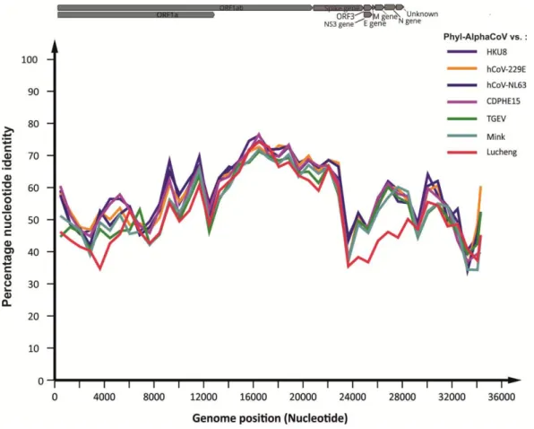
Figure 13 - Nucleotide sequence identity between Phyl-AlphaCoV and other alphacoronavirus. ……….….….37
Figure 14 - Nucleotide sequence identity between Pte-BetaCoV and one sequence from each betacoronavirus’ clade. ………..…..38
viii
Table index
Table 1 - Cycling protocol of the first round of the betacoronavirus screening assay. ….………..…13
Table 2 - Cycling protocol of the second round of the betacoronavirus screening assay. ………..….14
Table 3 - List of the primers used for the elongation of the screening fragment. ………..………15
Table 4 - Cycling protocol for the first round of the extension of the screening fragment. ………..……...16
Table 5 - Cycling protocol for the second round of the extension of the screening fragment. ………..…16
Table 6 - List of samples used in the project. ...21
Table 7 - Putative transcription regulatory sequences (TRS) of the Phyl-AlphaCoV. ………...27
Table 8 - Putative transcription regulatory sequences (TRS) of the Pte-BetaCoV. ………..….27
Table 9 - Prediction of the putative pp1a/pp1ab cleavage sites of Phyl-AlphaCoV based on the comparison with the ICTV reference sequences……….………33
Table 10 - Prediction of the putative pp1a/pp1ab cleavage sites of Pte-BetaCoV based on the comparison with the ICTV reference sequences. ……….……….34
Table 11 - Comparison of amino acid differences of the Phyl-AlphaCoV genome... ……….……….35
Table 12 - Comparison of amino acid differences of the Pte-BetaCoV genome. …..………36
ix
Abbreviations
°C Celsius degrees 3Clpro 3C-like main protease
µL Microliter= 10-6 Liter a.a Amino acid
ADRP ADP-ribose 1-phosphatase BCoV Bovine coronavirus
BEAST Cross-platform program; Bayesian Evolutionary Analysis Sampling Trees BLAST Bioinformatic tool; Basic Local Alignment Search Tool
bp basepairs
BSA Bovine Serum Albumi
CoV Coronavirus
DNA Desoxyribonucleic acid
dNTP 2’-Desoxyribonucleoside-5’-triphosphate
E Envelope Protein
et al. From Latin: et alii/et aliae (meaning: and others)
ExoN 3’-to-5’ exoribonuclease
FIPV Feline Infectious Peritonitis Virus HE Hemagglutinin-esterase
HEL Helicase
Hz Hertz
IBV Infectious Bronchitis Virus
ICTV International Committee for the Taxonomy of Viruses IFN Interferon
x
M Membrane glycoprotein
MAFFT Multiple Alignment program; Multiple Alignment using fast Fourier transform
MERS-CoV Middle East Respiratory Syndrome coronavirus Mg milligram=10-3 Kilo
MgCl2₂ Magnesium Chlorate
MgSO4₄ Magnesium Sulfate
Min Minute
mM Milimole=10-3 molar
mRNA Messenger Ribonucleic Acid N Nucleocapsid Protein
NCBI National Center for Biotechnology Information NendoU Nidoviral uridylate-specific endoribonuclease NMT N7 methyltransferase
No. Number
NSP Nonstructural protein
Nt Nucleotide
NTPase Nucleoside triphosphatase O-MT Ribose-2’-O-methyltransferase ORF Open Reading Frame
PBS Phosphate-Buffered Saline PCR Polymerase Chain Reaction PEDV Porcine Epidemic Diarrhea Virus
xi Plpro Papain-like protease
Sec Second
SADS Swine Acute Diarrhea Syndrome
SARS-CoV Severe Acute Respiratory Syndrome coronavirus
SSE Sequence Editor, Database and Analysis Platform; Simple Sequence editor
RBD Receptor-binding domain
RDP4 Recombination Detection Program RdRp RNA-dependent RNA polymerase RGU RdRp Grouping Units
RNA Ribonucleic Acid
RT-PCR Reverse transcriptase Polymerase Chain Reaction
S Spike glycoprotein
SARS Severe Acute Respiratory Syndrome
ss Single-Stranded
TBE TRIS-borate
TGEV Transmissible Gastroenteritis Virus of Swine
Tm Melting Temperature. The temperature at which half of the DNA strand is in the random coil or single-stranded state
TRS Transcription regulatory sequence TRS-B Body transcription regulatory sequence TRS-L Leader transcription regulatory sequence
xii
UV Ultraviolet
V Volts
1
2
1.1. Family Coronaviridae
1.1.1. Taxonomy of coronaviruses
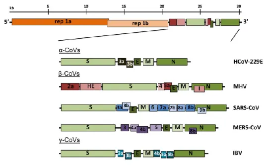
The order Nidovirales is divided into 3 families: Arteriviridae, Roniviridae and Coronaviridae. This last family is divided into 2 subfamilies: Coronavirinae and Torovirinae. Coronaviridae comes from the Latin word “Corona” which means “halo” or “crown” and refers to the appearance of projections on the surface that resemble a solar corona. This family, commonly known as coronavirus (CoV) is a monophyletic family of the order Nidovirales and includes four genera: alpha-, beta-, gamma- and deltacoronavirus. In addition, the betacoronavirus genus is furthermore divided into 4 clades: A to D (Figure 1) (De Groot et al., 2012).
Figure 1 - Diversity of CoV (Kaslow et al., 2014).
The genera Alpha- and Betacoronavirus mainly infect mammals while the genera Gamma- and Deltacoronavirus infect mainly birds. However, members of the genus Deltacoronavirus were also found in pigs and in Asian leopards and members of the genus Gammacoronavirus were found in the beluga whale and in other cetaceans (Drexler et al., 2014; Woo et al., 2014; Woo et al., 2012).
CoVs are usually related to respiratory, enteric and hepatic to central nervous system illness depending on the virus clade and host, in both birds and mammals including humans (Lai & C., 2007; Weiss & Navas-Martin, 2005).
3
1.1.2. Morphology and genome organization
CoVs possess a positive single-stranded, nonsegmented genome. Described as a large RNA virus, the genome can range between 26-32 kb with a 5’ terminal cap structure and a 3’ poly-A tail. Almost two-thirds of the genome holds 2 Open Reading Frames (ORFs): ORF1a and ORF1b which encode in total 16 non-structural proteins (NSP). The polyprotein pp1a encodes for the NSPs1 to 10 and pp1b for NSP12 to 16, which includes major proteins such as the RNA- dependent RNA polymerase (RdRp) (NSP12) and Helicase (HEL) (NSP13). The rest of the genome encodes for essential structural proteins such as Envelope, Spike, Membrane, Nucleocapsid and accessory proteins (Lai & Cavanagh, 1997) (Figure 2). The accessory proteins are frequently interspaced between the major ORFs and their number varies with a species and most of these proteins do not play a role in virus replication cycle. However, it has been demonstrated that some accessory proteins are important for the virus pathogenicity, like the Murine CoV ns2 protein (Zhao et al., 2012). These proteins were initially labeled as nonstructural proteins but later it was demonstrated that some proteins are components of the virion like for example the Hemagglutinin-esterase (HE) (Fehr & Perlman, 2015; Masters, 2006).
Figure 2 - Schematic representation of the genome organization of the main Open Reading Frames (ORFs) of
important CoV. Top to bottom: Human alphacoronavirus 229E (hCoV-229E); betacoronavirus: Murine hepatitis virus (MHV), Severe acute respiratory syndrome (SARS-CoV), Middle East respiratory syndrome (MERS-CoV);
4 The virion holds 4 structural proteins: Nucleocapsid (N) protein, Membrane (M) glycoprotein, Spike (S) glycoprotein, Small envelope protein (E) and in some cases, CoV like Betacoronavirus 1, Murine CoV and human CoV (hCoV) HKU11 (clade A betacoronavirus) possesses hemagglutinin-esterase (Figure 3).
Figure 3 - CoV morphology. (a) Morphology of SARS-CoV observed by electron microscopy. (b) Schematic
representation of a CoV virion: E, Envelope protein; N, Nucleocapsid protein; S, Spike protein; M, Membrane glycoprotein (Stadler et al., 2003).
The Spike glycoprotein accounts for the entry and fusion by recognition of the cell receptors, an important factor for the determination of viral host tropism. It contains 3 structural domains between the N-terminus and the C-terminus and 2 subunits. The S2 subunit is responsible for the membrane-fusion while the S1 receptor binding subunit, which contains the receptor-binding domain (RBD) is responsible for the recognition of cell receptors (Gallagher & Buchmeier, 2001). The membrane protein is the most abundant protein in the CoV virion and it is responsible for giving shape to the envelope structure. This triple spanning transmembrane protein has a short amino-terminal domain in the exterior of the virion and a long carboxy-terminal domain in the inside (Rottier, 1995). Interactions between M proteins allows excluding some host proteins from the host membrane of the viral envelope (de Haan et al., 2000; Neuman et al., 2008).
In the nucleocapsid, the N protein is an RNA binding phosphoprotein and it is bonded to the viral RNA. This protein is divided into three conserved domains, with hypervariable regions between these conserved regions and plays a role in the genome encapsidation, RNA synthesis and translation and also acts as a type I interferon antagonist (De Groot et al., 2012). The E protein is a small polypeptide and it is a minor constituent of the virion envelope. This integral membrane protein with an ion channel and/or membrane permeabilizing activities plays a role in virion assembly and it has been identified as a virulence factor for SARS-CoV.
5 Also, the N protein can have viroporin activities playing an important role in the virus progeny production and might also interfere in host cellular functions, contributing to viral pathogenicity (De Groot et al., 2012; Masters, 2006; Nieva & Carrasco, 2015).
Some CoVs such as Betacoronavirus 1, Murine CoV and hCoV HKU11, hold in their envelope an additional glycoprotein: hemagglutinin-esterase (HE). This protein possesses a sialate-O-acetylesterase receptor destroying enzyme (RDE) activity, which acting as a lectin or a sialate-O-acetylesterase allows the virion to reversibly attach to O-acetylated sialic acids (Pfleiderer et al., 1991; Schultze et al., 1991; Vlasak et al., 1988).
1.1.3. Replication Cycle
After the attachment of the virus to the host membrane and the release of the viral RNA into the host cytoplasm, the replicase gene that encodes the 2 major ORFs, ORF1a and 1b is directly translated by the host ribosomes (Sola et al., 2011) (Figure 4). To accomplish the correct translation, the virus appeals to a ribosomal frameshifting mechanism. Within the ORF1ab there is a slippery sequence (5’-UUUAAAC-3’) and an RNA pseudoknot which leads to a ribosomal stop of the elongation within the slippery sequence. This block makes the ribosome to move back one nucleotide and therefore changing the reading frame to -1 frameshift. Upon this, the host ribosome can continue the elongation of the rest of the ORF1ab and after the translation step, the poplyprotein1ab is cleaved into individual non-structural proteins (NSPs) (Fehr & Perlman, 2015; Lim et al., 2016).
After the translation of the major NSPs, the virus RNA is transcribed by the RdRp into an antisensenegative-strand RNA. This is later used for the generation of positive-antisensenegative-strand RNA molecules and sub-genomic mRNA which is used to generate several structural and accessory proteins (Fehr & Perlman, 2015).
6
Figure 4 - Replication cycle of CoV (Gorbalenya et al., 2015).
The generation of sub-genomic RNA is held by a discontinuous transcription mechanism (figure 5). At the 5’ end of the CoV genome and at the end of each mRNA coding sequence, there is a transcription regulatory sequence (TRS) which helps to mark the location of each mRNA coding sequence. The RdRp starts the replication in the 3’ end of the positive-strand RNA and pauses at every TRS which in this case, is called a body TRS (TRS-B). When the RdRp encounters the TRS-B there are 2 options: the first option is to continue the elongation until it finds the next TRS-B or amplifies the leader TRS (TRS-L) located at the 5’ end of the genome adding it to the 3’ end of the newly synthesized strand. This process is repeated for all genes that code for the structural and accessory proteins. Following the translation of the structural and accessory proteins, the new virion is assembled and released into the host plasma (Fehr & Perlman, 2015; Sawicki et al., 2007; Sola et al., 2011).
7
Figure 5 - Scheme of the discontinuous transcription of CoV. The short lines in the upper figure represent the nested
set of sub-genomic mRNAs and the dark part represents the TRS-L. The dark grey area represents the gene to be translated. In the lower figure: CS-L, core sequence in the TRS-L; CS-B, core sequence of the TRS-B (Sola et al., 2015).
The classification of CoVs by the International Committee for the Taxonomy of Viruses (ICTV) analyses the amino acids (a.a) encoded by the following set of NSPs allowing the classification of a new CoV species: NSP3, NSP5, NSP12, NSP13, NSP14, NSP1 and NSP16. If the a.a sequence identity within these concatenated NSPs is inferior to 90% compared to species previously identified, a new species is classified. If the a.a similarity is inferior of 46% with the previous genera identified, a new genus is assigned. This method can have some problems such as the type of samples used like faeces which contain substances that contribute to RT-PCR assay inhibition making it difficult to sequence the NSPs and consequently, the full CoV genome to assign a new CoV species (Drexler et al., 2010).
Therefore, Drexler et. al., (2010) suggested an alternative method for the classification of CoV based on RdRp - grouping units (RGU), which is based on the comparison of 816 a.a sequence differences of the RdRp.
1.1.4. Diversity and evolution of coronaviruses
Within the 4 CoV genera, bats have been assigned as the ancestors of alpha and betacoronavirus whereas birds have been assigned with the ancestors of gamma and deltacoronavirus (Wong et al., 2019). A vast variety of pathogenic CoV have an evolutionary history of cross-species transmission events, as exemplified by several betacoronavirus such as SARS-CoV (clade B), MERS-CoV (clade C) and hCoV OC43 (clade A); and alphacoronaviruses such as: Porcine Epidemic Diarrhea Virus (PEDV), hCoVs 229E and NL63, Transmissible Gastroenteritis Virus of Swine (TGEV) and Feline Infectious Peritonitis Virus (FIPV) (Saif, 2004).
Genetic and evolutionary characteristics such as the large genome and high mutation rate of approximately 10 ¯⁴ substitution per year per site give to this viral family a high level of plasticity. All these characteristics can help to explain the ability of adaptation to a new host and ecological niches (Woo et al., 2009; Woo et al., 2006).
The detection and importance of CoV were reinforced after the outbreaks caused by emerging CoVs. In 2003, a new epidemic emerged in the "wet markets" (type of market where is sold fresh fish and meat and where they also have living animals) in the Guangdong province, South of China (Guan et al., 2003; Ksiazek et al., 2003; Lai & C., 2007; Xu et al., 2004). A novel virus named SARS-CoV (Severe Acute Respiratory Syndrome) was discovered. Epidemiological and phylogenetic studies revealed the origin of this virus and indicated a bat origin, with the participation of civets (Paguma larvata) as an intermediated host.
8 However, direct transmission of CoV between bats and humans cannot be discarded. Since this epidemic, bats have been associated with CoVs.
In 2012, in the Middle East, a new CoV emerged. MERS-CoV (Middle East Respiratory Syndrome) was identified as the cause of the epidemic and its transmission from dromedaries to humans was probably due to multiple and frequents events of cross-species transmission (Corman et al., 2014).
Other emerging CoVs also have a big importance for humans and livestock. The hCoV-NL63, for example, presents a close relationship with Kenyan bats indicating a possible bat zoonotic origin, but the intermediate host origin it is yet not known.
Theories indicate a possible cross-species transmission event from ancient bat CoVs allowing them the ability to infect and cause diseases to humans (Corman et al., 2018; Tao et al., 2017). Phylogenetic studies indicate that hCoV-229E also has a zoonotic origin.
The identification of 229E-related CoVs in bats from Africa, alpacas from South America and dromedaries from South Arabia indicate a zoonotic origin and possibly a route of cross-species transmissions which led to the infection and stabilization in the human host. Other hCoVs like OC43 and HKU1 are hosted in cattle and for example, hCoV OC43 presents a high similarity with the Bovine CoV (BCoV), suggesting a possible origin from this BCoV (Corman et al., 2018; Lim et al., 2016).
Recently, in 2018 a new emerging CoV, SADS (Swine Acute Diarrhea Syndrome) that killed approximately 25.000 piglets in China, has been connected with the Rhinolophus bat genera. This emerging CoV was related, with high similarity to HKU2 bat CoV, being this HKU2 like CoV later nominated as SADS (Gong et al., 2017; Zhou et al., 2018). The emergence of this new virus indicates once more an event of cross-species transmission between bats and new hosts, and points out the necessity of surveillance of new events like this, in order to avoid in the future economic, human health and environmental setbacks.
Also, small insectivorous mammals like the hedgehog and shrew have been associated with CoVs. These 2 mammals belong to the order Eulipotyphla which also includes other small insectivorous mammals such as moles and solenodons. The order Eulipotyphla is phylogenetically related to the Chiroptera order to which bats belong (Bininda-Emonds et al., 2007; Corman et al., 2014).
In sum, bats are important hosts for CoVs. Their ability to fly and migrate as well as the dimension of their social groups makes them an important host for the acquisition and maintenance of viruses (Moratelli & Calisher, 2015).
9
1.2. Neotropical bats
1.2.1. New and Old World bats
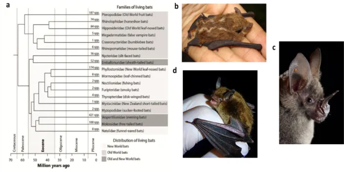
Bats emerged about 64 million years ago in the Cretaceous period (figure 6) (Teeling et al., 2005). They constitute approximately 20% of mammals species and are distributed through all continents except Antarctica, with more than 350 species divided into 3 superfamilies (Emballonuroidea, Noctilionoidea and Vespertilionoidea) (López-Aguirre et al., 2018; Simmons, 2005).
The Neotropical bats emerged about approximately 30 million years ago (Teeling et al., 2005). In the Neotropics (Biographic region that includes South and Central America, southern part of Florida and Mexico, as well as West Indies) out of 18 bats families that exist in the world, only six families are endemic in the region and only 3 occur in both the New and Old World as shown in Figure 6. The Neotropical bats are present in a vast number of ecological niches and have a wide feeding habits from insectivorous, frugivorous to nectarivorous and carnivorous (Masters, 2006; Rex et al., 2008; Simmons, 2005; Teeling et al., 2005).
Figure 6 - Phylogeny of bats. (a) The time of the bat origin is represented in million years ago and described with the
geological periods (Peixoto et al., 2018). (b) Emballonuroidae superfamily. In the image is represented the Lesser
Sac-winged bat. Access date and link: 16.07.2019 https://stricollections.org
/portal/taxa/index.php?tid=54181&taxauthid=1&clid=50. (c) Noctilionoidea superfamily represented by Lesser spear-nosed bat. Access date and link: 16.07.2019 https://www.inaturalist.org/taxa/41080-Phyllostomus-elongatus. d) Vespertilionoidea superfamily represented by Little brown bat. Access date and link: 16.07.2019 https://en.wikipedia.org/wiki /Little_brown_bat.
10 Bats are natural reservoirs for a vast number of viruses. For over 100 years, bats have associated with rabies virus and later with other human viruses, such as Hendravirus, Nipahvirus, Paramyxovirus, Filoviruses and CoV (Calisher et al., 2006). In the aftermath of the SARS- CoV epidemic, in particular, several bat CoVs have been described (Calisher et al., 2006; Chen et al., 2014). However, the number of studies yield in the Neotropical bats is very scarce and only partial sequence fragments have been acquired in all studies harboured in the Neotropics. Until now only a few studies were performed on the Neotropical bats. One was performed in bats from Trinidad and Tobago where two alphacoronavirus were described (Carrington et al., 2008), another one from Mexican bats (Anthony et al., 2013) where both beta- and alphacoronavirus clustered in 13 different clades. Other 2 studies were conducted in bats from Costa Rica, one which 5 alpha and 2 betacoronavirus were described (Corman et al., 2013) and another where several alphacoronaviruses were described (Moreira-Soto et al., 2015).
In addition, since 2009 was established a Consortium designed to address the global diversity of CoVs and over the years it has acquired partial genomes from bat CoVs from different parts of the world such as Africa, Latin America and Asia (Anthony et al., 2017; Consortium, 2014).
1.3. Aim of the Thesis
This thesis with the purpose of obtaining a Master degree in Medical Microbiology had as main goals the assessment of the CoVs diversity in South America throughout the analyzes of samples from Brazil. In addition, 2 full genomes were analyzed and characterized in order to examine the diversity of CoV from South America. One unpublished full genome from a Costa Rica Pteronotus parnellii bat CoV and another one from a Brazilian Phyllostomus discolor bat CoV obtained during the study of CoVs’ diversity.
11
12
2.1. Bat sampling and processing
2.1.1. Origin and capture of bat samples
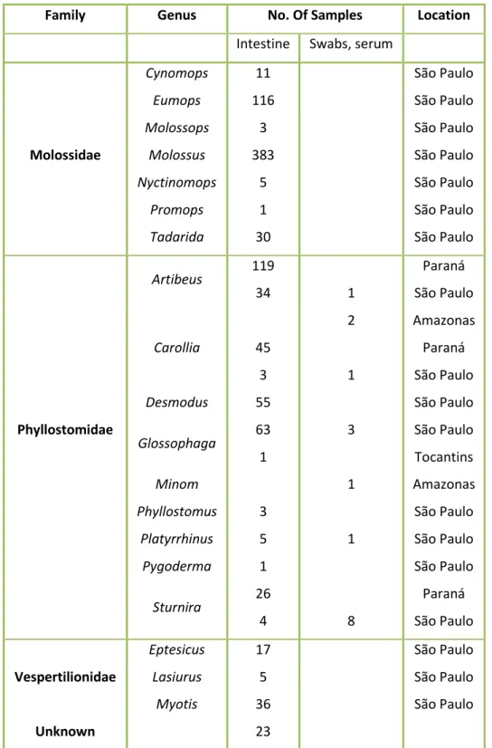
In a previous study (Goés et. al. 2016), 1004 bat samples (n=987 Intestines; n=15 rectal swabs and n=2 serum) were sampled in 4 different locations of Brazil. Out of the 1004 bat samples, 32 samples were previously screened for CoV in (Góes et al., 2016). All sampling, capture and sample transfers were done with the proper wildlife permits and ethics clearance and complied with the current laws of host countries. The samples were transported from Brazil to the Institute for Virology-Charité (Berlin, Germany) in styrofoam boxes with dry ice to prevent the samples to thaw. After receiving the samples they were stored at -80°C before further study.
2.2. Sample extraction
From 972 intestine samples that were not previously screened, approximately 30 mg was cut with the help of a sterile scalpel, Petri dishes and inside a hood, to prevent contamination of the outside of the hood and the samples. The samples were placed in a safety lock Eppendorf with 500 µL of PBS (Phosphate-Buffered Saline) and a metallic bead. The samples were homogenized using the TissueLyser II equipment (Qiagen, Hilden, Germany) at 30 Hz for 3 min. After the samples were centrifuged, pools of 8 samples were prepared whereas 35 µL of each sample was transferred to a new tube.
The pools were mixed and centrifuged using a vortex and a centrifuge, and 250 µL of the homogenate was transferred to a plate with 250 µL of Tissue Lyser buffer. The purification of the pools was performed in the MagNA Pure 96 System (Roche, Prenzberg, Germany) with elution of 100 µL. The intestine pools were purified using a Large volume kit (Roche, Prenzberg, Germany) and the swab and serum already screened using a Small volume kit (Roche, Prenzberg, Germany).
2.3. Screening assays for coronavirus
The CoV screening was divided into two parts more specifically the screening for betacoronavirus and alphacoronavirus due to the high variability and diversity among this 2 different genus.
Regarding the alphacoronavirus screening assay, all screening assays available (Anindita et al., 2015; Anthony et al., 2017; Chu et al., 2011; de Souza Luna et al., 2007; Falcón et al., 2011; Hu et al., 2018; Lacroix
13 et al., 2017; Lelli et al., 2013; Moreira-Soto et al., 2015; Quan et al., 2010; Vijgen et al., 2008; Wacharapluesadee et al., 2015; Watanabe et al., 2010) and other primers present in the Institute were evaluated using a bioinformatics approach in order to verify which assay could properly cover all diversity of alphacoronavirus. All ICTV reference sequences from the 4 CoV genera (alpha-, beta-, gamma- and deltacoronavirus) and other relevant alphacoronavirus sequences, from which complete genomes were available, were aligned using MAFFT alignment, on Geneious Prime software. The primers from each screening assay were annotated in the sequences and the primer binding regions and their mismatches were compared to all reference sequences and alphacoronavirus sequences.
The presence of betacoronavirus RNA was detected by using a heminested RT PCR (Reverse transcription PCR). A 228nt fragment from the RdRp gene was amplified using the following primers (5’-3’) (Annan et al., 2013) : Pan2cRdRP-R: GCATWGCNCWGTCACACTTAGG; Pan2cRdRp-Rnest: CACTTAGGRTARTCC CAWCCA; Pan2cRdRp-FWD: TGCTATWAGTGCTAAGAATAGRGC.
For the first PCR round and for each reaction, 2,5 µL of the purified RNA was used as a template and added to a master mix constituted by 1,55 µL of RNase free water; 6,25 µL of 2X Reaction Mix (buffer containing 0,4 mM of each dNTP; 3,2 mM of MgSO₄); 0,2 µL of MgSO₄ (50mM); 0,5 µL BSA (Bovine Serum Albumin) (1mg/mL); 0,5 µL of each Pan2cRdRp-FWD and Pan2cRdRP-R primers and 0,5 µL of Superscript III RT Mix. For the second PCR round and for each reaction, 1 µL first round product was added to the following master mix constitution: 17,15 µL of RNase free water; 2,5 µL of 10X PCR Buffer without MgCl₂; 0,5 µL of dNTP Mix (10 mM of each dNTP); 1,25 µL of MgCl₂ (50 mM); 1 µL of Pan2cRdRp-FWD; 1,5 µL of Pan2cRdRP-Rnest and 0,1 µL of Platinium Taq DNA Polymerase. The cycling protocols used for the first and second steps of the heminested RT-PCR are shown in table 1 and 2.
Step Temperature ( °C) Duration Number of cycles Reverse transcription 50oC 20 min
Initial denaturation 95oC 3 min
Denaturation 94oC 15 sec 20X Annealing 60oC* 15 sec Elongation 72oC 30 sec Denaturantion 95oC 15 sec 30X Annealing 50oC 15 sec Elongation 72oC 30 sec
Final Elongation 72oC 1 min
Table 1- Cycling protocol of the first round of the betacoronavirus screening assay.
14
The second round PCR product was analyzed and separated by gel electrophoresis. The samples were loaded into agarose gels. 2% agarose gels were done using 1x TBE buffer (TRIS-borate) and added 5 µL of Midori Green Advance DNA staining. 3 µL of the PCR product was mixed with in 2 µL of 5x gel loading dye pH 7.0. To determine the size of each band, a DNA Ladder was used as a standard reference. The gels were placed in a chamber with 1X TBE buffer and the run was done with 230V during approximately 20min. The gel with the separated bands was observed under UV-light. The PCR products that had the correct amplicon size band were sent for sequencing as explained further in section 2.5 of the Materials and Methods.
2.4. Extension of PCR product with specific primers
The 394nt screening fragment of the positive samples which were previously screened for CoV was extended to achieve the 816 nt fragment of the RdRp. Based on sequence similarities, samples were divided into 4 groups: Group I (G1) Alpha-CoV, Group II (G2) Alpha-CoV, Sturnira and Beta-CoV. For each group reverse primers were designed specifically. All primers used are listed in table 3 and were annotated in aligned sequences of the screening fragments and full genomes of the closely related CoV. The primers were verified on oligocalc online software (http://biotools.nubic.northwestern.edu/OligoCalc.html) to analyze the melting temperature and self-complementary properties.
Step Temperature ( °C) Duration Number of cycles Initial denaturation 95oC 3 min
Denaturation 94oC 15 sec 20X Annealing 60oC* 15 sec Elongation 72oC 30 sec Denaturation 95oC 15 sec 30X Annealing 50oC 15 sec Elongation 72oC 30 sec
Final Elongation 72oC 1 min
Table 2- Cycling protocol of the second round of the betacoronavirus screening assay.
15 A heminested RT-PCR was performed using two specific reverse primers, one designed in the beggining of the 394nt screening fragment achieved in the previous study of Góes et al., (2016) using the an adapted screening protocol published by Chu et al., (2011), and the second one in a region downstream of the same fragment (100nt). The consensus forward primer was previously designed by Drexler et. al. (2010). The PCR rounds for each group of samples consisted of the same quantities of each reagent, however, each assay had specific primers. The cycling protocols used for the first and second steps are shown in table 4 and 5.
Name Sequence (5'-3') Assay Tm
AlphaCoVG1 RGU R1 (10mM) TTGCAMARACACTARTACYTCTCACT Hemi-Nested G1 AlphaCoV; 1st round 56,8°C-67,9°C AlphaCoVG1 RGU R2 (10mM) GAACCMAGWATCATGGCDGAGATCAT Hemi-Nested G1 AlphaCoV; 2nd round 64,6°C-67,9°C AlphaCoVG2 RGU R1 (10mM) TAGAAWCCACCATTDGARTAMACAAC Hemi-Nested G2 AlphaCoV; 1st round 60,1°C-64,6°C AlphaCoVG2 RGU R2 (10mM) GAMCCYAAWAYCATSGCAGAGATCAT Hemi-Nested G2 AlphaCoV; 2nd round 62,9°C-67,9°C SturniraCoV RGU R1 (10mM) CCACCCGTACAATGAACAACTTC Hemi-Nested Sturnira CoV; 1st round 62,9°C SturniraCoV RGU R2 (10mM) CTTTGCACCYAGTATCATCGCTG Hemi-Nested Sturnira CoV; 2nd round 62,9°C-64,6°C BetaCoV RGU R1 (10mM) TAGAAWCCACCATTDGARTAMACAAC Hemi-Nested BetaCoV; 1st round 62,9°C-64,6°C BetaCoV RGU R2 (10mM) GAMCCYAAWAYCATSGCAGAGA CAT Hemi-Nested BetaCoV; 1st round 62°C-62°C
SP3080 (10mM) CTTCTTCTTTGCTCAGGATGGCAATGCTGC Hemi-Nested BetaCoV;
1st and 2nd rounds 72,1°C GrISP1 (10mM) TTCTTTGCACAGAAGGGTGATGC Hemi-Nested G1 and G2 AlphaCoV; 1st and 2nd rounds 62,9°C GrISP2 (10mM) CTTTGCACAAAAAGGTGATGCWGC Hemi-Nested Sturnira CoV; 1st and 2nd rounds 63,6°C-63,6°C Sequencing RGU sturnira R (10mM) TCATATTAGGCAATGCACGG Reverse primer
specific for sequencing 56,4°C
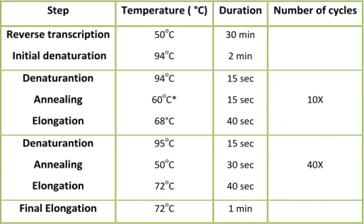
16 Step Temperature ( °C) Duration Number of cycles
Reverse transcription 50oC 30 min
Initial denaturation 94oC 2 min
Denaturantion 94oC 15 sec 10X Annealing 60oC* 15 sec Elongation 68°C 40 sec Denaturantion 95oC 15 sec 40X Annealing 50oC 30 sec Elongation 72oC 40 sec
Final Elongation 72oC 1 min
For the first round of the heminested PCR and for one reaction, 2,5 µL of the extracted RNA was added to a master mix constituted by the following reagents: 0,05 µL of RNase free water; 6,25 µL of 2X Reaction Mix; 0,2 µL of MgCl₂; 0,5 µL of BSA (1mg/mL); 0,2 µL of MgSO₄ (50mM); 0,5 µL of BSA (1mg/mL); 0,5 µL of Superscript III RT Mix and 1,25 µL of each forward and reverse primer assigned in the table 4.For the second round and for each reaction, 1 µL of first round PCR product was added to the following master mix: 17,65 of RNase free water; 2,5 µL of 10XPCR Buffer without MgCl₂; 0,5 µL of dNTP mix (10mM each); 0,75 µL of MgCl₂ (50 mM); 0,1 µL of Platinium Taq DNA Polymerase and 1,25 µL of each second-round primers assigned for each group assay. The second round PCR products were analyzed through gel electrophoresis as described in section 2.2 of the Materials and Methods.
Step Temperature ( °C) Duration Number of cycles Initial denaturation 95oC 3 min
Denaturation 94oC 15 sec 10X Annealing 60oC* 20sec Elongation 72oC 40 sec Denaturation 95oC 15 sec 40X Annealing 50oC 20sec Elongation 72oC 40 sec
Final Elongation 72oC 1 min
Table 4 - Cycling protocol for the first round of the extension of the screening fragment.
fragment.
Table 5 - Cycling protocol for the second round of the extension of the screening fragment.
fragment.
* Touchdown of -1 °C per cycle. fragment.
* Touchdown of -1 °C per cycle. Temperature decreases 1°C per cycle.
17
2.5. DNA Sequencing
The samples with bands that correspond to the expected amplicon size were sent for sequencing to Microsynth SEQLAB Sequence Laboratories Goettingen GmbH. The DNA sequencing method was based on the dye terminator sequencing method (Sanger et al., 1977). The cleanup of the samples consisted on the addiction 5 µL of the second round PCR product to 2 µL of the reagent ExoSAP and incubation during five minutes at 37°C and finally inactivation by heat at 80°C during 10 minutes. 3 µL of the ExoSAP reaction was added to 5 µL of RNase free water and 2 µL of the second round reverse or forward primer. The results were analyzed in Geneious prime software version 2019.0.4 and compared to a public nucleotide database, using BLAST from the National Center for Biotechnology Information (NCBI) (https://www.ncbi.nlm.nih.gov/).
2.6. Genome analysis
2.6.1. Genome assembling and phylogeny
A full genome from a betacoronavirus acquired from a Pteronotus parnelli bat present in the Institute and not published was characterized along with an alphacoronavirus from a Phyllostomus discolor bat acquired by Gustavo Góes during his stay at the Institute. The characterization included the prediction of the genome organization and their respective non-structural proteins (NSPs), phylogenetic analysis and species delineation to classify the viruses.
2.6.2. Prediction of the genome organization and phylogenetic analysis
For the prediction of the presumed ORFs, each genome was aligned using the MAFFT alignment (Katoh et al., 2002; Katoh & Standley, 2013). For the betacoronavirus alignment, the ICTV reference species of betacoronavirus was used and for the alphacoronavirus in addition to the ICTV reference sequences used, all alphacoronavirus sequences related to the hCoV and bat-related NL63 and 229E and the Wencheng Sm shrew CoV were used. The ORFs were predicted using the tool “Find ORF” in Geneious Prime software.
18 The predicted main ORFs were annotated and extracted along with the ORFs from the other sequences. The nucleotide sequences were translated into an aminoacid alignment. Amino acid Bayesian phylogenetic analysis for each ORF was calculated by Mr Bayes v3.1 software (Ronquist & Huelsenbeck, 2003), using WAG amino acid substitution model for amino acid Bayesian trees and 2,000,000 generations sampled every 100 steps. The amino acid trees were annotated in the TreeAnnotator v1.5 program, using a burn-in of 5000 and a posterior probability limit of 0.5. The annotated trees were visualized with FigTree v1.4 from the BEAST package (Drummond et al., 2012). The amino acid sequences were furthermore compared by BLAST comparison (Blastp) program for the analyze of the homologies. In addition, the unknown ORFs were predicted using the online software Phyre (Kelley et al., 2015).
The prediction of the TRS (Transcription Regulatory Sequences) start and end codons of each ORF and slippery sequences were done by comparison with the reference sequences.
2.6.3. Prediction of nonstructural proteins (NSPs)
For the NSPs prediction of the new genomes, the OFR1ab of both genomes were aligned with the references species sequences and the NSPs were annotated based on the amino acid comparison of the first and final amino acid residues and their position. Each NSP was extracted and translated into amino acids and realigned using MAFFT alignment. Each NSP and the prediction of the functional putative domains were compared using the Blastp feature for confirmation of the new NSPs.
2.6.4. Genome similarity plots, species delineation and detection of recombination
events
According to the ICTV, in order to assign a new species or a new genus the following criteria are used: a) A rooted phylogeny and calculation of pair-wise distances for the conserved domains of the replicase proteins pp1ab: ADRP, NSP5 (3CLpro), NSP12 (RdRp), NSP13 (Hel), NSP14 (ExoN), NSP15 (NendoU) and NSP16 (O-MT); b) The new member can be considered a representative of a new genus when it does not cluster with any of the current genera and share less the 46% amino acid sequence identity in the replicase domains mentioned above, with any other member of the family. C) New members that share more than 90% amino acid sequence identity of the replicase domains are considered to belong to the same species. Therefore, similarity plots were generated using the full genome of the new viruses and compared with the reference sequences on the SSE software version 1.3 (Simmonds, 2012) using a sliding window of 900 and a step size of 800 nucleotides.
19 The pairwise identities were calculated using MEGA 6.0 (Tamura et al., 2013) using the domains of the predicted NSPs and also the domains that were previously concatenated using Geneious Prime software. For the detection of recombination events, the nucleotide alignment of the new viruses and the reference sequences was uploaded to the RDP4 software (Martin et al., 2015) and analyzed.
20
21
3.1. Coronavirus detection in Neotropical bats
3.1.1. Screening for coronavirus
A total of 978 bat samples were collected by Gustavo Góes in different regions of Brazil: Amazonas (n=3), Paraná (n=190), São Paulo (n= 789) and Tocantins (n=1). Figure 7 and Table 6 represents the location and number of samples collected in each area and a list of samples used for this study.
Family Genus No. Of Samples Location
Intestine Swabs, serum
Molossidae
Cynomops 11 São Paulo
Eumops 116 São Paulo
Molossops 3 São Paulo
Molossus 383 São Paulo
Nyctinomops 5 São Paulo
Promops 1 São Paulo
Tadarida 30 São Paulo
Phyllostomidae Artibeus 119 Paraná 34 1 São Paulo Carollia 2 Amazonas 45 Paraná 3 1 São Paulo
Desmodus 55 São Paulo
Glossophaga 63 3 São Paulo
1 Tocantins
Minom 1 Amazonas
Phyllostomus 3 São Paulo
Platyrrhinus 5 1 São Paulo
Pygoderma 1 São Paulo
Sturnira 26 Paraná
4 8 São Paulo
Vespertilionidae
Eptesicus 17 São Paulo
Lasiurus 5 São Paulo
Myotis 36 São Paulo
Unknown 23
Table 6 - List of samples used in the project
aThe unknown samples correspond to intestines where the identification of the species and the
22
Figure 7 - Location and number of the samples collected as well as the bat species in which the full genomes where
detected in the study. (a) Locations and number of the samples collected in Brazil. The number in parenthesis corresponds to the number of samples collected from bats. Amazonas n=3; Paraná n=189; São Paulo n=785 and Tocantins n= 1. (b) Phyllostomus discolor bat. Also known as Pale spear-nosed bat from the Phyllostomidae family is
present in South and Central America. Access date and webpage: 15.07.2019
https://en.wikipedia.org/wiki/Pale_spear-nosed_bat. (c) Pteronotus parnellii bat. Also known as Parnell’s mustached bat is a bat species that belongs to the Mormoopidae family is present in North, Central and South America. Access date and webpage: 15.07.2019 https://www.flickr.com/photos/23556954@N07/8709342848 (Simmons, 2005; Solari, 2016).
972 intestine samples were pooled into 122 pools of 8 and were screened for betacoronavirus. Out of the 122 pools, only 4 pools had a band with the corrected amplicon size after visualization in agarose gel. The PCR products were sent for sequencing and all were negative for betacoronavirus.
Recently many novel bat alphacoronavirus were detected. Regarding the alphacoronavirus screening assays, all published screening assays were analyzed in order to observe if some screening assay could cover all diversity of alphacoronavirus. An alignment was built with all ICTV reference sequences from the 4 CoV genera (alpha-, beta-, gamma- and deltacoronavirus) and with other important alphacoronavirus sequences for which complete genomes were available.
The primers from each screening assay were annotated and the primer binding regions of each screening assay and their mismatches to all reference alphacoronavirus were compared with the references. However, no screening assay was able to cover the alphacoronavirus’ diversity due to the fact that the
23 primers used in the published screening assays only matched with some sequences and not the majority used in the allignment. Therefore, I started to develop a new assay but due to lack of time, the samples were not tested in this study.
3.1.2. Elongation of the screening fragment
In order to achieve the 816nt fragment from the RdRp, 35 already positive samples for CoV that were obtained by in Góes et al. (2016) were divided into 4 groups: Group I (G1) CoV, Group II (G2) Alpha-CoV, Sturnira and Beta-CoV (Figure 8) and the RdRp fragment of 394bp was amplified in a heminested RT-PCR. For the Group 1 AlphaCoV (G1) (n=14), the 816nt fragment was obtained only from 11 samples. For the Group 2 AlphaCoV (G2) (n= 7), 5 samples had the correct amplicon size. Regarding the Sturnira Group (n=11), 3 RdRp fragments were amplified with the correct amplicon size and from the Beta Group (n=2) no amplification was achieved. Regarding the sequencing step for the samples from which the RdRp fragment was amplified (n=17), the sequencing results did not show results for CoV except for one sample from G1 AlphaCoV: Phyllostomus discolor intestine sample.
24
BatCoV Phy dis CCZ50 2014
JQ731782 BtCoV KP816 Phy dis PAN 2011 BatCoV Mim cre 2111 BRA 2014 DF
BatCoV Glo sor 100 BRA 2014 EU769558 BatCoV 1CO7BA Glo sor TRI 2007
BatCoV Glo sor 163 DEPAVE 2013 BatCoV Glo sor 137 DEPAVE 2013 BatCoV Glo sor 158 DEPAVE 2013
BatCoV Stu lil 150SR DEPAVE 2013
JQ731787 BtCoV KP534 Art lit PAN 2010
BatCoV Art lit 157 DEPAVE 2013
JQ731785 BtCoV KP524 Art jam PAN 2010 JQ731793 BtCoV KCR216 Car per CRC 2010
JQ731794 BatCoV 100 Car per BRA 2009 EU769557 BatCoV 1FY2BA Car per TRI 2007
BatCoV Car per 1599 BRA 2012
BatCoV Car per 2079 BRA 2014 DF BatCoV Car per 2087 BRA 2014 DF
BatCoV Car per 1514 BRA 2011 BatCoV Car per 1516 BRA 2011
NC005831 HCoV NL63
KT253296 229E-related BtCoV/KW1E-F77 Hip aba GHA 2011 NC002645 HCoV 229E
JQ410000 Alpaca respiratory coronavirus isolate CA08-1 2008 FJ710046 BatCOV Hip Gha 19 2008
KT253269 229E-related BtCoV/KW2E-F151 Hip cf. rub GHA 2011 KT253272 229E-related BtCoV AT1A-F1 Hip aba GHA 2010
BatCoV Mol ruf 092 BRA 2013 BatCoV Mol ruf 63 BRA 2014 KC886321 BatCoV 28 Mol ruf BRA 2010
JQ731800 BatCoV 103 Mol cur BRA 2009 JQ731799 BatCoV 182 Mol ruf BRA 2009 HQ184057 BatCoV I Myo myo SPA 2007
EU834951 BatCoV CoV34 Myo mac AUS 2008
BatCoV Art lit 1816 BRA 2010
JQ731784 BtCoV KP256 Art jam PAN 2010
BatCoV Art lit 2294 BRA 2012
DQ249224 BatCoV Myo ric HKU6 CHN EU375866 BatCoV D7 Myo dau GER 2007 DQ648858 BatCoV 512 Sco kuh CHN 2005 KF430219 BatCoV CDPHE15 Myo luc USA 2006
NC003436 PEDV
KC110771 BatCoV POA127 Mol Tad BRA 2012
BatCoV Myo nig CCZ17 BRA
BatCoV Myo nig 117 BRA 2013 BatCoV Myo rip 259 BRA 2013 NC010437 BatCoV 1A
DQ249226 BatCoV Min mag HKU7 CHN 2006 NC010438 BatCoV Min pus HKU8 CHN 2006
AF124987 FIPV AF124986 Canine CoV DQ811787 PRCV ISU 1 DQ811789 TGEV
NC023760 MinkCoV WD1127
BatCoV Stu lil 1613 BRA 2012 BatCoV Stu lil 314 DEPAVE 2014 BatCoV Stu lil 313 DEPAVE 2014
BatCoV Stu lil 1573 BRA 2012
BatCoV Stu lil 1617 BRA 2012 BatCoV Stu lil 150SW DEPAVE 2013 BatCoV Stu lil 152 DEPAVE 2013 BatCoV Pla lin 162 DEPAVE 2013 BatCoV Car per 171 DEPAVE 2013
DQ249235 Bat CoV HKU2-1 AF220295 BCoV Quebec NC005147 HCoV OC43 AY597011 HCoV HKU1 A EF065513 BatCoV Rou lec HKU9 CHN
NC004718 SARS CoV
KC881005 BatCoV SARSlike RsSHC014 KC886322 BatCoV 49 Pte dav MEX 2012
BatCoV Art lit 2064 BRA 2011
KC633197 BatCoV KCR260 Car per CR 2012 KC545386 CoV Erinaceus VMC DEU 2012
DQ249214 BatCoV Tyl pac HKU4 CHN DQ249217 BatCoV Pip abr HKU5 CHN HQ184062 BatCoV M Ept isa ESP 2007 KF312399 BatCoV Ept ser 384 ITA 2012
KJ473821 BatCoV Ves sup SC2013 CHN 2013 HQ184059 BatCoV Hyp sav J ESP 2007
KF500950 BatCoV 33037515 Pip kuh ITA 2012 BatCoV Eum gla 242 BRA 2013 KC243390 BatCoV 8724 Pip pyg ROU 2009 KC869678 BatCoV PMLPHE1 Neo cap RSA 2011
JX869059 CoVMERS EMC 2c 2012
KJ477102 CoVMERS Cam drom NRCEHKU205 EGY 2013
Phyllostomus α-CoV Glossophaga α-CoV I Glossophaga α-CoV II Artibeus α-CoV I Carollia α-CoV Molossus α-CoV Artibeus α-CoV II Myotis α-CoV Mimon α-CoV
25
Figure 8 - Phylogenetic tree of the 394bp of the RdRp of positive samples. The positive samples are coloured according
to their groups. The Phyllostomus discolor partial sequence from de RdRp is represented in green. Group I AlphaCoV (G1) (n=14), Group II AlphaCoV (G2) (n= 7), Sturnira Group (n=11), Beta Group (n=2). This tree was kindly supplied by Gustavo Goés.
3.2. Genome annotation
For this study, 2 full genomes were annotated and analyzed: A betacoronavirus unpublished previously discovered in the bat species Pteronotus parnellii (Pte-BetaCoV) (Figure 9) and another one, corresponding to an alphacoronavirus found in Phyllostomus discolor (Phyl-AlphaCoV) the sequence of which was completed in the course of this study. Phyl-AlphaCoV was obtained by Gustavo Goés using Illumina sequencing method whereas the Pte-BetaCoV kindly made available by Andrea Rasche, was acquired by Illumina HiSeq2500.
26
Figure 9 - Bayesian tree of the 816nt gap free of the translated RdRp fragment sequences. Pte-BetaCoV is represented
in a blue underline (Corman et al., 2013).
3.2.1. ORF prediction
The Pte-BetaCoV genome was aligned with the ICTV reference sequences and the Phyl-AlphaCoV was aligned with the ICTV reference sequences and with all alphacoronavirus related to the hCoV NL63 and 229E, related bat CoV 229E and Nl63 and Wencheng Sm shrew CoV. The ORFs were predicted using the Geneious prime software and by comparison with the found ORF with the annotated ORFs from the reference sequences.
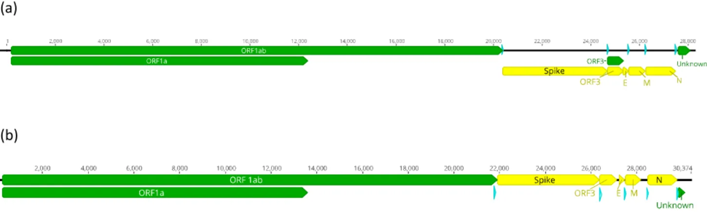
For the Phyl-AlphaCoV genome, 7 ORFs were predicted (Figure 10a): ORF1ab, Spike gene, ORF3, E, M, N and 1 unknown ORF. The ORFs were compared with the starting and end amino acid positions of the reference sequences and also with the presence of a TRS (Transcription regulatory sequence) upstream of the beginning of each ORF. The ORFs sequences were analyzed by BLAST to observer there homology with other sequences. For the Pte-BetaCoV genome (Figure 10b), the ORFs were predicted using the same methodology for the prediction of the Phyl-AlphaCoV ORFs. The ORF 1ab, Spike gene, ORF3, E, M, N and also 1 unknown ORF were predicted. The ORF3 of this virus presented similarities with an Eidolon bat coronavirus (accession number: ADX59467) (Tao et al., 2012).
The amino acid sequences of the unknown ORFs in both genomes were blasted (blastp) and predicted using the online software Phyre and no result consisting of the putative OFRs function appeared.
(a)
(b)
Figure 10 - Prediction of the genome organization of the complete genomes. In blue is represented the TRS. (a)
Prediction of the organization of the Phyl-AlphaCoV genome. (b) Prediction of the organization of the Pte-BetaCoV genome.
27
3.2.2. TRS and ribosomal frameshift prediction
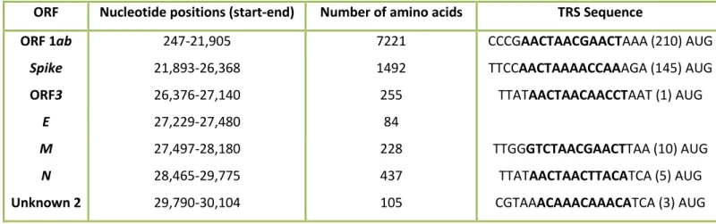
The Transcription Regulatory Sequence (TRS) was annotated based on the comparison of the TRS of the reference sequences. All the TRS found in both genomes were located upstream of each ORF. Table 7 and 8 contains the position and sequence of the putative TRS of both genomes. The leader TRS was only predicted for the Pte-BetaCoV and the TRS-B was found in 6 locations upstream of the Pteronotus ORFs and in 5 locations upstream of the Phyl-AlphaCoV ORFs.
ᵃ Number in brackets represents the number of nucleotides to the putative start codon.
ᵇ The blank spaces in the table represent the absence of a putative TRS for the ORF in question. In ORF1ab no TRS was found due to the missing of part of the initial genome sequence.
c
The TRS is represented in bold.
ORF Nucleotide positions (start-end) Number of amino acids TRS Sequence
ORF 1ab 179-20,376 6732 No data
Spike 20,383-24,681 1433 TCTCAACTAAGTGAAA(9) AUG
ORF3 24,684-25,340 219 CGTCAACTAAACATGT(0)AUG
E 25,324-25,545 74
M 25,552-26,244 231 ACGTCTAAACGAAGA(4)AUG
N 26,261-27,493 411 AATCAACTAAAAACA(6) AUG
Unknown 1 27,496-27,984 161 TCTCAACTAAAAATGC (1) AUG
ORF Nucleotide positions (start-end) Number of amino acids TRS Sequence
ORF 1ab 247-21,905 7221 CCCGAACTAACGAACTAAA (210) AUG
Spike 21,893-26,368 1492 TTCCAACTAAAACCAAAGA (145) AUG
ORF3 26,376-27,140 255 TTATAACTAACAACCTAAT (1) AUG
E 27,229-27,480 84
M 27,497-28,180 228 TTGGGTCTAACGAACTTAA (10) AUG
N 28,465-29,775 437 TTATAACTAACTTACATCA (5) AUG
Unknown 2 29,790-30,104 105 CGTAAACAAACAAACATCA (3) AUG
Table 7 - Putative Transcription Regulatory Sequences (TRS) of the Phyl-AlphaCoV.
28
ᵃ Number in brackets represents the number of nucleotides to the putative start codon.
ᵇ The blank spaces in the table represent the absence of a putative TRS for the ORF in question. In ORF1ab no TRS was found due to the missing of part of the initial genome sequence.
c
The TRS is represented in bold.
In both genomes, the ribosomal frameshift sequence present in the ORF1ab was predicted based on the slippery sequence found in the literature and in the reference sequences. The ribosomal frameshift sequence was annotated by the comparison of the amino acid frame of each ORF and the position of the stop codons in the reference and annotated sequence.
In the Phyl-AlphaCoV genome, the slippery sequence 5’-UUUAAAC-3’ is located at the nucleotide positions 12,322 to 12,328 and in the Pte-BetaCoV genome, the slippery sequence with the same nucleotides was located in the nucleotide positions 13,570 to 13,575.
3.2.3. ORF phylogenetic analysis
Bayesian analysis of the ORFs 1ab, S, E, M and N were performed including all ICTV reference sequences. For the alphacoronavirus alignment, all viruses related to the hCoV NL63 and 229E and Wencheng Sm shrew CoV were additionally included. The addition of these viruses was due to the fact that preliminary phylogenetic analysis of the S ORF demonstrated that the Spike protein of Phyl-AlphaCoV clustered with NL63 and 229E related viruses. Therefore, all 229E and NL63 viruses were added. The Wencheng Sm shrew virus was also added to complement the diversity of alphacoronaviruses due to the fact that this virus is a divergent virus within this genus.
For the Phyl-AlphaCoV analysis, the gammacoronavirus Infectious bronchitis virus (IBV) was used as the outgroup for all the ORF analysis. The IBV was used instead of a betacoronavirus due to the fact that the S ORF of Rhinolophus bat CoV HKU2 presents similarities with SARS-related CoVs due to recombination events (Lau et al., 2007). The amino acid trees are presented in Figure 11. The amino acid trees of the ORFs 1ab and S showed supported basal nodes. However the amino acid trees of the ORFs E, M and N were not supported. In the ORF1ab phylogeny, the ORF1ab from Phyl-AlphaCoV clusters distantly with the other sister's clades of this group. However, The ORF S phylogeny shows that the Spike protein clusters with the group of the hCoV NL63 and 229E along with their respective related bat CoV.
30
Figure 11 - Bayesian phylogenies of the major ORFs of the Phyl-AlphaCoV (Green). All bat CoV are shown in bold. The
gammacoronavirus Infectious bronchitis virus (IBV) was used for rooting the trees. The collapsed branches include all complete genomes related to the clade NL63/229E. All complete genomes and their accession number are displayed in Table S1 in the Supplementary section. The Bayesian trees were calculated using a WAG amino acid substitution model. Values with a posterior probability above 0.95 are shown in black dots and values between 0.90 and 0.95 are shown in grey dots.
31 For the Pte-BetaCoV, the human alphacoronavirus NL63 was used to root the Bayesian phylogenetic trees as shown in Figure 12. OFRs E and M were statistically supported (not all nodes present a posterior probability superior to 0.90) whereas the other major ORFs were fully supported and the Pte-BetaCoV clustered distantly with clade C betacoronavirus group where the hCoV MERS, Erinaceus BetaCoV, HKU5 and HKU4 belong.
32
Figure 12 - Bayesian phylogenies of the ORFs of the Pte-BetaCoV (Blue). All bat CoV are shown in bold. The human
alphacoronavirus NL63 was used for rooting the trees. The Bayesian trees were calculated using a WAG amino acid substitution model. Values with a posterior probability above 0.95 are shown in black dots and values between 0.90 and 0.95 are shown in grey dots.
3.2.4. Prediction of nonstructural proteins (NSPs)
To further delineate the characterization of the Phyl-AlphaCoV and Pte-BetaCoV genomes, the prediction of the putative polyprotein pp1a/1ab cleavage sites and their non-structural proteins (NSPs) was performed. Both genomes were aligned with the correspondent reference sequences. The locations of the 16 NSPs was done by comparison to the locations of the reference NSPs sequences and by the locations of other NSPs of genomes characterized in the literature. In addition, the N- and C-terminal amino acid residues of each new coding sequence were compared to the N- and C-terminal amino acid residues of the NSPs of the reference sequences. All NSPs from both genomes present a similar amino acid length to the reference NSPs however the nucleotide position of the NSPs- coding sequences differs in some cases. The prediction of the putative functional domains of the NSPs was done by associating the putative NSPs to the main function of the NSPs of the reference sequences and the function described in the literature. Table 9 and 10 provides the information about the protein length, location of the first and end amino acid residues and their putative functional domains.
33
Table 9 - Prediction of the putative pp1a/pp1ab cleavage sites of Phyl-AlphaCoV based on the comparison with the
ICTV reference sequences. The putative function of each domain is based on the ICTV.
ᵃ Superscript number represents the positions of the nonstructural proteins in the polyprotein pp1a/pp1ab with the assumption of the ribosomal frameshift based on the slippery sequence (5’-UUUAAAC-3’) present in CoV. The underlined NSPs indicate the NSPs used for the speciation criteria of the ICTV.
ᵇ IFN, Interferon; ADRP, ADP-ribose 1-phosphatase; PL1 pro, papain-like protease 1; PL2 pro, papain-like protease 2; 3CLpro, 3C-like main protease; RdRp, RNA-dependent RNA-polymerase; RTC, replicase/transcriptase complex; HEL, helicase; NTPase, nucleoside triphosphatase; ExoN, 3’-to-5’ exoribonuclease; NMT, N7 methyltransferase; NendoU, nidoviral uridylate-specific endoribonuclease; OMT, ribose-2’-O-methyltransferase.
c
The blank spaces represent the unknown function of the corresponded NSPs.
NSP 1st amino acid residue-last amino acid residue
Protein size (no. of amino
acids)
Putative functional domain(s)
NSP 1 Met¹-Gly110 110 IFN antagonist; Degradation of host mRNA; Inhibition of translation; Cell cycle arrest
NSP 2 Asn111- Gly777 667
NSP 3 Gly778-Gly2449 1,672 ADRP;PL1pro; PL2pro
NSP 4 Ala2450-Gln2934 485
NSP 5 Ser2935-Gln3237 303 3CLpro
NSP 6 Cys3238-Gln3515 278
NSP 7 Ser3516-Gln3598 83 ssRNA binding
NSP 8 Ser3599-Gln3793 195
Noncanonical "secondary" RdRp with putative primase activity; forms hexadecameric
supercomplex with NSP7 NSP 9 Asn3794-Gln3902 109 ssRNA binding; associates with RTCs
NSP 10 Ala3903-Met4036 134
Dodecameric zinc finger protein; associates with RTCs, simulates NSP 16 methyltransferase
activity
NSP 11 Ser4038-Asp4056 19 Short peptide at the end of ORF1a
NSP 12 Gln4037-Gln4966 930 RdRp
NSP 13 Ser4967-Gln5563 597 Hel, NTPase
NSP 14 Ala5564-Gln6082 519 ExoN,NMT
NSP 15 Gly6083-Gln6429 347 NendoU