Nuno José Prego Ramos
Licenciado
Crosslinked Hyaluronic Acid nanoparticles
as delivery vehicles for dendritic
cell-targeted vaccines
Dissertação para obtenção do Grau de Mestre em
Bioquímica para a Saúde
Orientadora: Paula Videira,PhD, NOVA Medical School e
Faculdade de Ciências e Tecnologia, Universidade NOVA de Lisboa Co-Orientador: Vítor Espirito Santo, PhD, Instituto de Biologia Experimental e Tecnológica
(iBET)
Março 2016
Nuno José Prego Ramos
Licenciado
Crosslinked Hyaluronic Acid nanoparticles
as delivery vehicles for dendritic
cell-targeted vaccines
Dissertação para obtenção do Grau de Mestre em
Bioquímica para a Saúde
Orientadora: Paula Videira,PhD, NOVA Medical School
e
Faculdade de Ciências e Tecnologia Universidade NOVA de Lisboa Co-Orientador: Vítor Espirito Santo, PhD, Instituto de Biologia Experimental e Tecnológica
(iBET)
Júri:
Presidente: Prof. Doutor(a) Nome Completo Arguente(s): Prof. Doutor(a) Nome Completo
Vogal(ais): Prof. Doutor(a) Nome Completo
[Local de realização das provas públicas de defesa da dissertação]
Março 2016
Acknowledgements
First and foremost I would like to thank Professor Paula Videira for the possibility of being part of a team of such brilliant people. I am very thankful for her guidance and orientation.
I am very thankful to Dr. Vítor Espiríto Santo, ITQB/iBET for the support and mentoring provided.
I cannot thank enough to Dr. Zélia Silva (CEDOC) for her time, patience and encouragement.
I owe my gratitude to the coordination of the Masters in Biochemisty for Health, specially to Professor Teresa Catarino, Professor Sebastião Rodrigues and Professor Pedro Matias, for the opportunity given.
I wish to acknowledge Eva Balslev Jørgensen (Novozymes). Her cooperation was fundamental.
I would like to single out my colleague Francisca Arez for the availability and friendship. Her support in the hardest times was very important.
I would like to thank to all my colleagues for the encouragement and motivation, you were very fun but also inspiring.
I am very blessed and grateful for having a family that encourages me always and is willing to go the path: my children, Vasco e Beatriz for the love and understanding; my wife who is my other half in all dimensions; my mother and in laws for always believing and being there for me; my brothers Sérgio e André for the confidence instilled.
Abstract
The immune system has been the target of the most recent advances in cancer treatment. Immunoconjugates, targeted therapies and immunotherapies are the most researched class of products for the treatment of cancer with many drug candidates currently in pre-clinical and late clinical stages of development. Therapies focused on Dendritic cells (DCs) are very promising since DCs are antigen presenting cells and that turns them into interesting targets for controlling the immune response in the presence of cancer cells.
Considering that antigens overexpressed by cancer cells could be presented to DCs inducing a protective anticancer immune response, it is important to assess the vehicle with affinity to DCs, as well as finding the right biomaterial to transport the antigens (or fractions of the antigen) to the surrounding environment where DCs can be found and where maturation can be induced.
We have hypothesized the development of a nanoparticle system for the delivery and targeting of the drug candidate, mainly an antigen or fractions thereof, which can be formulated using a biodegradable polymer, a non-toxic vehicle that can improve bioavailability of the biologic agent, increasing the targeting capabilities of a pharmaceutical formulation. Hyaluronic acid (HA) was the biopolymer chosen due to its viscoelastic properties, affinity to the surface receptors of DCs such as CD44 and to Toll-like receptors inducing migration and activation of T lymphocytes in the region nodes. The main goal of this thesis was to find an effective method of producing HA nanoparticles through a novel crosslinking method, characterizing the nanoparticles by Nanoparticles Tracking Analysis, measuring particles size, concentration and size distribution. Later, the toxicity of the nanoparticles system was evaluated using Peripheral Blood Mononuclear Cells (PBMCs) upon nanoparticles presentation (by Neubauer chamber and Flow cytometry analysis).
Our findings show that it is possible to produce nanoparticles according to the proposed methodology and that the nanoparticulate system does not induce any cytotoxic activity as shown in the assays performed.
Resumo
O sistema imunitário tem sido o alvo dos mais recentes avanços no tratamento do cancro. Imunoconjugados, terapias direcionadas e imunoterapias são as classes de medicamentos mais investigados no tratamento do cancro, com muitos candidatos a fármacos atualmente em fase de desenvolvimento pré-clínico e fase final de desenvolvimento clínico. Terapias focadas em células dendríticas (CDs) são muito promissoras, uma vez que as CDs são células apresentadoras de antigénios, o que as transforma em alvos interessantes para controlar a resposta imune aquando da presença de células cancerígenas.
Considerando que os antigénios sobre expressos pelas células cancerígenas podem ser apresentados às CDs induzindo uma resposta imune protetora contra o cancro, é fundamental investigar um veículo de transporte com afinidade para as DCs, bem como definir o biomaterial certo para transportar os antigénios (ou frações do antigénio) para o ambiente circundante, onde as CDs existem e onde pode ser induzida a sua maturação. Hipotetizámos o desenvolvimento de um sistema de nanopartículas para a entrega e direcionamento de um candidato terapêutico, nomeadamente um antigénio ou suas frações, numa formulação utilizando um polímero biodegradável, um veículo não tóxico que pudesse melhorar a biodisponibilidade do agente biológico, aumentando as capacidades de segmentação de uma formulação farmacêutica. O ácido hialurónico (AH) foi o biopolímero escolhido devido às suas propriedades viscoelásticas, a sua afinidade para os recetores de superfície das CDs, como o CD44 e para os recetores TLR induzindo assim a sua ativação e consequente migração para os linfócitos T nos gânglios regionais. O principal objetivo desta tese foi encontrar um método eficaz de produzir nanopartículas de AH através de um novo método de reticulação, procedendo à sua caracterização por Nanoparticle Traking Analysis, obtendo informação relativa à dimensão das partículas, concentração e distribuição das dimensões. A toxicidade do sistema de nanopartículas foi avaliada posteriormente utilizando PBMCs e analisada por câmara de Neubauer e citometria de fluxo.
A nossa pesquisa demonstra que é possível produzir nanopartículas de AH de acordo com o método proposto, e que este sistema não parece induzir qualquer efeito citotóxico, tal como demonstram os resultados dos ensaios realizados.
1 INTRODUCTION 1
1.1 The immune system – Overview 3
1.1.1 Dendritic Cells - Background 4
1.2 DCs Targeted Vaccines 5
1.3 Use of nanotechnology for targeting Dendritic Cells 7
1.4 The development of nanodrugs – Background and review 9 1.4.1 Methods of production of nanoparticles systems 10
1.5 Polymer based nanoparticles delivery systems 11
1.5.1 Hyaluronic Acid 11
1.5.2 HA capability of inducing immune activation 13 1.5.3 Methods of producing HA-Nanoparticles 13
1.6 Context and aims of the project 15
2 MATERIALS AND METHODS 17
2.1 Materials and equipment 19
2.2 Optimization of the method of preparation of HA-Nanoparticles 20 2.2.1 Method of preparation of Hyaluronic Acid-OVA-FITC-Nanoparticles 20 2.2.2 Method of preparation of Hyaluronic Acid – BSA -Nanoparticles 21
2.3 Nanoparticles Tracking Analysis – Method introduction 22 2.3.1 NTA – HA Nanoparticles characterization assays 23
2.4 Assays to test cell viability in PBMCs using HA-NPs 24 2.4.1 Cytotoxicity assessment of HA-NPs in cell culture using Neubauer chamber 25 2.4.2 Cytotoxic assessment of HA-NPs in cell culture by Annexin V/7AAD and
analysis by flow cytometry 25
3 RESULTS 27
3.1 Nanosystem characterization using Nanoparticles Tracking Analysis 29
3.2 Influence of HA-NPs in cell viability during 24h and 48h periods measured by trypan blue staining through cell counting method using Neubauer chamber 33
3.3 Influence of HA-NPs in cell viability during 24h and 48h periods through labelling with Annexin and 7AAD analysed by flow cytometry 34
XII
4 DISCUSSION 37
4.1 General discussion of the results 39
5 CONCLUSION 41
5.1 Conclusion and Future Perspectives 43
Index of Figures
Figure 1. MUC1 protein expressing several types of novel cancer-associated antigenic epitopes in cancer cells (adapted from Acres and Limacher 2005) ... 6 Figure 2. The size range of nanoparticles used in nanovaccinology. Adapted from Zhao et al Vaccine 32 (2014) 327– 337 ... 7 Figure 3. Time line of clinical stage nanomedicines. Adopted from Chem Soc Rev. Kamaly,N. et al. (2012) ... 10 Figure 4. Hyaluronic acid molecular structure (Adapted from Novozymes) ... 12 Figure 5. 1.3-Dihydroxyacetone dimer molecule (Retrieved from Sigma Aldrich MSDS) ... 15 Figure 6. NTA analysis of particle size of HA-OVA-FITC- NPs for the samples containing 0,150ml of protein in a ratio of 1:1 to HA. 20% of crosslinking agent was used. ... 29 Figure 7. NTA analysis of particle size of HA-OVA-FITC- NPs, for the samples containing 0,150ml of protein in a ratio of 1:1 to HA. 40% of crosslinking agent was used. ... 29 Figure 8. NTA analysis of particle size of HA-BSA- NPs for the samples containing 0.150ml of protein in a ratio of 1:1 to HA. 20% of crosslinking agent was used. ... 30 Figure 9. - NTA analysis of particle size of HA-BSA- NPs, for the samples containing 0.150ml of protein in a ratio of 1:1 to HA. 40% of crosslinking agent was used. ... 30 Figure 10. NTA analysis of particle size of HA- NPs, for the samples without protein. 40% of crosslinking agent was used ... 30 Figure 11. NTA analysis of particle size of HA- NPs, for the samples without protein. 20% of crosslinking agent was used. ... 30 Figure 12. Cell counting using Neubauer chamber. Results show no significant influence in cell viability of HA-NPs containing samples over the periods of 24h and 48h. ... 33 Figure 13. Cell viability analysis performed by flow cytometry (n=1). This graph shows that HA-NPs produced with our crosslinking agent (DHA 40%) does not induce any significant cytotoxic effect when compared with the control group in 24h and 48h samples ... 34
XIV
Figure 14. Representative dot plots of cell viability assays performed by Flow cytometry. A and B FSC vs SSC dot plots of PBMCs where it is shown the gate acquired cells, excluding and debris after 48h. C and D. annexin V vs 7AAD staining of R1 gated cells. A) and C) control sample B and D) 100ug/mL HA-NPs sample 48h.. ... 35
Index of Tables
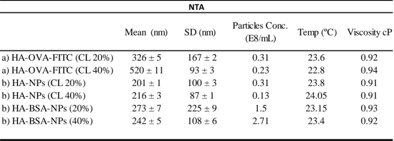
Table 1. Mean Size and Size Distribution of NPs from NTA measurements
... 32
Abbreviations
APC(s) Antigen presenting Cell(s)
BSA Bovine serum albumin
CCD Charge-coupled device
CL Crosslink(ed)
DC(s) Dendritic cell(s)
DDS Drug delivery system(s)
DHA Dihydroxyacetone
DVS Divinyl sulfone
ECM Extracellular matrix
FITC Fluorescein isothiocyanate
GAG(s) Glycosaminoglycans
HA Hyaluronic acid
HPMA N- (2-hydroxypropyl) methacrylate
ISCOM Immunostimulating complex
MHC Major histocompatibility complex
MLR Mixed lymphocyte reaction
MRI Magnetic resonance imaging
NOD-like nucleotide-binding oligomerization domain receptors
NP(s) Nanoparticles
NTA Nanoparticle tracking analysis
OVA Ovalbumin
PAMP(s) Pathogen associated molecular pattern(s)
PBMC(s) Peripheral blood mononuclear cell
PEG polyethylene glycol
PKR Protein kinase receptor
PLA Poly lactic acid
PLGA Poly (D, L-lactic-co-glycolic acid)
PRR(s) Pathogen recognition receptors
RHAMM Hyaluronan-mediated motility receptor
RNA Ribonucleic acid
XVIII
SPR Surface plasmon resonance effect
AFM Atomic force microscopy
TEM Transmission electron microscopy
TLR(s) Toll-like receptor(s)
TNFIP6 Tumor necrosis factor-induced protein-6
1.1The immune system – Overview
The immune system has the ability to protect the body against different aggressions, in particular against external antigenic threats, either by microorganisms or macromolecules, or against internal aggressions. The immune system response can be divided into two categories: innate immunity and adaptive immunity. The induction of an adaptive response begins when a pathogen is ingested by an immature Dendritic cell (DC). DCs are professional antigen presenting cells (APCs) specialized in triggering and regulating the adaptive response (Banchereau and Steinman, 1998). They have the ability to recognize antigens through surface receptors such as Toll-like receptors (TLRs) (Akira, 2011). When immature DCs take up a pathogen they become activated and migrate into the regional lymph nodes in order to activate pathogen-specific lymphocytes (Benencia et al, 2012;. Banchereau et al., 2000).
By identifying the antigens overexpressed in cancer cells it might be possible to isolate certain parts of the antigen (eg.peptides) with the objective of presenting them to DCs in order to develop immune responses to cancer cells. These features have led DCs to have become a possible target for tumour treatment (Benencia et al., 2012) mainly in cancer immunotherapies. Cancer immunotherapies use the immune system to eliminate or neutralize tumor cells, targeting the factors that promote tumour survival. Several strategies focusing on adaptive immunity have been developed in order to stimulate antitumor immune responses, such as anti-tumor vaccination, adoptive transfer of immune cells (lymphocytes) and inhibition of immune suppression (Sakaguchi, Shimon et al. 2009). Anti-tumour vaccines are based on the mobilization of immune cells, amplifying the immune response that recognizes and neutralizes cancer cells. Despite the different types of vaccines, our work will be focused on Dendritic cell- targeted vaccines. They represent a method in which the DCs are used as APCs specific for tumour antigens (Engleman, 1997).
4
1.1.1 Dendritic Cells - Background
The first time that cells with dendritic morphology present in the skin were observed, dates back to 1868 by Paul Langerhans, a german medical student. At that time it was thought that these cells were part of the nervous system when in fact they are currently known as epidermal dendritic cells or Langerhans cells. Despite this discovery, the true nature and function of these cells, remained a mystery for more than 100 years. In the theory of clonal selection proposed by Frank Macfarlane Burnet in 1957 it was postulated that the cells proliferate in response to antigen only when the antigen binds to its receptor, but this theory did not explain how the presented antigen triggered the immune response. Ralph M. Steinman, set out to investigate which agent enabled the presentation of antigen to initiate the lymphocytic immune response as he observed that by adding specific antigens to lymphocytes no immune response was achieved. Robert Richard and Dutton Mishell published in 1966 a study showing the first successful antibodies primary response in vitro by adding sheep red blood cells to a suspension of mouse spleen cells (Dutton et al. 1967). In 1970 Ralph M. Steinman and Zanvil A. Cohn began researching these cells in mouse spleen and discovered, using phase contrast microscopy, a sparse population of cells with multiple ramifications, mobile and rich in mitochondria. Given its peculiar morphology, both researchers first used in 1973 the term "dendritic cells" (DCs) to refer to these cells (Steinman et al. 1974). During the 70s, most immunologists considered macrophages as the primary antigen presenting cell (APC) of the immune system and the hypothesis proposed by Steinman on the key role of DCs in the generation of the immune response was not widely considered. Steinman and other researchers characterized the proteins expressed on the surface of the DCs (Steinman et al. 1974), which proved to be key to determining the role of DCs. One of the most significant findings was the high protein expression of the major histocompatibility complex (MHC), which later proved to be necessary for antigen presentation to T lymphocytes. After a series of experiments based on the mixed lymphocyte reaction (MLR), Steinman was able to show that DCs were 100 to 1000 times more efficient to initiate the immune response than generic spleen cells (Nussenzweig and Steinman et al. 1980).
Since DCs were so rare, initial studies were difficult to perform and it was not until the 1980s when it was accepted that DCs were considered “professional APCs”. Later studies described the maturation process of DC through which immature DCs capture antigens
in peripheral tissues to further become efficient initiators of the immune response (Inaba and Steinman et al. 1983). The shortage of DCs was an impediment to its study and only in the 1990s did researchers discover how to generate large quantities of in vitro DCs derived from bone marrow precursors or monocytes, eliminating the hard and complex task of its purification from lymphoid organs (Roman and Steinman et al. 1989)
1.2
DCs Targeted VaccinesActing as the sentinels in the non-lymphoid peripheral tissues, immature DCs are specialized in the capture and processing of antigens, recognizing the pathogen
associated molecular patterns (PAMPs) or molecular patterns associated with pathogens.
These structures highly preserved, include microbial lipids, polysaccharides, nucleic acids and viral RNA, being recognized via pattern recognition receptors (PRRs). These receptors are diversified, which in turn include TLR, nucleotide-binding oligomerization domain (NOD-like) receptors, activated protein kinase (PKR) receptors and RIG-18 type helicases. After the antigenic recognition, the capture includes micropinocytosis, endocytosis and phagocytosis mediated by surface receptors. The internalization is mediated by a large number of receptors that processes endocytosis and phagocytosis, including Fc receptors, complement receptors, scavenger receptors, C-type lectin receptor and integrins. DCs process the endogenous and exogenous antigens, presenting them to T lymphocytes in the form of antigenic peptides bound to MHC molecules. This processing is different, taking into account the origin and the molecular nature of the antigen. Three different display mechanisms are described: i) via MHC class I or cytosolic (endogenous); ii) via MHC class II or endocytic (exogenous); iii) presentation of lipid antigens coupled to CD1 molecules. DCs also have the unique ability to present in vivo exogenous antigens to CD8 + T lymphocytes via MHC-1, in a process called cross-presentation (Bonifaz, Laura C et al. 2004).
This modulation of the immunogenic response and peripheral tolerance makes them a highly attractive target for the development of innovative immunotherapeutic strategies, especially for increasing the anti-tumor response in vaccine development using TAAs (Smyth et al. 2001; Schirrmacher, Volker et al. 2005)
6
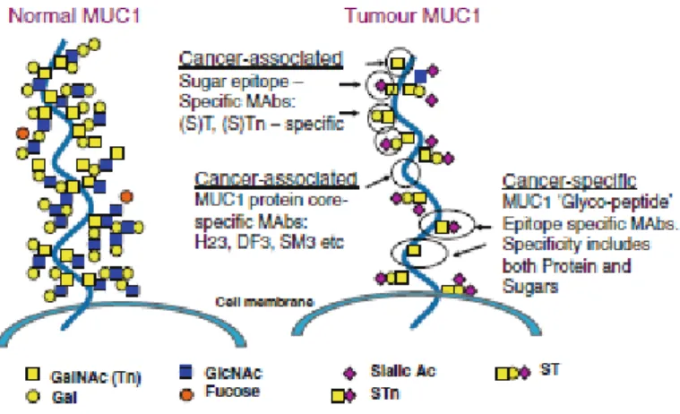
TAAs can be classified in seven categories: 1) Cancer germ cell lines; 2) Novel antigen encoded by tumour-specific transcripts; 3) Viral antigens; 4) Post-translational modified antigens; 5) Genetic mutated antigens; 6) Differentiation antigens; 7) Antigens overexpressed in tumours. An example of a candidate for the development of a targeted therapy is MUC1 that was found to be a tumour associated antigen abnormally expressed on a variety of cancer cells, mainly in tumour-associated form (in approx. 72% of the new cases in 1999). MUC1, a large epithelial glycoprotein of the mucin family is encoded in the gene 1q21. MUC1 is a membrane-bound mucin, a glycoprotein with more than 50% of O-linked glycans that expresses several cancer-associated antigenic epitopes in cancer cell lines (Acres and Limacher et al. 2005), as represented in Figure 1.
Figure 1. MUC1 protein expressing several types of novel cancer-associated antigenic epitopes in cancer cells (adapted from Acres and Limacher 2005)
MUC1 influences the transcriptional regulation of genes “with immune regulation,
inflammation and drug resistance, apoptosis, proliferation, angiogenesis, metastases and tumour cell invasion “(Nath and Mukherjee, 2014). Immune tolerance to MUC1 in
transgenic mice has been demonstrated (Rowse et al. 1998; Acres et al 2000) and evidence of T cell response after vaccination was observed in hMUC1-vaccinated hMCU1 transgenic mice.
Since Medawar (Billingham et al.1953) and Burnet in 1960, that the medical and scientific community became more aware to the possibility of manipulating the immune responses to some antigens. When in 1975 Kohler and Milstein created developed a “hybridoma”, the whole paradigm shifted and the conditions were then set to discover and purify antigens.
Therapeutic vaccines have a growing importance in the development of present and future treatments and Dendritic Cells as antigen presenting cells, are very interesting targets in the development of anticancer vaccines. There are several types of vaccines: total tumor cell vaccines, proteins, peptides, cytokines or costimulatory molecules, anti-idiotypic antibodies, vectors, and finally, DCs vaccines. (Arosa et al, 2012;. Riether et al, 2013). For the treatment of cancer diseases, DCs vaccines were shown to be very promising in several clinical studies (Zhang and Engleman, 2006). They can be loaded with tumour lysates, peptides, and proteins from tumour cells (Arosa et al., 2012). Studies in mice have demonstrated that DCs loaded with tumor antigens are able to induce a protective response inducing cancer immunity (Yu and Restifo, 2002). The immunogenicity of the antigens delivered by DCs was also studied in healthy humans (Dhodapkar et al., 2002) and clinical and immunological responses have been demonstrated, showing no significant toxicity (Davis et al. 2003; Hsu et al., 1996).
1.3Use of nanotechnology for targeting Dendritic Cells
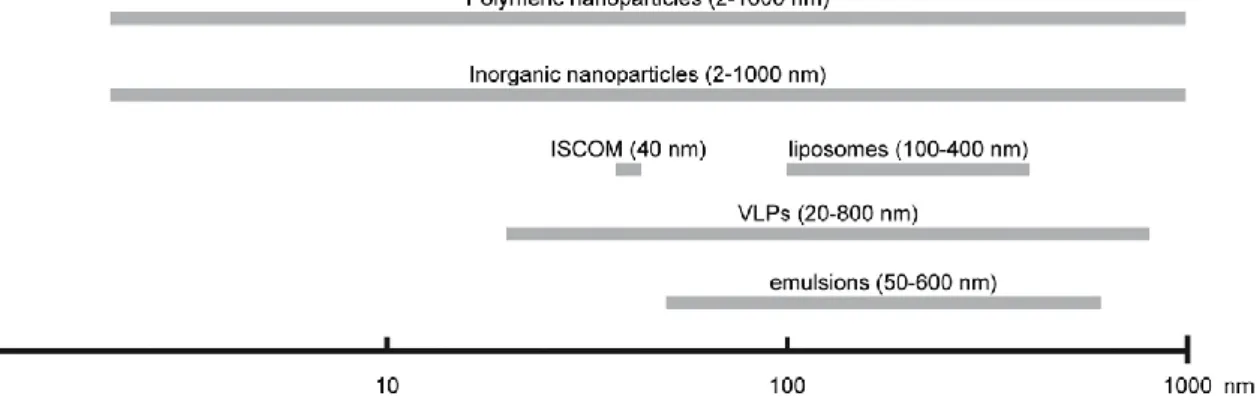
In the development of Dendritic cells-targeted vaccines, different types of nanosystems are being used: polymeric nanoparticles; inorganic nanoparticles; Immunostimulating complex (ISCOM), Virus-Like Particles (VLPs); liposomes; and emulsions.
Figure 2. The size range of nanoparticles used in nanovaccinology. Adapted from Zhao et al Vaccine 32 (2014) 327– 337
8
Nanoparticles have a comparable size to pathogens and can be easily recognized and consequently uptaken by APCs, subsequently inducing an immune response (Xiang et al.2006). DCs usually uptake particles with an average size of 20–200 nm, while for instance macrophages uptake bigger size particles (0.5–5m)(Xiang et al. 2006)]. The mean size for an optimal uptake of NPs by the DC´s is below 500 nm as shown in an
in-vitro study using polystyrene particles (Foged, Camilla et al. 2005). The degree of
internalization as well as the degree of activation is greater in NPs with a size of 300nm compared with 17,7m and 1m particles (Foged, Camilla et al. 2005). Nevertheless there are differences in uptake efficiency of vaccines according to the material used. This is why the material used has been the focus of intense research and now, it is commonly accepted that surface size, shape and affinity to in situ receptors are very important aspects to consider when developing dendritic cells-targeted vaccines using nanoparticles. NPs with cationic surfaces showed a higher uptake by APC because of the electrostatic interactions with anionic cell surfaces (Foged, Camilla et al. 2005). Additionally, the shape also influences the process by which NPs and DCs interact. Elongated and “worm” shaped particles showed residual phagocytosis compared with spherical shaped NPs (Champion, Julie A et al 2009).
But what is in fact considered nanotechnology?
Nanotechnology is “the science of understanding and control of matter at dimensions
ranging from 1 to 1000 nm” (The US National Nanotech Initiative, s.d.), comprising the
engineering and technology, and involving areas such as imaging, measuring, modelling and manipulation of the nanoscale field. It is a cross-sectional area to various scientific fields such as Engineering, Energy, Biotechnology, Electronics and Computing. There are several debates that aim to standardize the understanding of nanotechnology, however the complexity of defining what is in fact a nanomaterial, and the exact size of the nanoscale, has hindered its implementation. Some consider that a nanomaterial, in order to be classified as such, has its dimensions ranging between 1 nm and 100 nm.
Nanotechnology (and in particular bionanotechnology) is a very important tool in nanomedicine since it can be used in the diagnosis, prevention and treatment of diseases also contributing for the increase of the knowledge and understanding of the biological processes. In this field, the objective is to manipulate matter in order to obtain
nanostructures of the same size of biomolecules that can interact with human cells, achieving a range of solutions for the diagnosis and treatment by focusing, directly or indirectly, in the repair of the biological mechanisms. The use of micro- and nanosystems for the administration of drugs has been increasing, and its development implies continuous research and the design of new materials as well as improved delivery methods for vectoring and releasing the therapeutic agents of interest.
Nanotechnology can be divided into three categories, regarding healthcare applications:
1. Diagnosis, biosensors and surgical tools 2. Imaging agents and monitoring technologies
3. Innovation technologies and biomaterials (often combined with cellular therapy) that may be used in drug delivery and placement, as well as promoting tissue repair and engineering.
1.4 The development of nanodrugs – Background and
review
In the second half of the 20th century, the first nanodrugs began to emerge surrounded by great scepticism by the medical and scientific community that continued questioning the efficacy of such small molecules and its effective large scale industrial application. The discovery of colloidal mechanisms for drug delivery in the 1960’s and 1970’s, led to the development of drug delivery systems (DDS) which include liposomes (Bangham, Gregoriadis, Papahadjopoulos, Barenholz), nanoparticles and nanocapsules (Speiser, Couvreur, Kreuter), drug-DNA complex (de Duve, Trouet) and drug-polymer conjugates (Davis). For these last type of NPs, the use of polyethylene glycol (PEG) to modify a polymer surface, prevented opsonisation, which is the nonspecific binding of NPs to blood components. Thus it reduced their rapid absorption and in vivo clearance by the mononuclear phagocytic system cells, allowing for a prolonged half-life of the nanosystems.
In the 1990s, Iron Oxide NPs began to be clinically applied in solutions for parental infusion used to treat anaemia and also used as imaging agents in magnetic resonance imaging (MRI). Antibody-drug conjugates (Wilchek, Arnon Sela), albumin-drug
10
(Trouet), and micellar block copolymers (Ringsdorf, Kataoka, Kabanov) were the technologies that followed.
Over the years, intensive research led to the ability of potentially increasing the specificity of the NPs through bioconjugation of affinity ligands such as antibodies, antibody fragments, peptides, aptamers, sugar and small surface molecules, in order to obtain targeted NPs.
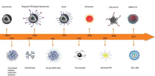
Figure 3. Time line of clinical stage nanomedicines. Adopted from Chem Soc Rev. Kamaly,N. et al. (2012)
1.4.1 Methods of production of nanoparticles systems
There are two basic approaches used in the production of nanosystems: the bottom-up techniques and the top-down techniques. The first one is developed upwards, that is., it is a process through which ordered structures are developed starting at an atomic and molecular scale. This approach requires the least amount of material, and it is generally associated with lower losses in relation to production in comparison with the top-down method. However, it is a more time consuming technique. In the second technique nanosystems are generated from the top down, that is, starting with large material, which is being lowered successively until the desired nanoscale is achieved. This approach requires larger amounts of material and can lead to waste (usually the excess is discarded
and not reused). Depending on the material used in the development of the nanosystem, one can choose from one or both of these approaches using different production methods.
1.5Polymer based nanoparticles delivery systems
Several polymeric materials have been used for over 40 years in the medical field and in the pharmaceutical industry. These materials have evolved since their initial implementation resulting into products with acceptable biodegradability such as: reabsorbable sutures, orthopaedic implants, drug delivery systems and matrices which can act as drug deposits able to exercise their controlled release, to the development of multifunctional NPs with vectoring and controlled release properties used in drugs and imaging agents. Polymeric NPs have the capacity to carry various drugs, releasing them in a controlled way by diffusion of drug molecules, either through a polymer matrix, by the dissolution of differential layers of surface coating, specific enzyme degradation or erosion of the particles. These can be target-oriented, incorporating target ligands in formulations led to increase absorption rate and therapeutic efficacy.
Currently, the most commonly used polymers for controlled drug delivery applications include poly (D, L-lactic-co-glycolic acid) (PLGA), poly (lactic acid) (PLA), poly (glutamic acid) (PGA) poly (caprolactone) (PCL), copolymers of N- (2-hydroxypropyl) methacrylate (HPMA), polysaccharides (Hyaluronic Acid, Chitosan, etc) and poly (amino acids). Due to its biocompatibility and biodegradability, PLA, PLGA and PGA have been used extensively in an impressive amount of formulations. This is due, in part, to the clearance of the polymeric matrix by the metabolic system.
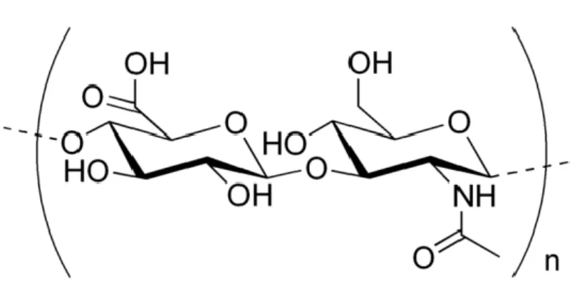
1.5.1 Hyaluronic Acid
One of the most important biopolymers used in the development of nanoparticles, as mentioned before, is Hyaluronic acid (HA). HA is a linear, natural mucopolysaccharide containing alternating units of D-glucuronic acid and N-acetyl-glucosamine linked by β
-12
1,3 and β-1,4 glycosidic bonds. It belongs to a group of substances called
glycosaminoglycans (GAGs).
Figure 4. Hyaluronic acid molecular structure (Adapted from Novozymes)
However, HA is very different from other GAGs in most respects. Its primary structure is simpler, it contains peptides and it is the only non-sulfated glycosaminoglycan. It is constituted by one polysaccharide chain, differently from the other glycosaminoglycans (Figure 1), but its molar mass is usually of the order of MDaltons. At a first glance, the uniformity of its structure seems to restrict its biological functions, but this limitation is overcome by the number of HA specific binding sites existing in other molecules or cell surfaces (Fraser et. Al., 1997).
In the body, HA is widely present in the extracellular matrix, (ECM), abundant in the vitreous of the eye, in the umbilical cord, in synovial fluid, in heart valves and skeletal tissues. It is produced mainly by mesenchymal cells and almost all cell types are responsive to the stimulation by HA, a stimulation able to modify secretory and behavioural cell properties.
In animals and in man, the half- life of HA in tissues, range from one to several days. It is catabolized by endocytosis mediated by receptors and is degraded by lysosomes on site or after transportation through lymph nodes. The remaining enter the general circulation and blood, being later removed primarily by the endothelial cells of the hepatic sinusoids with a half-life of 2 to 5 mins (Fraser et. Al., 1997).
Due to its physicochemical properties and its biocompatibility, HA has wide range of applications in the pharmaceutical, cosmetic and medical fields, with an emphasis in ophthalmology, orthopaedics and oncology.
1.5.2 HA capability of inducing immune activation
HA has been recently studied as one of the most rapidly-growing platform for intracellular delivery of anticancer agents, exploring receptor-mediated active targeting strategies (Mangla, Bharti et al. 2015). HA covalently binds to different proteins and is able to influence their activity (Toole BP. 2000). Proteins that bind to HA include: Toll-like receptors (TLR2 and TLR4), CD44, RHAMM, TNFIP6, brevican, LYVE1, among others. A set of different biological and disease processes are regulated by HA, mediated by the interaction of proteins and the location of HA-binding proteins. HA-CD44 interactions are known to influence the recruitment and homing of leucocytes. Additionally, HA-TLR interactions trigger HA inflammatory signalling and the first is involved in the regulation of tumour growth and metastasis through the interaction between CD44 and RHAMM.
Many studies indicate that HA signalling takes place though a CD44-dependent tyrosine kinase pathway, however recent research has shown that HA stimulates the production of chemokines even in the absence of CD44 (Teriete, Peter et al. 2004). If CD44 is expressed by T cells, through which the interaction with HA occur, with DCs this interaction is mediated by TLR4 (Voelcker, Verena et al.2008). It has been demonstrated that HA has the ability to influence DCs maturation and is capable of initiating alloimmunity. HA is known to induce DC maturation, promoting dendritic and endothelial cell release of pro-inflammatory cytokines such as TNFα, IL-1β, and IL-12 through TLR4. Promoting DCs activity with HA improves their capacity to stimulate allogeneic and antigen-specific T cells (Termeer, Christian C., et al. 2000)
1.5.3 Methods of producing HA-Nanoparticles
HA chemical modification can occur either by crosslinking or by conjugation and it can be performed on two functional sites: the carboxylic acid group - and the hydroxyl group. There are several types of nanosystems composed of HA, nevertheless we will be focusing on crosslinked HA-NPs.
14
One of the most used methods of preparation of crosslinked HA-NPs is through carbodiimide chemistry, a method of producing NPs that was initially developed by Fakhari (Fakhari, Amir et al.2003). Using this method, covalent binding NP´s were first described by amidation with a bifunctional amine as the crosslinker. In another method using carbodiimide chemistry, the formation of interpolyelectrolyte complexes was obtained via the interaction between polysaccharides with opposite charges (HA and poly-L-lysine). Szarpak, A. also developed biodegradable capsules through covalent crosslinking using HA and poly(allylamine). Another method for producing photo-crosslinked micelles was developed by Xu (Xu, Jing et al. 2011), using negatively charged HA and positively charged styrylpyridinium (SbQ) through self-assembly. During recent years, bioresponsive CL-HA-NPs were investigated by several groups and Baier et al. was able to obtain stable nanocapsules using polyhexanide through an inverse miniemulsion technique (Baier, Grit et al. 2013). A new method was described by Yu-Hsien in 2012 using an electrostatic field system in an aqueous phase. Glutaraldehyde is also used as a crosslinking agent using a solvent-non solvent technique, a method which results in particles with higher efficiency in xenograft tumour sites due to the surface plasmon resonance effect (SPR). A different method for producing HA-Polyethylene glycol transdermal NPs trough crosslinking by inverse suspension polymerization (water in oil technique) was developed by Lim, H.J. in 2012. Ilgin in 2010 described the production of a HA-NPs crosslinked using divinyl sulfone (DVS). Besides its use in monopolymer based nanosystems, Hyaluronic Acid as also been used in crosslinked preparations in conjunction with other polymers such as Chitosan. In a crosslinked nanoparticles system the agent used to mediate the chemical bonding between natural or synthetic polymers is called a crosslinking agent. These polymers can be linked by covalent or ionic chemical bonds.
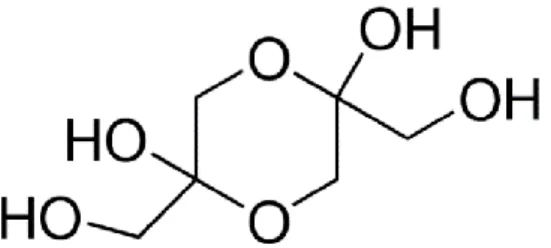
Taking into consideration the information available regarding research previously performed using 3 to 5 carbon atoms ketose in the tanning of proteins, we have decided to test 1,3-Dihydroxyacetone dimer as the crosslinking agent in the preparation of our novel HA-NPs biodegradable delivery system. Dihydroxyacetone is a 3 carbon atoms ketose usually used as a tanning agent in dermatological topical formulations. DHA is a substitute for aldehyde tannins (eg. glutaraldehyde) which bonds to HA and proteins by covalent bonding, resulting in condensation products. DHA forms imines following the condensation of lysine and arginine, double condensation will result in crosslinked products. The hydroxyl group in the alpha-position of the carbonyl function of
Dihydroxyacetone, results in an imine obtained after condensation on a terminal amino acid being able to cyclize with the carboxylic function of this acid forming a δ-lactone.
Figure 5. 1.3-Dihydroxyacetone dimer molecule (Retrieved from Sigma Aldrich MSDS)
1.6Context and aims of the project
Considering the state of the art in cancer treatment and recent advances in immuno-oncology, our first aim was to obtain hyaluronic acid (HA) nanoparticles (NA), so we first tested the feasibility of the methodology. We decided to test several formulation under different conditions in order to achieve a viable delivery vehicle for presenting Tumor Associated Antigen (TAAs) to Dendritic Cells, inducing T-cell activation in oncological disease scenarios. Following MUC1 antigen research of our laboratory group, we have theorized the future use of this antigen in a DC vaccine entrapped in a HA nanoparticles system.
For the development of this HA nanosystem, we have decided to use a polysaccharide with affinity to DCs surface receptors, non-toxic, non-immunogenic and biodegradable, using a non-solvent technique and a novel crosslinking method. We have decided to research and develop a crosslinked hyaluronic acid (CL-HA). A crosslinked version of a biopolymer such as Hyaluronic Acid is suitable for the delivery of bioactive ingredients in pharmaceutical formulations by its ability of entrapping the therapeutic agents with controlled release and adequate mechanical properties.
Firstly we tested different methods of producing Hyaluronic Acid nanoparticles through the use of 1.3-Dihydroxyacetone dimer as the crosslinking agent methods and proceeded with a characterization of the produced batches of particles. Secondly, after successfully producing the HA-based nanoparticles, we intended to pursue with characterization of the
16
system, namely by the analysis of the particle size, size dispersion and concentration of the nanoparticles. Lastly if the first two objectives were achieved we intended to assess cell viability of our nanoparticles testing our nanoparticles in PBMCs.
This project was developed in the Glycoimmunology Group of Chronic Diseases Research Center (CEDOC), NOVA Medical School of Universidade NOVA de Lisboa and in the Animal Cell Technology Unit of ITQB, Universidade NOVA de Lisboa/iBET.
2.1Materials and equipment
Materials
i. Hyaluronic Acid (Novozymes) ii. Dihydrocyacetone (Sigma) iii. Ultra-purified Milli-Q water iv. Ovalbumin-FITC (Invitrogen)
v. Bovine Serum Albumin (Sigma) vi. PBMCs (fresh from healthy
humans)
vii. RPMI 1640 (Sigma) viii. Tripan blue
ix. Annexin V/7AAD x. 1M Na2HPO4 (Sigma) xi. 1M NaH2PO4 (Sigma)
Equipments:
i. NS500 (NanoSight, iBET); ii. Centrifuge (Eppendorf
Termomixer C)
iii. UltraCentrifuge (Beckman Coulter)
iv. Neubauer chamber
v. Flow cytometer (Life Technologies)
vi. CO2 incubator
vii. Microscope viii. Vortex
ix. Heating Magnetic Stirrer x. Magnets
xi. T flasks xii. Auto pipettes xiii. Sterile Tips
xiv. 96 wells plaque, 200µ xv. Sterile Eppendorfs
Sodium Hyaluronate (Hyasis 850P, Novozymes) with a molecular weight between 0.6-1.1 MDa was initially obtained from Sigma. During the process of optimization, samples of Sodium Hyaluronate were kindly provided by Novozymes. The choice of this specific HA was made according to safety issues and purity. Hyaluronic Acid we used in this project is produced by fermentation of the bacterial strain Bacillus subtilis, without the use of animal-derived raw materials and organic solvents. It is biocompatible, nontoxic, non-immunogenic and biodegradable, differently from HA produced by Streptococcus fermentation.
20
Initially, green fluorescent fluorescein ovalbumin conjugate (OVA-FITC) was the protein chosen in the development of the first method of preparation of nanoparticles. OVA-FITC is a protein with a relatively low molecular weight (~45.000 daltons), labelled with fluorescein. The decision of using OVA-FITC was made with the purpose of pursuing a further stage characterization of HA-OVA-FITC-NPs in cell cultures (PBMCs) by which the degree of NPs internalization in the cells could be assessed. Bovine Serum Albumin fraction V was later tested in the development of optimized formulations. The choice of this protein was related with the size (mW ~66.000 daltons) and availability of the said protein in the laboratory.
2.2Optimization of the method of preparation of
HA-Nanoparticles
During the process of optimization of HA-NPs, several formulations were prepared in different conditions in an attempt to obtain crosslinked nanoparticles. Different HA and DHA concentrations, addition timings, temperatures, centrifuge force and techniques were tested before crosslinked NPs were successfully achieved as described below in this thesis.
2.2.1 Method of preparation of Hyaluronic
Acid-OVA-FITC-Nanoparticles
10 mg of HA powder was dissolved overnight in 4ml 10mM Phosphate buffer, pH 7.4 solution previously prepared, by stirring slowly heating. From this HA solution, different samples of HA were prepared in different concentrations. OVA-FITC was previously dissolved in 10 mM Phosphate buffer, pH 7.4 solution in a concentration of 2.5 mg/mL. A stock solution of DHA was prepared by dissolving the crosslinking agent in a 10 mM phosphate buffer, pH 7.4 in a concentration of 45 mg/mL and left to rest at room temperature during 35 minutes until DHA dimers stabilized in the form of monomers.
With the objective of testing different concentrations and ratios of HA, protein and DHA, several 1mL samples were equally prepared in 1.5mL sterile Eppendorfs.
HA was first pipetted to the Eppendorfs in volumes varying from 0.1mL to 0.15mL and to these solutions OVA-FITC was added in different volumes in a ratio of 1:1 and 1:2. Later, the solution containing DHA was added to the solutions containing HA and Ova-FITC in a ratio per sample of HA to DHA of 1:2 and 1:4.
In a solution of nanoparticles without OVA-FITC, HA in volumes varying from 0.1mL to 0.15mL was first pipetted to the Eppendorfs and to this solution DHA was added in a ratio per sample of HA to DHA of 1:2.
Samples were then mixed and stirred heating at 500 rpm for 90 minutes. Solutions in the Eppendorfs were then centrifuged using a Beckman Coulter high speed centrifuge at 30.000 g for 40 minutes at 24ºC to settle. The sediment with the nanoparticles was then collected, washed with buffer and resuspended for characterization.
2.2.2 Method of preparation of Hyaluronic Acid – BSA
-Nanoparticles
In an optimized method, HA was dissolved overnight in 10 mM Phosphate buffer, pH 7.4 solution, by stirring slowly heating in a concentration of 2.5 mg/mL. BSA was dissolved in 10mM Phosphate buffer, pH 7.4 solution, to obtain BSA in a concentration of 2.5 mg/mL. DHA in this experimental assay was not previously dissolved in the buffer. With the objective of testing different concentrations and ratios of HA, protein and DHA, equal 1mL samples were prepared in 1.5 mL sterile Eppendorfs.
In this optimized method, 0.1 mL and 0.15 mL of HA was first pipetted to the Eppendorfs, to these solutions and BSA was added in a ratio of 1:1 in relation to the amount of HA per sample. Later, DHA powder was directly added to the solutions containing HA and BSA in a ratio per sample of HA to DHA of 1:2 and 1:4.
In a solution of nanoparticles without BSA, HA in volumes varying from 0.1mL to 0.15mL was first pipetted to the Eppendorfs and DHA powder was directly added to the solution in a ratio per sample of HA to DHA of 1:2 while stirring.
22
Samples were then mixed and stirred heating at 500 rpm for 90 minutes. Suspensions in the Eppendorfs were then centrifuged using a Beckman Coulter high speed centrifuge at 30.000g for 40 minutes at 4ºC to settle. The sediment with the nanoparticles was then collected, washed with phosphate buffer and resuspended for characterization.
2.3Nanoparticles
Tracking
Analysis
–
Method
introduction
An adequate characterization of nanoparticles is very important during the development process. The choice of the right characterizing techniques poses a huge challenge, since results are influenced by the size and nature of the particles, the concentration of the sample, the properties of the solutions and the fundamental principles of the methods applied (Domingo et al., 2009). Amongst the various existing methods of NPs characterization, we decided to use Nanoparticles Tracking Analysis (NTA) to identify particles, calculate the concentration of the population of particles as well as particle size. This process allows us to characterize the nanosystems in liquid formulations, determining the hydrodynamic diameter and concentration present in the sample.
Despite very accurate and robust, this characterization technique is still not widely described in the literature as some of the other well-known techniques. This characterization technique is based on the Brownian motion of the nanoparticles and their light scattering properties. This technique allows an analysis of particles with a size range between 10 nm and 2000 nm. The Brownian motion of the particles is analysed in real time by a scientific digital camera, through which each particle is simultaneously, but individually, displayed and monitored (screened) by an image monitoring software (Carr & Wright, 2012; NanoSight, 2013th). The visualization of the particles in the sample is possible due to light scattered by the nanoparticles when they are illuminated by the laser. The intensity of light scattered by nanosystems is captured by a video camera and the motion of the particles is monitored frame by frame. The average distance travelled by each particle is calculated automatically. From this value, it is possible to calculate the particle diffusion coefficient (Dt), identifying solvent viscosity (η), the hydrodynamic diameter (d) and knowing the sample temperature (T). Once the Brownian motion of the particles is done in three dimensions, the particle size is calculated by analysis in 2
dimensions using a variation of the Stokes-Einstein equation where KB is the Boltzmann
constant (Carr & Wright, 2012; NanoSight, 2013b), as represented in the following formula (Equation 1):
NTA software allows the visualization of nanoparticles in real time, counting and measuring the size of particles and providing histograms of the size distribution of the analysed particles (including a file with data from the analysis and a video in .avi format of the same sample) ( NanoSight, 2013th). Considering that the results are related with samples previously diluted, initial concentrations can be calculated using the following formula (Equation 2):
C
1V
1= C
2V
22.3.1 NTA – HA Nanoparticles characterization assays
Evaluation of the concentration and average diameter of the particles was accomplished by particle location analysis (NTA - Nanoparticle Tracking Analysis) using the equipment NS500 (Nanosigth, UK) at iBET.
The equipment apparatus includes a CCD camera (30 frames per second), a laser with 40 mw of continuous power, a wavelength of 638 nm and "NTA Software Suite" software. In each analysis the software identifies and monitors the center of each particle in each frame which is then captured by a CCD camera during 30 to 60 seconds (900 to 1800 frames per analysis). The use of this technique in this project aimed to determine the formation of HA-NPs, HA-BSA-NPs and HA-OVA-FITC-NPs, allowing us to determine: a) particle formation; b) concentration, mean size and size distribution of the nanoparticles.
NTA assays were conducted only on the samples prepared where it was possible to identify effective crosslinked sediment after the centrifugation. During the optimization process of the nanosystem, samples were obtained using two preparation methods.
Dt y x 2 4 , h B d T K 324
Samples resulting from both preparation methods were analysed in triplicate for each one of the samples in each one of the concentrations. In the first method tested, HA-NPs without protein and with OVA-FITC prepared with 20% and 40% crosslinker were tested. Since NTA is a method of detecting particles in liquids, it was necessary to dilute our samples containing a polysaccharide, 1/100 in phosphate buffer pH 7.4 in order to avoid any clog formation in the flow, allowing for a better characterization of the nanoparticles. For these experimental assays, we used the established protocol for the NS500 equipment, running 1.5 ml liquid samples.
2.4 Assays to test cell viability in PBMCs using HA-NPs
PBMC (Peripheral blood mononuclear cells) are mainly composed of lymphocytes and monocytes. Purified PBMC are used in vitro to assess several lymphocyte functions. Activation of monocyte / macrophages by small molecules, cytokines and pathogen components may also be monitored. PBMCs assays can also be used to perform structural and functional immunological studies, analysing factors such as apoptosis and the production of cytokines and other mediators in vitro. In the following assays we used PBMCs to assess the cytotoxicity of HA-NPs.
To address the general effects of HA on human immune cells we used cryopreserved peripheral blood mononuclear cells (PBMCs) that had been isolated from healthy individuals and cultured them in the presence of different doses of HA for 48h.
PBMCs were first thawed and incubated in RPMI 1640 medium at 37ºC, 5% CO2
overnight. Cells were then collected by centrifugation at 800rpm (104 rcfs) for 5 min at room temperature. A stock solution of nanoparticles with a concentration of 400 ug/mL HA-NPs was initially prepared in culture medium. From that, sequential dilutions were made to prepare solutions at different HA-NPs concentrations (200, 100, 50 and 10 µg/mL). Equal volumes of the solutions containing the HA-NPs were mixed with a cell suspension containing 2x10⁶células /mL. In this way, cells suspension with (1x10⁶células /mL) and different HANPS concentrations (200, 100, 50 and 10 µg/mL) were obtained. Cells were seeded in triplicate in a 96 wells plate with 200 µL per well and incubated with the nanoparticles at 37ºC, 5% CO2 for 48h. Control wells, without HA-NPS were
2.4.1 Cytotoxicity assessment of HA-NPs in cell culture
using Neubauer chamber
Cytotoxicity upon exposure to drugs can be measured by the exclusion of supravital dyes such as trypan blue. The principle of this test consists in the indirect measurement of cell viability through the analysis of membrane integrity, in which dead cells with damaged membrane by the process of dye absorption, consequently stain blue. On the other hand living cells remain intact and no staining is observed (PERES & CURI, 2005). After the incubation period of 24h and 48h, 25 µL samples of cell suspension were collected, centrifuged at 2000gs for 2 minutes and supernatant was discarded. To the samples we added 50 µL of trypan blue, and from this solution an aliquot was taken for cell counting using a Neubauer chamber.
2.4.2 Cytotoxic assessment of HA-NPs in cell culture by
Annexin V/7AAD and analysis by flow cytometry
During apoptosis certain morphologic features occur such as loss of plasma membrane asymmetry and attachment, condensation of both the cytoplasm and of the nucleus as well as internucleosomal cleavage of DNA. In our experimental assays we assessed cell viability using Annexin V which is a 35-36 kDa Ca2+ dependent phospholipid-binding protein with a high affinity for PS that binds to PBMCs with exposed PS.
Annexin V staining precedes the loss of membrane integrity which occurs during the latest stages of apoptosis. Therefore, staining with APC Annexin V is usually used together with a vital dye such as 7-Amino-Actinomycin (7-AAD) allowing the identification of dead cells. PBMCs that are viable and with intact membranes exclude 7-AAD, whereas the membranes of dead and damaged cells are permeable to 7-AAD. For the assays all content of the wells was collected and centrifuged at 1300rpm for 7 min at room temperature. Supernatant was removed and later discarded. PBS buffer was then added to the pellet.
26
For the staining we added Annexin V/7ADD to samples. 5 µL of 7AAD and 5 µL of annexin were added to cells suspensions and stored for 10 minutes at room temperature in the dark. Flow cytometry was later performed.
3.1Nanosystem characterization using Nanoparticles
Tracking Analysis
Our first goal was to produce hyaluronic acid (HA) nanoparticles (NP), so we first tested the feasibility of the methodology.
The first histograms represent the results obtained considering particle size and concentration, showing: a) the presence of nanoparticles; b) the influence of the applied NPs preparation methodology in the formation of nanoparticles.
Two methods were tested during the optimization process. One of the differences in the methods was the preparation and addition conditions of DHA in the formulation. When preparing fresh DHA the stability of the dimer is of a critical importance. In a solution it turns into a monomer very rapidly and we found we can obtain better results if the reaction took place in the final solution containing HA and HA-NPs by adding the powder directly in the exact quantities by continuously maintaining the agitation and stirring.
The ultracentrifuge temperature was reduced from 24ºC to 4ºC. This decision was made based considering phase transition temperature of crosslinked HA, which is around 20ºC (Collins et al. 2008), in order to increase the pellet and improve production yield.
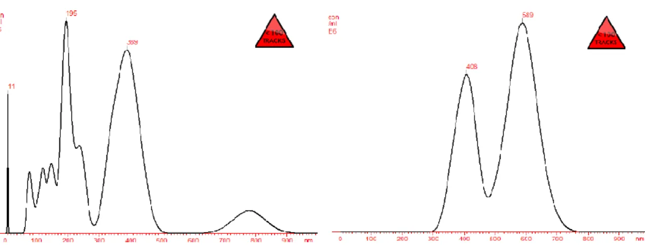
Figure 7. NTA analysis of particle size of HA-OVA-FITC- NPs, for the samples containing 0,150ml of protein in a ratio of 1:1 to HA. 40% of crosslinking agent was used.
Figure 6. NTA analysis of particle size of HA-OVA-FITC- NPs for the samples containing 0,150ml of protein in a ratio of 1:1 to HA. 20% of crosslinking agent was used.
Particle size / Concentration Particle size / Concentration Particle
30
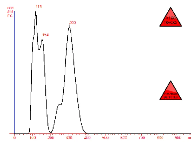
Figure 8. NTA analysis of particle size of HA-BSA- NPs for the samples containing 0.150ml of protein in a ratio of 1:1 to HA. 20% of crosslinking agent was used.
Figure 9. - NTA analysis of particle size of HA-BSA- NPs, for the samples containing 0.150ml of protein in a ratio of 1:1 to HA. 40% of crosslinking agent was used.
Figure 11. NTA analysis of particle size of HA- NPs, for the samples without protein. 20% of crosslinking agent was used.
Figure 10. NTA analysis of particle size of HA- NPs, for the samples without protein. 40% of crosslinking agent was used
Particle size / Concentration Particle size / Concentration Particle
size /
Particle size / Concentration Particle size / Concentration Particle
The software used for capturing and analysing data was NTA 2.2 Analytical Software. Samples were measured for 40 s with manual shutter and gain adjustments. Three measurements of the same sample were performed for all nanoparticles formulations in order to verify the influence of the crosslinker and remaining conditions in the nanoparticles obtained. Mean size and size distribution values obtained correspond to arithmetic values calculated with the sizes of all the particles analysed by the software. Samples in Figure 6 and Figure 7 were prepared by the first production method tested, revealing polydispersity, where we can observe two major curves in the graphics, representing two main clusters / groups of nanoparticles differentiated according to size. Figure 6 reflects a concentration of 0.31x10⁸ particles / mL and mean particle size of 328 nm with a size distribution of 167 nm. In Figure 7 we can observe nanoparticles with a concentration of 0.23 x10⁸ particles / mL with a mean size of 524 nm and a size distribution of 93 nm.
After having optimized the protocol we can observe an increase of the nanoparticles obtained as showed in the Figures 8 to 11. Nanoparticles are in a concentration of 1.44x10⁸ particles / mL with a mean size of 180 nm and a size distribution of 225 nm.
Figure 9 shows a concentration of 2.71 x10⁸ particles / mL and mean particle size of 223 nm, with a size distribution of 108 nm. Nanoparticles were also obtained in formulations using only HA without protein as seen in the last two Figures. Figure 10 shows nanoparticles in a concentration of 0.31 x10⁸ particles / mL and a mean size of particles of 202 nm with size distribution of 100 nm. In Figure 11 we can observe nanoparticles in a concentration of 0.13x10⁸ particles / mL, a mean size of particles of 218 nm and size distribution of 87 nm. In these last samples (optimized method), nanoparticles were present in substantially higher concentration than the samples previously tested.
32
Table 1. Mean Size and Size Distribution of NPs from NTA measurements
a) Nanoparticles produced by the first method (n =3); b) nanoparticles produced by the optimized method (n =3). SD standard deviation calculated by NTA software; Conc. concentration in particles E/mL as measured by NTA; temperature in Celsius (ºC) and viscosity in centipoise (cP) was measured by NTA software.
We can observe that smaller size nanoparticles were obtained in the formulations produced using the second production method (optimized), mainly in the formulation using HA without protein (Table 1) and bigger size particles were obtained when using higher concentrations of crosslinker. We can also observe that inversely, formulations with more volume of crosslinker (40%) resulted in lower levels of size distribution obtained. Nanoparticles concentration was higher in the samples produced according to the second method in the formulations including protein (Table 1), therefore we can presume that crosslink reaction is benefited by the presence of proteins in the formulations.
Mean (nm) SD (nm) Particles Conc.
(E8/mL) Temp (ºC) Viscosity cP
a) HA-OVA-FITC (CL 20%) 326 ± 5 167 ± 2 0.31 23.6 0.92 a) HA-OVA-FITC (CL 40%) 520 ± 11 93 ± 3 0.23 22.8 0.94 b) HA-NPs (CL 20%) 201 ± 1 100 ± 3 0.31 23.8 0.91 b) HA-NPs (CL 40%) 216 ± 3 87 ± 1 0.13 24.05 0.91 b) HA-BSA-NPs (20%) 273 ± 7 225 ± 9 1.5 23.15 0.93 b) HA-BSA-NPs (40%) 242 ± 5 108 ± 6 2.71 23.4 0.92 NTA
3.2Influence of HA-NPs in cell viability during 24h and
48h periods measured by trypan blue staining through
cell counting method using Neubauer chamber
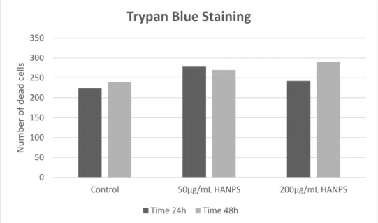
In order to investigate the influence of HA nanoparticles in cell viability, we assessed the samples by Trypan Blue staining according to the number of stained cells counted on the microscope using the Neubauer chamber. When analysing the number of stained cells, we could observe no significant difference in the number of cell death in the samples containing HA-NPs in the concentrations tested (200 µg/mL and 50 µg/mL, CL 40%) in comparison with the control samples for the determined periods of 24h and 48h. The two concentrations tested were chosen as representative of an accepted interval for analysing the influence of the nanoparticles in cell culture, considering 100 µg/mL as the average dose of HA determined as ideal in previous internalization assays (Yang, R et al. 2002).
Figure 12. Cell counting using Neubauer chamber. Results show no significant influence in cell viability of HA-NPs containing samples over the periods of 24h and 48h.
0 50 100 150 200 250 300 350
Control 50µg/mL HANPS 200µg/mL HANPS
N u m b er o f d ead cel ls
Trypan Blue Staining
34
3.3Influence of HA-NPs in cell viability during 24h and
48h periods through labelling with Annexin and 7AAD
analysed by flow cytometry
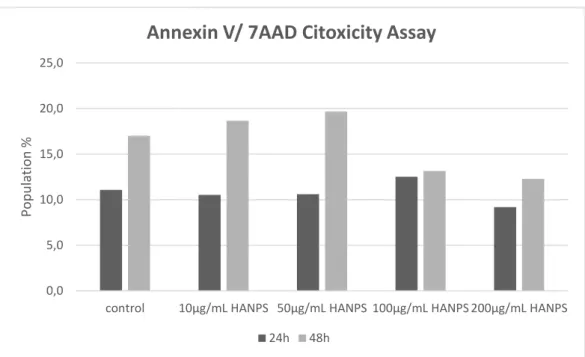
As shown in Figure 13 we observe no influence of the HA-NPs in the increase of the percentage of apoptotic cells for either 24h or 48h versus the control sample. In fact a non significant decrease of cell death was observed in the samples including higher concentration of HA-NPs, which might be explained by the presence of higher concentration of HA, an ECM component that might have influenced the reduction of cell apoptosis by an eventual stimulatory effect.
Figure 13. Cell viability analysis performed by flow cytometry (n=1). This graph shows that HA-NPs produced with our crosslinking agent (DHA 40%) does not induce any significant cytotoxic effect when compared with the control group in 24h and 48h samples
0,0 5,0 10,0 15,0 20,0 25,0
control 10µg/mL HANPS 50µg/mL HANPS 100µg/mL HANPS 200µg/mL HANPS
Pop
u
lation
%
Annexin V/ 7AAD Citoxicity Assay
A
B
C
C
D
Discussion
As we can observe in Figure 13, HA-NPs did not induce relevant apoptosis when comparing different HA-NPs concentrations (200, 100, 50 and 10 µg/mL, CL 40%) and with control at 24 and 48h. Dot plots show no interference of the HA-NPs in PBMCs apoptosis. In the dot plots represented in Figure 14 we can visualize no significant changes in cell population in the dot plots as confirmed in images B and D, consistent with the results obtained with control samples as represented in images A and C. Dot
Figure 14. Representative dot plots of cell viability assays performed by Flow cytometry. A and B FSC vs SSC dot plots of PBMCs where it is shown the gate acquired cells, excluding and debris after 48h. C and D. annexin V vs 7AAD staining of R1 gated cells. A) and C) control sample B and D) 100ug/mL HA-NPs sample 48h..
36
plots represent a comparison of HA-NPs in 100µg/mL at 48h, a concentration chosen as a reference average value for assessing HA internalization and toxicity as described in the literature (Yang, R et al. 2002).
4.1
General discussion of the resultsIn this project we have hypothesized the possibility of developing a novel method of producing crosslinked Hyaluronic Acid nanoparticles using Dihydroxyacetone as the crosslinking agent in order to evaluate the possibility of generating a viable delivery system for the uptake of antigens in the development of immunotherapies, mainly in the development of anticancer vaccines.
After a long and repeating task of development of a nanoparticles system, we could finally obtain nanoparticles as confirmed through the characterization made by NTA in which we could observe that the introduction of changes in the production process during the optimization of the methodology, positively influenced nanoparticles obtained. Since the design of the first method, several changes were added to the process and we can observe a significant difference in the results obtained in the first experiments in comparison with the last experiments as shown in the Figures 6 to 11 of NTA and Table 1 where we can observe relevant differences in particle size, concentration of nanoparticles and size dispersion.
We could verify a significant difference in the amount of nanoparticles obtained by NTA analysis in the formulations in which the crosslinker was added directly to the solution containing HA and proteins, instead of adding DHA after previous dissolution in buffer. It is our understanding that due to the instability of DHA, it should be added directly to the solution during the stirring and mixing process in order to promote the reaction in contact with HA and the proteins. Also, the temperature reduction during centrifugation at 30.000g was reduced below the liquid/gel transition temperature of Hyaluronic Acid, which might have influenced the sediment collection and concentration.
Particle sizes obtained through the optimized method, either with BSA or with HA only, are in general of a size below 300nm, that is, with particles lower in size than the ones obtained with OVA-FITC in the first method. The formulations using higher concentrations of the crosslinker (40%) were the ones that produced nanoparticles in higher concentration, which might be associated with a higher degree of crosslinking obtained. It is possible that a formulation using 40% of DHA, might be more suitable to develop future formulations. A reduction in the particles size using this last formulation might be obtained if the stirring and mixing conditions are modified (e.g. increase rpm).