UNIVERSIDADE DE LISBOA
FACULDADE DE CIÊNCIAS
DEPARTAMENTO DE BIOLOGIA VEGETAL
DECIPHERING HOST FACTORS FOR
MYCOBACTERIA INTERNALIZATION DURING
PHAGOCYTOSIS
DISSERTAÇÃO
Pedro Manuel Dias Timóteo
MESTRADO EM MICROBIOLOGIA APLICADA 2013
UNIVERSIDADE DE LISBOA
FACULDADE DE CIÊNCIAS
DEPARTAMENTO DE BIOLOGIA VEGETAL
DECIPHERING HOST FACTORS FOR
MYCOBACTERIA INTERNALIZATION DURING
PHAGOCYTOSIS
Dissertação orientada pelas Professoras Doutoras: Elsa Anes (Faculdade de Farmácia da Universidade de Lisboa)
Margarida Gama Carvalho (Faculdade de Ciências da Universidade de Lisboa)
Pedro Manuel Dias Timóteo
MESTRADO EM MICROBIOLOGIA APLICADA 2013
DECIPHERING HOST FACTORS FOR
MYCOBACTERIA INTERNALIZATION DURING
PHAGOCYTOSIS
Pedro Manuel Dias Timóteo
MASTER THESIS
2013
This thesis was fully performed at Centro de Patogénese Molecular – Unidade de Retrovírus e Infecções Associadas (CPM-URIA) of the Faculty of Pharmacy of the University of Lisbon under the direct supervision of Prof. Dr. Elsa Anes.
Prof. Dr. Margarida Gama Carvalho was the internal designated supervisor in the scope of the Master in Applied Microbiology of the Faculty of Sciences of the University of Lisbon.
Table of contents
Acknowledgements ... I Communications in Scientific Meetings ...III Relevant abbreviations ... IV Resumo ... V Abstract ... X
1. Introduction ... 1
1.1 General aspects of tuberculosis ... 1
1.2. Biology of Mycobacterium tuberculosis ... 3
1.3. Host-pathogen relationship ... 3 1.4. Phagocytosis ... 4 1.4.1 Macrophages ... 5 1.4.2 Internalization ... 5 1.4.3. Phagosome maturation ... 7 1.4.4. Vesicular traffic ... 8
1.4.5. Cathepsins and Cystatins ... 8
1.5. Objectives ... 9
2. Materials and Methods ...11
2.1. Mycobacteria cultures ...11 2.2. Cell cultures ...11 2.3. Infection protocol ...12 2.3.1. Preparation of macrophages ...12 2.3.2. Preparation of bacteria ...12 2.3.3. Infection ...13
2.3.4. Processing samples after infection ...13
2.3.5. Alternate protocol for screening of knockdown cells ...13
2.3.6. Confirming the absence of extracellular bacteria ...14
2.4. Protein knockdown ...14
2.4.1. Knockdown lentiviral libraries ...14
2.4.2. Building a stable THP-1 knockdown cell line ...15
2.4.3. Confirming protein knockdown via Western Blot ...15
2.5. Data acquisition and analysis ...16
2.5.1. Quantification of internalization by flow cytometry ...16
2.5.2. Statistical treatment ...16
2.5.3. Labelling hits in the knockdown screen ...17
3.1. Rates of internalization ...18
3.2. Effects of selective protein knockdown on internalization ...22
3.3. Evaluating protein knockdown of cathepsin L ...22
3.3. Inside or outside the macrophage? ...22
...23
4. Discussion ...26
4.1. Experimental factors influencing internalization ...27
4.2. Internalization of mycobacteria occurs rapidly ...27
4.3. Human monocyte-derived macrophages are efficient phagocytes in vitro ...28
4.4. Internalization is dependent on the species of mycobacteria ...29
4.5. Mycobacterium tuberculosis may inhibit its internalization by macrophages ...29
4.6. Host factors influence mycobacteria internalization ...30
4.7. Only positive regulators of internalization were found ...31
4.8. Rab Guanosine Triphosphatases are positive regulators of internalization...32
4.9. Some cathepsins have different roles in pathogenic and non-pathogenic mycobacteria ...33
4.10. Membrane-associated proteins STX4A, VAMP2 and SNAP23 are involved in internalization ...34
4.11. Implications on in vivo infections ...34
4.12. Drawbacks and future work ...35
4.13. Closing remarks ...36
Table of Figures
Figure 1. Estimated tuberculosis incidence rates ... 1
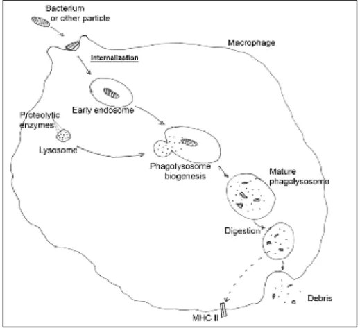
Figure 2. Simplified diagram of phagocytosis………....…6
Figure 3. Example populations of uninfected and infected macrophages...18
Figure 4. Internalization by J774 mouse macrophages ...18
Figure 5. Internalization by THP-1 macrophages ...19
Figure 6. Internalization by human monocyte-derived macrophages ...20
Figure 7. Internalization efficiency of different macrophages ...21
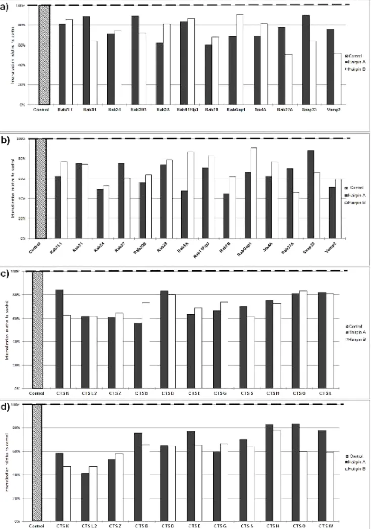
Figure 8. Efficiency of internalization for knockdown THP-1 macrophages ...23
Figure 9. Overview of the screen performed on vesicular traffic proteins. ...24
Figure 10. Overview of the screen performed on cathepsins and cystatins ...25
Figure 11. Western Blot for cathepsin L ...26
Figure 12. Images of macrophages with/without adherent extracellular bacteria ... 26
I
Acknowledgements
Pais. Não podia começar com outra palavra. É a eles que devo tudo o que tenho e tudo o que sou. É indescritível todo o apoio e amor que me têm dado, estou eternamente grato por me terem criado tão bem e me terem dado todas as oportunidades para ser quem eu quero ser... Eu assino a tese, mas o mérito é todo deles.
Só há outra pessoa no mundo com pais tão bons quanto os meus: o meu irmão. Somos tão parecidos que até chateia. Ele faz parte do que eu sou, e apoia-me e defende-me sempre, é um verdadeiro irmão mais velho.
Aos meus avós devo mais do que conseguiria enumerar. Criaram-me desde sempre, ensinaram-me como os verdadeiros professores que são e se consegui chegar longe na vida, é em muito graças a eles.
Aos Timóteos e aos Dias, primos e tios. São família, são insubstituíveis, e são o meu núcleo e o meu ADN.
À Professora Elsa Anes devo um enorme agradecimento, mas também uma grande admiração. A amabilidade com que me acolheu na equipa foi incrível, e todo o apoio que me tem dado nestes anos, particularmente nesta etapa final foi extremamente valioso e demonstrou uma enorme dedicação. Se eu vier a ser um grande cientista, já sei de quem é a culpa.
Aos colegas de equipa. Nuno, David, Paulo, João e Joanas. Foi um privilégio poder fazer parte de uma aliança tão acolhedora, e que tanto me ensinou. Este trabalho não passaria de um folheto se não fosse por eles. Obrigado por serem o meu meio de cultura!
Aos amigos, porque eu não seria o mesmo sem eles.
À Raquel, que me deste o juízo para ser uma pessoa como deve ser. Sei que posso contar contigo para tudo, e quer eu queira quer não queira vais estar a apoiar-me sempre.
Ao Tiago. Simultaneamente amigo e mentor. Sempre me puxaste na direcção certa e preocupas-te mais comigo do que eu próprio. Um bom modelo a seguir, é um privilégio ter a tua amizade. Por outras palavras, és melhor que comida para gatos.
Ao Pedro H, que faz parte desde que somos. Um Timóteo honorário, nem é preciso dizer mais.
II À Marta, a primeira amiga que fiz na Faculdade. E calhou-me a sorte grande. Percebes-me bem, acompanhaste-me nestes anos ainda melhor. Guardo muitas boas memórias dos tempos de faculdade, muito graças a ti, que estás lá sempre.
A toda a malta. Flávio, que é meu amigo mas não quer saber; Messias, que foi para longe mas está sempre perto; Margarida, que é minha amiga e já quer saber; Luís, mano mais velho que bem merece esse estatuto; Stufu, indescritível e insubstituível; Tiago-inho, a voz da razão e sempre com a cabeça no sítio; Splinter, um homem às direitas, como ele há poucos; Xangue, que me acompanha desde os tempos em que era um pequeno adolescente; Ricardo, o padrinho que sempre me mostrou que escolhi bem; Nathalie, parce que elle est toujours jolie et sympa, valeu?; Teresa, que me escuta sempre bem, mesmo quando digo disparates; Poças, a pessoa mais gentil e generosa a sul do Pólo Norte; Gameiro, honesto e sincero, de uma nobreza extraordinária, devia ser Sir; Diana, que põe o ‘di’ em ‘divertida’; Fred, sempre disposto a ajudar; Wilson, companhia impecável para pensar, filosofar e discutir cenas; Goofy, que de pateta tem muito pouco; Inês P, que ainda me atura sabe-se lá bem porquê; Rita, que bem tenta gozar comigo mas não consegue, é uma querida; Peixoto, disposto a tudo e feito de boas intenções; e a Sara, que teve de ler esta lista toda à procura do nome dela, não te preocupes, sabes que és relevante.
Tive a sorte de apadrinhar indivíduos realmente impecáveis na minha passagem pela Faculdade. Obrigado por me fazerem crescer, por me encorajarem, por me darem de volta setecentas vezes mais do que eu dei. Telma, a primogénita, António, Tiago, Dora, Bruno, Quina, Luísa, João, Marta, M.N. (assim está bom?), Sofia, Yann e já agora a Sara. Até mereciam um parágrafo para cada um, mas assim está tudo junto em família.
Estendo ainda o meu agradecimento à Faculdade de Farmácia da Universidade de Lisboa e ao CPM-URIA, pelo acolhimento que me permitiu levar este trabalho avante. Igualmente agradeço à Fundação para a Ciência e Tecnologia pelo financiamento que tornou tudo possível.
Agradeço à Professora Margarida Gama Carvalho o ter aceitado a orientação da minha tese e a disponibilidade que mostrou desde o início.
Dirijo ainda um sincero e respeitoso agradecimento ao Professor Rogério Tenreiro por me ter ajudado a entrar no mundo da ciência e por ter sempre demonstrado uma impressionante dedicação aos seus alunos. Se o mestrado é obra sua, esta tese também.
III
Communications in Scientific Meetings
Nuno Carmo, David Pires, Joana Bugalhão, Joana Marques, Paulo Bettencourt, Pedro Timóteo and Elsa Anes (2012). “Host factors affecting Mycobacterium tuberculosis macrophage infection”. Presented at the EMBO Conference – Tuberculosis 2012: Biology, Pathogenesis, Intervention and Strategies. Institute Pasteur, Paris, France. [Poster communication]
D. Pires, N. Carmo, J. Marques, J. Pombo, P. Timóteo, P. Bettencourt and E. Anes (2013). “Cut or be Cut: Cathepsins and their Inhibitors during mycobacterial infection of Macrophages and Dendritic Cells” presented at the 5th iMed.UL Postgraduate Students Meeting. iMed.UL, Faculty of Pharmacy of the University of Lisbon, Portugal. [Poster communication]
Pedro Timóteo (2013). “Deciphering host factors for mycobacteria internalization during phagocytosis”. Presented at the VI Cycle of Scienceshops: Tuberculosis: development of new strategies to treat tuberculosis. Faculty of Pharmacy of the University of Lisbon, Portugal.
IV
Relevant Abbreviations
HIV / VIH – Human Immunodeficiency Virus BCG – Bacillus Calmette-Guérin
MOI – Multiplicity of Infection
HMDM – Human Monocyte-derived Macrophages GFP – Green Fluorescent Protein
Rab GTPase – Guanosine triphosphatase belonging to the Rab family of proteins THP-1 – Human monocyte cell line THP-1 (ATCC®
TIB-202™) J774 – Mouse macrophage cell line J774A.1 (ATCC®
TIB-67™) shRNA – Small hairpin RNA
siRNA – Small interfering RNA
CD40 / CD17 – Cluster of differentiation receptor 40 / 17 MHC II – Major Histocompatibility Complex class II DMEM – Dulbecco’s Modified Eagle Medium FBS – Fetal Bovine Serum
RPMI – Roswell Park Memorial Institute (where this culture medium was developed) HEPES - 4-(2-hydroxyethyl)-1-piperazineethanesulfonic acid
M-CSF – Macrophage Colony Stimulating Factor EDTA – Ethylenediamine tetraacetic acid
HEK 293T – Human Embryonic Kidney cell line ATCC® CRL-11268™ PMA – Phorbol 12-myristate 13-acetate
PBS - Phosphate Buffered Saline OD – Optical Density
FITC - Fluorescein isothiocyanate
U-87MG – Human glioblastoma cell line U-87 MG (ATCC® HTB-14™) ESAT-6 – Early Secreted Antigenic Target Protein 6
V
Resumo
A tuberculose é uma doença infecciosa que tem acompanhado historicamente a Humanidade. Caracteriza-se tipicamente por uma infecção pulmonar, manifestando-se através de tosse persistente, febre e especialmente por uma fraqueza generalizada. Apesar da sua ancestralidade, é ainda hoje a doença infecciosa responsável pelo maior número de mortes a nível mundial.
O agente etiológico da tuberculose é uma bactéria, Mycobacterium tuberculosis, descrita pela primeira vez por Robert Koch no século XIX. Pertence à ordem Actinomycetales e à família das Mycobacteriaceae; são bacilos, Gram-positivos, e apresentam por norma um crescimento lento e uma membrana com uma composição invulgar, rica em lípidos particulares como o lipoarabinomanano.
M. tuberculosis é um patogénio particularmente resistente por se encontrar extremamente adaptado ao hospedeiro humano, e o problema agrava-se com a actual epidemia do vírus de imunodeficiência humana (VIH). Indivíduos infectados com VIH não só têm uma probabilidade maior de desenvolver tuberculose, como apresentam morbilidades mais elevadas, sendo a tuberculose a principal causa de morte entre doentes de VIH. Outro dos obstáculos mais significativos é a resistência a antibióticos, que nas últimas décadas se tem tornado mais proeminente em M. tuberculosis. Várias ocorrências de resistência aos antibióticos de primeira e segunda linha foram já documentados, e o surgimento de estirpes multirresistentes representa um sério problema de saúde pública. Continua a não existir uma vacina eficaz para substituir a BCG (Bacilo Calmette-Guérin, estirpe atenuada do agente da tuberculose bovina Mycobacterium bovis), que está em uso desde 1921, e cuja eficácia na protecção contra a tuberculose pulmonar em adultos é manifestamente insuficiente.
Muita da dificuldade em combater a tuberculose está também relacionada com a sua prevalência e persistência. Segundo a Organização Mundial de Saúde, um terço da população mundial pode ser portadora de M. tuberculosis no seu estado latente, ou seja, sem desenvolver doença activa. Ainda que a doença não seja contagiosa neste estado, a sua presença no hospedeiro pode vir a manifestar-se de forma oportunista em casos de enfraquecimento imunitário, como por VIH, drogas imunossupressivas, doença concomitante ou simplesmente pelo envelhecimento.
VI Por estas razões a investigação no combate à tuberculose está cada vez mais a dirigir-se no dirigir-sentido de compreender os sistemas de imunidade do hospedeiro e a sua relação com este patogénio.
A pedra angular da interacção patogénio-hospedeiro é a fagocitose. É um processo de imunidade inata altamente conservado evolutivamente através do qual células eucarióticas são capazes de internalizar e digerir partículas externas ou microorganismos, eliminando-os. Certas células do sistema imunitário humano servem-se deste processo para neutralizar patogénios invasores, denominando-se células fagocitárias, ou fagócitos, entre os quais se destacam os macrófagos. M. tuberculosis é tipicamente inalado por aerossóis e nos pulmões entra em contacto com os macrófagos alveolares residentes, que oferecem a resposta fagocitária inicial.
Da fagocitose fazem parte várias etapas. Tem início pela internalização das partículas, que consiste na captura e envolvimento do alvo numa vesícula endocítica. Esta é depois transportada pelo citoplasma, via elementos do citoesqueleto, e vai fundindo com outras vesículas endocíticas. Quando o grau de maturação é compatível funde-se com lisossomas, cujo conteúdo enzimático assegura uma degradação proteolítica do alvo. A este processo dá-se o nome de biogénese do fagolisossoma, e nele inicia-se a acção de proteases denominadas catepsinas, bem como óxidos nítricos e superóxidos de efeito bactericida. Depois de digerido o conteúdo fagocitado, os antigénios resultantes serão utilizados para despoletar uma resposta imunitária adquirida, levada a cabo por células T.
Quando a partícula a fagocitar é o M. tuberculosis, este é capaz de subverter os mecanismos de defesa do fagócito, conseguindo sobreviver e multiplicar-se no interior da célula hospedeira. Sendo complexo o arsenal molecular de que o fagócito dispõe para combater invasores, também o é o modo como esta micobactéria o evita. O M. tuberculosis consegue impedir a maturação do fagossoma, controlando o hospedeiro através de processos que ainda não são totalmente compreendidos. Sabe-se, porém, que influencia a função de proteínas de tráfego vesicular e transporte membranar, bem como as referidas catepsinas. Sendo estes alguns dos alvos da sua virulência, tornam-se também importantes alvos de investigação biomédica, pois podem ajudar a combater a estratégia de infecção de M. tuberculosis, fortalecendo as defesas do hospedeiro.
A fase de internalização das bactérias por macrófagos é um ponto crítico na infecção, uma vez que representa um dos primeiros contactos entre o patogénio e o hospedeiro, e é através dela que têm início todos os restantes mecanismos. Interessa pois compreender como é regulada a internalização, e que influência terá M. tuberculosis nessa regulação.
VII Para estudar devidamente factores de internalização, é necessário em primeiro lugar definir o modelo de internalização de partículas inertes para estabelecer o que ocorre na fagocitose como fenómeno abrangente.
Criámos um desenho experimental que nos permitiu explorar um grande número de variáveis. Fizemos variar o tipo de micobactéria: destruída no fagócito (caso de M. smegmatis) versus sobrevivente intracelular (caso de M. tuberculosis), o tempo, a multiplicidade de infecção (MOI, representa o número de bactérias por cada macrófago) e o tipo de macrófago. Para este trabalho foram utilizados três tipos de macrófagos: macrófagos derivados de monócitos humanos (HMDM), macrófagos humanos da linha celular THP-1 e macrófagos de ratinho da linha celular J774. Utilizámos também três espécies dentro do género Mycobacterium: M. tuberculosis, M. bovis BCG e M. smegmatis, sendo as duas últimas consideradas não-patogénicas. Obtendo os valores de internalização ao longo de três pontos no tempo (30 minutos, 1 hora e 4 horas) tornou-se possível traçar perfis de internalização para cada combinação de variáveis.
As estirpes à nossa disposição expressavam proteína verde fluorescente (GFP) por meio de um plasmídeo replicativo, o que permitiu que os macrófagos que internalizaram micobactérias pudessem ser identificados em citometria de fluxo através da fluorescência no comprimento de onda do verde.
Armados desse desenho experimental e de um protocolo de infecção eficiente que permitiu testar várias variáveis em simultâneo, descrevemos curvas de internalização para as várias combinações de macrófago-micobactéria. Destes resultados foi possível chegar à conclusão esperada de que a internalização é fortemente influenciada pelo tempo e pela multiplicidade de infecção, ou seja, estes factores condicionam fortemente a resposta fagocitária dos macrófagos. Tornou-se também evidente que os HMDM são os agentes fagocitários mais eficientes de entre os macrófagos testados, tendo sido capazes de atingir elevados níveis de internalização mais rapidamente que os restantes. Uma observação que se nos revelou pertinente foi a de que ao fim de apenas 30 minutos, todos os tipos de macrófagos foram capazes de realizar uma considerável internalização das micobactérias na amostra. Põe-se assim em causa muito do background experimental em micobactérias, que prevê infecções de duração igual ou superior a 3 horas. Este nosso trabalho sugere que infecções mais breves são não apenas funcionais, mas até preferíveis para o estudo de reguladores das fases iniciais de fagocitose, como é o caso da internalização. Infecções demasiado prolongadas podem levar à perda desta informação e a uma alteração do estado fisiológico dos macrófagos, particularmente com cargas bacterianas elevadas.
VIII Foi também importante observar que as diferentes espécies de Mycobacterium apresentam diferenças ao nível da internalização. M. smegmatis foi internalizada mais rapidamente e em maiores quantidades, seguida por M. bovis BCG e esta por M. tuberculosis. Ora, a esta ordem de internalização corresponde a ordem de inversa de patogenicidade, ou seja, com o aumento da patogenicidade diminui a internalização. Levanta-se imediatamente a sugestão de que a diminuição da internalização será um dos processos de virulência de M. tuberculosis. Parece haver uma contradição, pois sendo M. tuberculosis um patogénio intracelular esperar-se-ia que promovesse a sua internalização o mais possível. No entanto, uma carga microbiana demasiado elevada sobre as células do hospedeiro leva a processos de morte celular, privando assim M. tuberculosis do seu habitat. Força-se então um equilíbrio cuja regulação importa compreender.
Para investigar o papel de factores do hospedeiro que influenciem a internalização, realizámos uma triagem sobre 71 proteínas envolvidas em tráfego vesicular e na maturação do fagossoma, a maioria pertencente aos grupos das Rab guanosina trifosfatases (Rab GTPases), catepsinas e cistatinas. Pelo silenciamento de cada uma das proteínas individualmente torna-se possível avaliar a sua contribuição para o processo de internalização, ou seja, descobrir a sua função por redução ou ausência-de-função.
O silenciamento individual das proteínas foi feito por intermédio de bibliotecas lentivirais contendo plasmídeos que codificam para um pequeno RNA de interferência (hairpin), complementar ao RNA mensageiro de um gene específico. Graças a essa complementaridade o RNA mensageiro é destruído, impedindo-se assim a produção da proteína correspondente. Assim construímos uma linha knockdown de macrófagos THP-1, a qual submetemos a ensaios semelhantes aos que desenvolvemos na primeira parte dos trabalhos. Na abordagem experimental, as variáveis de tempo e MOI foram restringidas a apenas 1 hora e uma MOI de 10, pois estes valores representam um estímulo mais moderado e permitem assim realçar as eventuais diferenças provocadas pelos silenciamentos. Utilizámos nesta fase apenas M. tuberculosis e M. smegmatis na qualidade de controlo não-patogénico.
Nestas condições, a grande maioria dos silenciamentos provocou um decréscimo na internalização de micobactérias, o que levou a que as suas proteínas-alvo fossem classificadas como potenciais reguladores positivos da internalização. Aplicando limites para aquilo que deveria ser considerada uma diferença significativa face aos controlos não-silenciados, 26 proteínas emergiram como reguladores, sendo por isso denominados os hits da triagem. Este é um número surpreendentemente elevado, tendo em conta que as
IX bibliotecas lentivirais não foram desenhadas especificamente para ensaios de internalização.
Destes hits destacam-se especialmente aqueles que se manifestaram apenas em infecções com uma das micobactérias, pois permitem inferir uma relação directa com a patogenicidade. É o caso da catepsina L, identificada como hit apenas para M. smegmatis. Considerando que a ausência desta proteína levou a que M. smegmatis fosse internalizado em menor nível e, tendo presente a correlação demonstrada entre baixa internalização e patogenicidade, pode dizer-se que nesta situação M. smegmatis se comportou como uma micobactéria patogénica. Daqui se retira que o silenciamento da expressão de catepsina L pode ser uma das vias através das quais M. tuberculosis manifesta a sua patogenicidade.
Três outras proteínas demonstraram um papel regulatório na internalização apenas em infecções com M. tuberculosis: Rab37, Rab38 e catepsina W. Desta última são conhecidas funções na regulação da citotoxicidade de linfócitos, mas não é claro o seu mecanismo de acção. No caso de Rab37 e Rab38, este trabalho representa a primeira associação destas à infecção por M. tuberculosis. Tratando-se de uma triagem abrangente, só será possível definir o modo de funcionamento destas proteínas com novas experiências no futuro.
Os restantes 22 hits não são menos importantes; apesar de conhecida a sua associação à tuberculose, demonstraram pela primeira vez ser reguladores positivos da internalização.
Há limitações ao estudar infecções in vitro, pois as conclusões a retirar não são necessariamente igualmente válidas para o contexto real de infecções por M. tuberculosis. As diferenças acentuam-se se considerarmos que neste trabalho experimental se simulou um cenário de uma primo-infecção, ou seja, o primeiro contacto entre M. tuberculosis e células humanas, sendo este um cenário artificial que não corresponde à maioria dos casos de tuberculose em humanos. É uma concessão necessária para a investigação experimental e apesar de tudo não impede que estas descobertas tenham potencial terapêutico. As proteínas aqui identificadas poderão servir para controlar mais eficazmente a infecção, se aprofundado o conhecimento sobre a sua função.
Esta tese apresenta pois uma nova abordagem experimental, na forma de infecções breves e eficientes e de novos intervenientes nos primeiros passos do equilíbrio dinâmico entre patogénio e hospedeiro.
Palavras-chave: Mycobacterium tuberculosis, macrófago, fagocitose, internalização, triagem, citometria de fluxo, regulador positivo
X
Abstract
Mycobacterium tuberculosis is a very successful pathogen that is able to survive and multiply inside the host’s immune cells, thus triggering the active disease of tuberculosis or alternatively entering into an asymptomatic latent state. Its success lies on a powerful subversion of the fundamental innate immunity process of human-host cells: phagocytosis.
M. tuberculosis is known to resist destruction by phagocytes through a complex molecular sabotage of phagosome maturation, an integral part of the phagocytic process. On the other hand, internalization, the first and arguably most important step of phagocytosis, has not been so thoroughly investigated.
In this work we outline a method for quantifying internalization of green fluorescent mycobacteria through flow cytometry. With it we provide profiles of internalization-over-time for J774, THP-1 and human monocyte-derived macrophages (HMDM), which were infected with M. tuberculosis, M. bovis BCG and M. smegmatis. From these experiments we gathered that HMDM are the most efficient in internalizing mycobacteria, that there is considerable internalization even after short time periods such as 30 minutes and also that M. tuberculosis is internalized to a lesser extent than its non-pathogenic counterparts. It is suggested that M. tuberculosis manipulates its own internalization. In order to decipher which are the host factors targeted by this subversive strategy, we performed an internalization-based screening on knockdown THP-1 macrophages. Protein knockdowns were achieved by shRNA interference from lentiviral libraries targeting a total of 71 proteins. Using the previously designed method, we identified 26 putative positive regulators of internalization including a large number of Rab GTPases and cathepsins, proteins associated with vesicular trafficking and proteolysis in the mature phagosome.
Through this novel approach we were able to quantify rates of internalization, which were previously only empirically estimated, and use them as a model to highlight new potential therapeutic targets in the ongoing battle against tuberculosis.
Keywords: Mycobacterium tuberculosis, macrophage, phagocytosis, internalization, screen, flow cytometry, positive regulator
1
1. Introduction
1.1 General aspects of tuberculosis
Tuberculosis has a record as one of the leading causes of death worldwide. It was known to be responsible for one in every four deaths in nineteenth century Europe, and is at present still a very widespread and burdensome disease, responsible for 1.4 million deaths and 8.7 million new infected individuals each year, as estimated by the World Health Organization 1.
It is a disease that primarily affects the lungs, but can also cause systemic infections, and its symptoms include persistent coughs, often with blood or sputum, fever, weakness and weight loss. Infections on other organs yield different symptoms and are often difficult to diagnose.
The etiological agent of tuberculosis is a bacterium, Mycobacterium tuberculosis, which was first identified by the microbiologist Robert Koch in the late nineteenth century. It has been widely studied since, and we now know of many particular molecular features that make it a very persistent threat.
Despite the complex cellular and molecular mechanisms underlying tuberculosis, it is to an extent a poverty disease, in that it is strongly correlated to low living standards. Poverty
Figure 1. Estimated tuberculosis incidence rates. Map and data from the Global Tuberculosis
2 and malnutrition can be considered the strongest risk factors for tuberculosis. Four out of every five cases of tuberculosis occur in sub-Saharan countries, and 80% of all cases can be attributed to just twenty-two low-income developing countries (Figure 1)1.
The sequencing of the M. tuberculosis genome in 1998 provided a valuable boost in tuberculosis research2, but current progress in combatting this age-old disease has been threatened in the past decades by the serious challenges of Human Immunodeficiency Virus (HIV) and resistance to antibiotics. HIV greatly increases the susceptibility to tuberculosis, such that the latter is responsible for almost 25% of deaths among people infected with HIV, which are also twenty times more likely to contract tuberculosis. The world is experiencing an HIV epidemic that is especially prevalent on sub-Saharan Africa, the region in which tuberculosis is more widespread, so this bacterium-virus synergy presents a difficult hurdle to overcome. Apart from HIV, there are other ways by which an individual’s immune system can become compromised, such as immunosuppressive drugs, concomitant disease or quite simply old age, and so it becomes even clearer that poverty inescapably exacerbates the issue1.
As for antibiotics, multidrug-resistant strains are becoming increasingly common, appearing in around 10% of tuberculosis infections worldwide. In some particular eastern countries, like India and China, the incidence is much greater. The strongest first-line antibiotics rifampicin and isoniazid are growing ineffective3 and as a consequence much of the research in the field is turning towards the search of new therapeutic targets.
Research on tuberculosis has been granted a large funding recently, in an effort to eliminate tuberculosis as a major health problem by the year 2050. Some hope lies on a novel vaccine. The current vaccine, Bacillus Calmette-Guérin (BCG) has been in use since 1921. It is the most used vaccine in the world, and is still administered to all newborns in Europe. Although it is capable of preventing tuberculous meningitis in children, its efficacy is severely low in protecting against pulmonary tuberculosis in adults. Moreover, there is a risk of disease for immunodeficient patients and children of HIV-positive mothers1.
Current research aims to develop treatments that rely on boosting the host’s immune response in a way that is fast and cost-effective. Antibiotic-based therapies can take up to several months, which increases the risk of antibiotic resistance and also struggle with accessibility towards developing countries and isolated populations.
Hopefully, more natural and effective treatments will stem from the study of the host-pathogen relationship, aided by the recent advances in proteomics and systems biology.
3
1.2. Biology of Mycobacterium tuberculosis
M. tuberculosis is a bacillus belonging to the family Mycobacteriaceae, within the phylum Actinobacteria. It is nonmotile, does not form spores and grows better on high-oxygen environments, hence its perceived preference towards the lungs. Mycobacteria have a particular type of cell wall that is rich in lipoarabinomannan and do not possess an outer cell membrane. Moreover, they are considered Gram-positive but recent electron microscopy associated with tomography showed an outer membrane similar to Gram-negative species. They are acid-fast bacilli, as they resist discoloration by acids. Its particular membrane structure is thought to play an important role in infection. Lipoarabinomannan is analogous to Lipopolysacharides, a common bacterial antigen known to trigger an aggressive immune response, but many other lipids from mycobacteria were shown to be involved in virulence. Another consequence of their unique cell wall is a resistance to several antibiotics that target cell wall biosynthesis, such as penicillin4.
The genus Mycobacteria is divided into two groups based on growth rate. M. tuberculosis is a slowly growing mycobacterium, with a generation time of between 15 and 20 hours. This slowness is relevant to its virulence because it forces the mycobacteria to be internalized into macrophages, where they encounter the conditions to multiply freely: they are facultative intracellular pathogens.
1.3. Host-pathogen relationship
Mycobacterium tuberculosis shows a marked adaptation to the human host, and its resistance to the natural human defense mechanisms suggests a very ancient relationship5. The association was thought to have begun upon the first domestications of cattle, in the Neolithic period, and M. tuberculosis was thought to have been a human variant of the bovine counterpart Mycobacterium bovis, the etiological agent of bovine tuberculosis. Recent studies instead suggest the pathogen’s affinity towards humans extends further back in time, to the first African populations that subsequently migrated to Europe, Asia, and the rest of the world around 125 000 years before present 6.
The known relationship between humans and M. tuberculosis actually challenges definition. The complex nature of their interaction is not a linear host-pathogen scenario, for in most cases of infection by M. tuberculosis, there is no active disease: the pathogen simply remains dormant inside the host’s macrophages, and can evade immune surveillance for years or even decades without causing any symptoms: this is called a latent
4 infection7. The World Health Organization estimates that one in every three people is a carrier for M. tuberculosis, while the prevalence of active disease is largely inferior1. The bacterium’s discreet dormancy state favors its continued existence8. It is only when the immune system is weakened that tuberculosis becomes active, and given that this is the only contagious stage of the disease, it can be seen simply as a way for the bacterium to seek a new host when the current one fails.
Only a small percentage (5-10%) of infected individuals develops the active and contagious stage of the disease throughout their lifetime. Another unknown number of people will have contracted and successfully eliminated the pathogen9. Given that the vast majority of the carriers of M.tuberculosis suffer no negative consequences from it, there are grounds to question the nature of this paradoxical host-pathogen relationship. At the very least, it can be one of commensalism (i.e. the bacterium benefits without harming the host) but it may be considered a case of mutualism, as the presence of the bacteria inside the host’s immune cells is likely to stimulate them via production of interferon-γ and other inflammatory cytokines, making them more effective in responses to other extraneous pathogens10.
The process that defines which path M. tuberculosis and indeed which path the disease will take is phagocytosis.
1.4. Phagocytosis
Phagocytosis is a highly-conserved process by which invasive particles are captured, internalized and neutralized by a host cell. These particles may be inert (for example, debris or latex beads) or living, such as bacteria or small eukaryotes. The term phagocytosis encompasses the steps of internalization, digestion, recycling and exocytosis of debris (Figure 2), although it may also be used to refer exclusively to the internalization step. Many human cells have the capacity to phagocytize, such as neutrophils, dendritic cells, monocytes and macrophages, all of which are considered professional phagocytes11.
M. tuberculosis is known to act upon phagocytosis: its success as a pathogen relies precisely on its sabotage of the otherwise very efficient phagocytic mechanisms, in a complex molecular tug-of-war.
5
1.4.1 Macrophages
In the context of an infection by M. tuberculosis, which commonly enters through the lungs in aerosols, the first host cells it encounters and invades are the alveolar resident macrophages12.
The alveolar macrophages in the lungs provide one of the first lines of defense against the invading pathogen. Peculiarly, macrophages are both the antagonist and the habitat of M. tuberculosis, thus it is crucial to understand the macrophages’ physiology and the equilibrium between the two.
Macrophages can be in either a resting or an activated state. Activation may be triggered by bacterial antigens of inflammatory signals and induce significant morphological and molecular changes on the macrophage, the most relevant of which is the release of pro-inflammatory cytokines13. During a pulmonary infection, M. tuberculosis is phagocytized by resting alveolar macrophages, which then become activated and promote an inflammatory response14. Following activation, the macrophages become primed and generally more aggressive against M. tuberculosis, achieving higher levels of intracellular killing of bacteria15.
1.4.2 Internalization
The first step in phagocytosis is internalization, but many events must take place before the bacteria are truly engulfed by the macrophages. Internalization is mediated by cell surface receptors and ligands, with varying degrees of specificity, which bind to and promote the uptake of M. tuberculosis. Identified receptors include complement receptors, scavenger receptors, surfactant protein receptors, C-type lectins, Tumor Necrosis Factor receptors (e.g. CD40) and glycosylphosphatidylinisotol-anchored receptors (e.g. CD14)16. Toll-like receptors are also relevant in the context of infection, bringing about the activation of the cytokine network and consequently the innate immune response17. A relevant and well-characterized ligand of M. tuberculosis is lipoarabinomannan, which often contains terminal mannose residues, thus becoming recognizable by the macrophage’s CD14 receptors as well as the mannose receptors18.
Internalization is an important step in determining the outcome of the host response to the bacilli, as it represents the first physical contact between host and pathogen19. However, the results of this contact are not limited to internalization. The activation of receptors, and their type, will ultimately steer the response of the immune system. For example, both macrophages and dendritic cells express T-cell co-stimulants, triggering a
6 more thorough immune response17. If phagocytosis is completely effective, processed M. tuberculosis antigens are presented to T cells via the Major Histocompatibility Complex of the macrophages, thus bridging the innate and acquired immune systems and neutralizing the invading pathogen20.
The vesicles formed by internalization of bacteria or particles can vary in composition and size depending on what is being internalized, but all fall under the designation of phagosome – a phagocytic endosome. The uptake of bacteria is certainly a complex process. The citoplasmic membrane at the phagocytic cup and the cytoskeleton require significant structural rearrangement in the early stages of phagocytosis in order to form the pseudopods, engulf the bacteria and encase them in the phagosome. Rather than a simple shift in position of membrane lipids, there is recruitment from the endocytic pathway21. As for the cytoskeleton, it has already been demonstrated that M. tuberculosis inhibits actin assembly22, suggesting that it is capable of interfering with its own internalization by macrophages and with the transport events leading to phagosome maturation.
7
1.4.3. Phagosome maturation
From early to late stages of phagocytosis, the phagosome containing the internalized bacterium suffers a series of maturation events, which consist in partial fusion and fission events with trafficking vesicles. This allows the exchange of luminal and membrane components that will turn the phagosome “older” or more mature, meaning more competent to fuse with lysosomes. This requires organized rearrangement of membrane and cytoskeletal components. After the fusion, the mature phagolysosome presents a very hostile environment for the internalized bacterium: it becomes rich in hydrolytic enzymes, nitric oxides and superoxides21.
The internal space of the phagosome, pH-neutral by default, is acidified, by way of strong adenosine triphosphatase activity in its membrane, and the lower pH promotes activation of proteases and their regulators. In standard cases as in phagocytosis of non-pathogenic bacteria or inert particles, this would be sufficient for the invading pathogen to be destroyed, its remains recycled and transported to the Major Histocompatibility Complexes II (MHC II) for antigen presentation.
However, it is upon these processes that M. tuberculosis is able to act, by inhibiting several of the host cell’s defenses: the acidification itself, the activation and action of proteases and the fusion of the early endosome with lysosomes. This leads to an incomplete and immature phagosome23, which has been referred to as the “four minute phagosome” precisely because it persists in a stage which would be commonly found at the four-minute mark in a standard phagoytic event24.
In this stage, M. tuberculosis may achieve its latent status, or it may remain active and even multiply inside the host cell. The entry into latency requires a major shift in the gene expression of the Mycobacterium, and that state may actually be enforced by the macrophage as a defense mechanism. In either case, the development is the result of biochemical dialogue between pathogen and host25.
There has been recent speculation on whether M. tuberculosis is capable of escaping the phagosome into the cytosol, in a way similar to Salmonella sp. or Listeria sp.26 . While the cytosol would provide a rich environment for the mycobacterium, there is still no conclusive evidence of this escape, and electron microscopy studies have so far failed to replicate the initial results that originated this hypothesis.
8
1.4.4. Vesicular traffic
Phagocytosis is, as a whole, a matter of intracellular traffic, with each step requiring biogenesis, transport or fusion of vesicles. These vesicles are not exclusively derived from the plasma membrane, as there is also transport to and from the endomembranes of the endoplasmatic reticulum, the trans-Golgi network and of course lysosomes27. For there to be an effective neutralization of the captured pathogen, the sequential processes of the endocytic route and phagosome maturation must be adequately coordinated. Rab guanosine triphosphatases (Rab GTPases) are a particularly important group of proteins that regulate membrane identity and direct the traffic and fusion events that play a major role in phagosome maturation28,29.
M. tuberculosis is able to interfere with these vesicular traffic events and as part of its survival strategy. Through down-regulation of several GTPases, M. tuberculosis blocks vesicles bound for the cytoplasmic membrane to form the phagocytic cup and also denies the fusion events leading to phagolysosome biogenesis30. Two examples of interest are Rab5 and Rab7. The first is involved in in the early stages of phagocytosis, coordinating endocytosis from the plasma membrane and fusion between endosomes31. Rab7 acts in the later stages, regulating transport from the early to the late endosomes32. It has been shown that M. tuberculosis and even M. bovis BCG can affect these processes, limiting the expression and recruitment of those proteins even from within the endosome33.
Rab GTPases are potential targets for protecting against infection, as they are crucial for the fusion events leading to the mature phagolysosome. Not only that, but some Rab GTPases have been reported to also recruit cathepsins for the later stages of phagocytosis34.
1.4.5. Cathepsins and Cystatins
Cathepsins are proteases that exist in all animals, and are more commonly found in lysosomes. There are several classes of cathepsins, based on the aminoacid residue in the enzyme’s active center and their targets; most are cysteine proteases, such as cathepsins B, S or L.
The functions of cathepsins are not restricted to phagocytosis; quite the opposite, as their study and applications are very extensive and wide-reaching, including cancer research. They are, however, also studied as potential targets in regulating the host-pathogen interaction in M. tuberculosis infections, because they play an important role in the maturation and degradation events on the phagosome35. Specifically, they are important
9 in order to degrade the phagocytized bacteria into small peptides which can be delivered to the Major Histocompatibility Complexes, which in turn trigger the T-cell-mediated immune response36.
The majority of cathepsins require a low pH in order to function: many are self-regulated, by folding or autocatalysis that inactivates them while the environment is not sufficiently acidic37. If M. tuberculosis is able to prevent acidification, it also protects itself from the proteolytic action of cathepsins38.
Cystatins are known to be regulators of cathepsin activity39; therefore their role in the phagosome environment is also important, as is any interaction they may have with M. tuberculosis.
1.5. Objectives
The study of Mycobacterium tuberculosis is, as we’ve shown, inseparable from the study of macrophages, and their interaction is the essential part of the disease of tuberculosis. The laboratorial standard to study this interaction is the infection experiment, which is typically performed as an “end-point experiment”, whereby the infections with M. tuberculosis are usually carried over long periods of time (3 to 4 hours40).
The internalization process itself, via the cell membrane, is a complex process reliant on numerous signaling pathways. The molecular mechanisms behind them must be taking place very early on, certainly before the 3 to 4 hours of contact predicted in most protocols.
We therefore seek to resolve this incongruence between the theoretical and practical aspects of the internalization process on what constitutes an adequate infection time. We aimed to measure the internalization rates with several combinations of different macrophages and different mycobacteria, to obtain fundamental information on how the infection scenario is proceeding since its very early stages.
Using three time points with which to quantify the percentage of infected macrophages, we provide means to determine internalization rates by flow cytometry, and this can be done for several combinations of macrophages and mycobacteria.
We are then able to set a base model to use as a tool in order to screen for host factors influencing internalization. By measuring changes in internalization brought about by protein knockdowns, we are able to study the roles of those proteins by decrease or absence-of-function.
10 We targeted familiar proteins, with known functions in mycobacterial infections, including cathepsins, cystatins and Rab GTPases. The focus on internalization is an effort to complement the ongoing study of mycobacteria-host interactions in our laboratory, which in the past has experimented extensively with these same targets, measuring the survival of internalized mycobacteria via counting of colony forming units. We hope to uncover a clearer picture of the early regulatory processes happening in phagocytosis by using different techniques, such as flow cytometry and new experimental conditions, such as brief infections with a minimum of 30 minutes.
With these experiments, we also set the stage for M. tuberculosis to interfere so that we can study what its influence is on internalization and on the host factors that control it.
To conclude, the goals of this work were:
To define the internalization rates of the several macrophages and mycobacteria that are used in tuberculosis research in order to compare their efficiency in the phagocytic process;
To describe novel roles of certain proteins with previously unknown functions in the internalization process.
To evaluate the influence of mycobacteria, particularly M. tuberculosis, on the internalization process.
11
2. Materials and Methods
2.1. Mycobacteria cultures
Mycobacterium tuberculosis strain H37Rv and Mycobacterium bovis BCG were cultured in 7H9 medium supplemented with 10% oleic acid albumin dextrose catalase and 0.05% tyloxapol. All manipulations of M. tuberculosis were performed in a Biosafety Level 3 environment.
Mycobacterium smegmatis (mc2 155) was grown in liquid culture medium containing 4,7g/l Middlebrook 7H9 broth and 5g/l Nutrient broth (DifcoTM), supplemented with 0.05% tyloxapol and 0.5% glucose.
Each of these species of mycobacteria harbors a plasmid constitutively expressing Green Fluorescent Protein (GFP) and a hygromicin resistance as a selection marker. The culture medium contained 50 µg/ml hygromycin (Sigma-Aldrich©) in order to maintain selective pressure on the plasmid carriers.
All cultures were kept at 37°C and were subcultured until achieving exponential phase before being used on experiments: 7-10 days beforehand for M. tuberculosis and M. bovis BCG, but only overnight for the fast-growing M. smegmatis.
2.2. Cell cultures
The murine macrophage cell line J774A.1 (ATCC® TIB-67TM), henceforth referred to as ‘J774’, was cultured in Dulbecco’s Modified Eagle Medium (DMEM, Gibco®) supplemented with 10% (v/v) fetal bovine serum (FBS), 1% (v/v) glutamine and 1% antibiotic – penicillin-streptomycin (v/v).
The human monocyte cell line THP-1 (ATCC® TIB-202TM) was cultured in RPMI 1640 medium supplemented with 10% (v/v) FBS, 1% (v/v) glutamine, 1% (v/v) antibiotic – penicillin-streptomycin, 1% (v/v) HEPES buffer, 1% (v/v) sodium pyruvate and 1% (v/v) non-essential amino acids.
The formulations described above are also referred to as ‘complete’ DMEM or RPMI media. If antibiotic is not included, they are referred to as ‘infection’ media.
Primary human monocyte-derived macrophages were obtained from blood from donors. Monocytes were first isolated according to the protocol from Miltenyi Biotec, using the MidiMACS kit and LS columns. For the differentiation of monocytes into macrophages, 300,000 cells were seeded on a 24-well plate with 300 µl RPMI medium per well and
12 allowed to adhere to the plate bottom. After 2 hours, 300 µl of RPMI with 20% (v/v) FBS, 2% (v/v) penicillin-streptomycin, 2% (v/v) sodium pyruvate, 2% (v/v) β-mercaptoethanol and 0.6 µl of macrophage colony stimulating factor (M-CSF 1000x) for a final concentration of 20 ng/ml in each well. After approximately 72 hours 600 µl of complete RPMI containing 20 ng/ml M-CSF were added. At the seventh day the macrophages were collected via resuspension with a trypsin-EDTA solution and were ready to be plated for the infection experiments.
The HEK 293T cell line (ATCC® CRL-11268TM) was used to amplify the viral constructs of the lentiviral knockdown libraries. They were maintained in DMEM supplemented with 10% (v/v) fetal bovine serum and 1% (v/v) antibiotic – penicillin-streptomycin.
All of the above were cultivated at 37°C in an atmosphere containing 5% carbon dioxide.
2.3. Infection protocol
2.3.1. Preparation of macrophages
Cells were seeded onto 24 or 12-well plates (approximately 500,000 cells per well) with the appropriate culture medium (complete DMEM or RPMI).
THP-1 cells were plated for 24 hours in contact with 20 nanomolar phorbol 12-myristate 13-acetate (PMA, Sigma©), which differentiates the monocytes into macrophages, followed by 24 hours of resting in complete culture medium without PMA.
J774 macrophages were plated 24 hours in advance at half the desired concentration, so that they would multiply to the intended final concentration on the day of the experiment.
(See section 2.3.5 for alternate protocol) 2.3.2. Preparation of bacteria
Disassembly of bacterial aggregates is necessary as large clumps may have a strong influence on phagocytosis. The steps involved are: washing with Phosphate Buffered Saline (PBS) at 1x concentration; resuspension in infection medium (DMEM or RPMI medium according to the cells to be infected); passaging several times with a 5ml syringe and a 21G needle; a 5-minute ultrasound bath followed by a short centrifugation at a low force: 1 minute, 300 G to pellet any remaining aggregates. After the last centrifugation, only the supernatant was collected.
13 By reading the optical density at 600 nanometers, bacterial suspensions were adjusted for concentrations corresponding to multiplicities of infection (MOI) of 1, 10 and 100 bacteria per macrophage. This calculation was based on the standard relationship between optical density (OD) and concentration of bacteria:
{OD 0.1 <=> 1*107 bacteria/ml}
2.3.3. Infection
The contact between macrophages and bacteria was initiated by removing the cell culture medium and replacing it with the bacterial suspension in infection medium.
To achieve the three different lengths of infection, the samples were infected at different times, so that afterwards they could all be stopped and processed at the same time. Considering the stopping time of the infection as the “time point –zero”, samples were infected at –4 hours, –1 hour and –30 minutes. Between manipulations, samples were incubated at 37°C in an atmosphere containing 5% carbon dioxide.
(See section 2.3.5 for alternate protocol) 2.3.4. Processing samples after infection
While remaining adhered to the plate, the cells were washed three times with warm PBS, thus removing the mycobacteria. Cells were then dislodged using 400µl trypsin with a 5-minute incubation at 37°C. Afterwards the cells were resuspended in 1 ml of infection (antibiotic-free) DMEM or RPMI medium to neutralize the proteolytic action of trypsin. This was followed by centrifugation for 10 minutes at 450 G. Fixation was then performed with 4% paraformaldehyde, for 30 minutes at room temperature, and the samples were then finally washed and resuspended twice in PBS with 1% fetal bovine serum and kept at 4°C until they were used for flow cytometry.
(See section 2.3.5 for alternate protocol)
2.3.5. Alternate protocol for screening of knockdown cells
The experiments regarding protein knockdown were restricted to the time point of 1 hour and the MOI of 10 mycobacteria per macrophage. This was necessary to accommodate the large number of samples – 181. The values were chosen because they present a more moderate burden on the macrophages and at the same time allow for more expressive differences across all cell types.
14 The experimental handling itself differs from the previous sections in that it was performed on 96-well microplates in proportionally smaller volumes. The infection and post-infection protocols were otherwise identical to those described before.
2.3.6. Confirming the absence of extracellular bacteria
Extracellular bacteria adherent to the macrophage, but not internalized, can compromise the readings of flow cytometry. To determine whether the post-infection protocol was effective in separating bacteria from the macrophages, a parallel experiment was conducted. Wild-type J774 macrophages were first plated in two different conditions: either onto glass coverslips placed on the plate wells (group A) or onto the plate wells’ bottom (group B). Both samples were infected with M. smegmatis with a MOI of 100 for 1 hour. Group B was subjected to the entire post-infection protocol, while group A was not. All samples were stained with 25µg/ml of ethidium bromide at the end of the infection. Extracellular mycobacteria stained red, while internalized mycobacteria emitted only green fluorescence from GFP.
Samples were placed on microscope slides and observed under a Zeiss Axioskop 40 fluorescence microscope (Göttingen, Germany) using a FITC filter. Besides qualitative observations of the relative abundance of red mycobacteria around the macrophages, quantification was also made: from a random sampling, we determined the percentage of macrophages with neighboring red mycobacteria (Figure 12).
2.4. Protein knockdown
2.4.1. Knockdown lentiviral libraries
Two different lentivirus libraries were used for the protein knockdowns. The first, obtained from The RNAi Consortium, contained hits from a previous screen performed in our laboratory, whereby knockdowns of several membrane-associated proteins were shown to affect intracellular survival of mycobacteria in macrophages (Carmo, N. et al, unpublished data). The second library includes target siRNAs for several cathepsins and cystatins and was built in partnership with Professor Luís Moita from the Institute of Molecular Medicine (Lisbon, Portugal) and by using MEROPS, a protease database from the Sanger Institute (Cambridge, United Kingdom).
15 Both libraries were contained in 96-well microplates with plKO.1 plasmids which coded for a different short specific RNA hairpin, lentiviral proteins and the resistance to puromycin as a selection marker. Each hairpin has a specific target on the host’s messenger RNA.
2.4.2. Building a stable THP-1 knockdown cell line
Part of this work was performed on knockdown THP-1 cells. Applying the lentiviral library to wild-type THP-1 cells is a multi-step approach.
The plasmids contained in the library were transfected into Escherichia.coli via electroporation. After overnight growth at 37°C with mixing (220 revolutions per minute), the DNA was extracted, in plasmid form, from the bacteria, using a PureLink™ Quick Plasmid Miniprep Kit and its respective protocol (Invitrogen).
The DNA was then quantified using picoGreen, a fluorescent nucleic acid stain, and its corresponding protocol (Life Technologies). The plasmids were co-transfected onto HEK 293T cells together with additional lentiviral proteins. These provide the necessary materials for the assembly of mature viral particles. The HEK 293T cells then began producing these particles containing the desired DNA construct, whilst being selected for transfection via the addition of 5µg/ml puromycin to the growth medium.
After 24 hours, the virus particles were passaged into DMEM containing 30% fetal bovine serum and collected 24 hours later. These virus particles were used to infect wild-type THP-1 monocytes, on a 96-well microplate. The monocytes were maintained in culture medium with 5 µg/ml puromycin and passaged or split into new microplates to obtain larger numbers of cells with a stable knockout.
Before infection, THP-1 cells were counted, normalized and plated on a fresh 96-well microplate at a concentration of 375 000 cells/ml.
The negative controls for the protein knockdowns were infected with viruses that contained an RNA hairpin with no target in the human genome, meaning that it would not bind to any messenger RNA of the host cell nor silence protein production. These hairpins contain a scrambled sequence and are called shScramble.
2.4.3. Confirming protein knockdown via Western Blot
A stable line of knockdown THP-1 monocytes were denatured at 95°C for 5 minutes in the presence of Laemmli buffer (Bio-rad laboratories©) and loaded onto 12% sodium dodecyl sulphate polyacrilamide gels. Electrophoresis was set at 200 Volt for 1 hour. Proteins were transferred onto nitrocellulose membranes with an electrical intensity of 30
16 Volt overnight at 4°C. On the following day, membranes were blocked for 1 hour at 20°C with a blocking buffer containing TBS-T (Tris-buffered saline with 0.05% (v/v) Tween 20) plus 5% (w/v) bovine serum albumin. Membranes were then incubated with primary antibodies diluted in TBS-T plus 1% (w/v) bovine serum albumin for 2 hours at 20°C. After three washes with TBS-T, the membranes were probed with horseradish peroxidase-conjugated secondary antibodies for 1 hour at 20°C. After three more washes with TBS-T, the membranes were stained for 5 minutes using the chemiluminiscent reagent Luminata Crescendo Western HRP Substrate (Milipore©). Chemiluminiscence was acquired with ChemiDoc XRS+ system (Bio-rad laboratories©)
2.5. Data acquisition and analysis
2.5.1. Quantification of internalization by flow cytometry
Samples were read on a Milipore EasyCyte H6 Guava flow cytometer, using its proprietary InCyte software, as well as the freeware Flowing Software (Turku Centre for Biotechnology, Finland). Fluorescence was measured using the excitation and emission wavelengths of 488 and 525 nanometers with a 75 miliwatt blue laser. The macrophage population was gated based on forward and side scatter values, in such a way that extracellular bacteria and debris were excluded from the event count. 5000 events from inside that gate were acquired with an average rate of 260 events per second.
The mycobacteria constitutively expressed Green Fluorescent Protein, while the macrophages did not, and so internalization was measured by quantifying the percentage of total macrophages that emitted intense green fluorescence (Figure 3). Uninfected macrophage samples were used as negative controls to calibrate the readings of green fluorescence intensity for each experiment.
Using the values from different time points, the kinetics of internalization were determined and plotted using a custom function in R software. Data points were connected to create a growth-like profile for the internalization rates, which include the average values as well as the standard deviation for each data point (Figures 4, 5 and 6). All experiments were performed in triplicate.
2.5.2. Statistical treatment
The software SigmaPlot 12.0 (Systat Software Inc.) was used for statistical analysis. Three-way ANOVAs were performed to evaluate the global differences in internalization
17 between different macrophages and different bacteria by taking into account the variables of time and MOI. Pairwise differences between types of macrophages and types of mycobacteria were evaluated using the Holm-Sidak method. Differences were considered significant for p<0.05.
2.5.3. Labelling hits in the knockdown screen
To evaluate whether the effect of knocking down the selected proteins was relevant for internalization, a criterion had to be fulfilled: two or more hairpins silencing each protein had to cause a difference in internalization of at least 2.5 times the standard deviation of the controls. This difference must necessarily have the same direction (positive or negative) among the considered hairpins.
18
3. Results
3.1. Rates of internalization
For this part of the experiment, the variables tested were: type of macrophage, type of mycobacteria, time and multiplicity of infection (MOI).
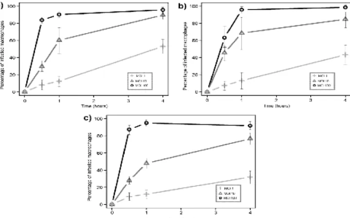
Figure 3. As described in section 2.5.1. Example populations of uninfected and infected macrophages, as obtained by flow cytometry and represented in histograms. Left: Uninfected control sample of THP-1 macrophages. Right: Sample of THP-1 macrophages infected with Mycobacterium smegmatis for 4 hours with a multiplicity of infection of 1. X axis represents intensity of green fluorescence in a logarithmic scale. Y axis represents number of events. The two samples are taken from the same experiment. With flow cytometry software we calculated the percentage of total events that are in each population.
Figure 4. Internalization by J774 mouse macrophages of different mycobacteria: a) M. smegmatis; b) M. bovis BCG; c) M. tuberculosis. The X-axis represents time, while the Y-axis represents percentage of infected macrophages. Each data point includes the average and standard deviation of three replicate experiments.
19 On Figure 4, detailing the internalization profiles of J774 mouse macrophages, we can see a marked difference between multiplicities of infection, as they maintain some graphical distance, with no overlap. With a MOI of 100 all of the mycobacteria achieved an almost ubiquitous infection of the macrophages, which is to be expected from such a heavy bacterial burden. The avirulent M. smegmatis is not only internalized faster, but also to a greater extent, reaching a higher percentage of infected macrophages than the other mycobacteria (although these differences were deemed non-significant by the Holm-Sidak pairwise comparison, p>0.05). M. tuberculosis appears to be internalized at a lower rate than the other mycobacteria during the first hour of contact with macrophages, which is particularly evident in the curve corresponding to a MOI of 1, which barely reaches 20% at the end point.
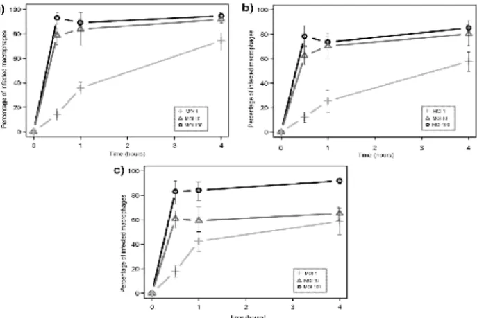
In Figure 5 we see no great difference between the species of mycobacteria, other than that M. smegmatis is slightly more internalized than its virulent counterparts. As will become evident in other experiments, the MOI of 100 tends to fall on extreme values, that is, extremely close to 100% of infected macrophages. M. tuberculosis shows a somehow
Figure 5. Internalization by THP-1 macrophages of different mycobacteria: a) M. smegmatis; b) M. bovis BCG; c) M. tuberculosis. The X-axis represents time, while the Y-axis represents percentage of infected macrophages. Each data point includes the average and standard deviation of three replicate experiments.
20 slower internalization when present in lower numbers (i.e. with a MOI of 1 or 10), indicated by a less steep curve, although it is able to recover the difference by the end of the experiment. Nonetheless, there is substantial difference in the kinetics of internalization of M. tuberculosis compared to the other mycobacteria at the early stages. Global differences were evaluated with the Holm-Sidak method, which determined the differences between M. tuberculosis and other mycobacteria were significant (p<0.05). The apparent decline on the 4 hour mark of MOI 100 in Figure 5.c is buffered by the standard deviation and is not likely to represent an actual decrease in internalization.
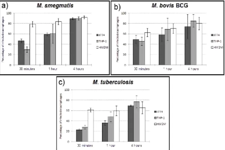
Human monocyte-derived macrophages (HMDM), represented in Figure 6, appear to reach a plateau earlier on, as we can see a shorter difference between the time points of 1 hour and 4 hours, which is all the more evident in the case of M. tuberculosis. The short decrease observed between the first and second time points is odd, although the values increase again towards the 4 hour mark; this will be addressed in the Discussion.
Figure 6. Internalization by human monocyte-derived macrophages of different mycobacteria: a) M.
smegmatis; b) M. bovis BCG; c) M. tuberculosis. The X-axis represents time, while the Y-axis
represents percentage of infected macrophages. Each data point includes the average and standard deviation of three replicate experiments.