Benign versus life-threatening causes of
pneumatosis intestinalis: differentiating CT
features
Sujin Ko1 Seong Sook Hong1 Jiyoung Hwang1 Hyun-joo Kim1 Yun-Woo Chang1 EunJi Lee1
1. Department of Radiology, Soonchunhyang University Seoul Hospital, Seoul, South Korea
http://dx.doi.org/10.1590/1806-9282.64.06.543
SUMMARY
OBJECTIVE: To assess the diagnostic performance of CT findings in differentiating causes of pneumatosis intestinalis (PI), including benign and life-threatening causes.
METHODS: All CT reports containing the word “pneumatosis” were queried from June 1st, 2006 to May 31st, 2015. A total of 42 patients with PI were enrolled (mean age, 63.4 years; 23 males and 19 females) and divided into two groups on based on electronic medical records: a benign group (n=24) and a life-threatening group (n=18). Two radiologists reviewed CT images and evaluated CT findings including bowel distension, the pattern of bowel wall enhancement, bowel wall defect, portal venous gas (PVG), mesenteric venous gas (MVG), extraluminal free air, and ascites.
RESULTS: CT findings including bowel distension, decreased bowel wall enhancement, PVG, and ascites were more commonly identified in the life-threatening group (all p<0.05). All cases with PVG were included in the life-threatening group (8/18 patients, 44.4%). Bowel wall defect, extraluminal free air, and mesenteric venous gas showed no statistical significance between both groups.
CONCLUSION: PI and concurrent PVG, bowel distension, decreased bowel wall enhancement, or ascites were significantly associated with life-threatening causes and unfavorable prognosis. Thus, evaluating ancillary CT features when we encountered PI would help us characterize the causes of PI and determine the appropriate treatment option.
KEYWORDS: Pneumatosis cystoides intestinalis. Pneumoperitoneum. Intestinal perforation. Mesenteric ischemia.
DATE OF SUBMISSION: 13-Sep-2017 DATE OF ACCEPTANCE: 25-Oct-2017
CORRESPONDING AUTHOR: Seong Sook Hong
Department of Radiology, Soonchunhyang University Seoul Hospital. 59, Daesakwan-ro, Youngsan-gu, Seoul, South Korea - 04408 Tel;82-2-709-9396 Fax 82-2-709-9066
E-mail: [email protected]
INTRODUCTION
Pneumatosis intestinalis (PI) is a radiographic or physical finding characterized by gas infiltration into the wall of the intestine. The clinical signifi-cance of PI can vary as it is the result of benign or life-threatening medical conditions and also can be an incidental finding1-3. Although the
pathophysiol-ogy of PI remains unclear, three mechanisms have been proposed as the cause of intestinal wall gas: (1) intraluminal gas entering the bowel wall through
mucosal breaks, which may cause gas spread along the mesentery4,5; (2) luminal bacteria producing
ex-cessive amounts of hydrogen gas, causing intestinal luminal pressure increase and resulting in direct-ly-forced gas trapped within the submucosa6,7; and
(3) pulmonary gas from alveolar rupture, coursing through the mediastinum to the retroperitoneum and mesentery8.
PI is traditionally considered a sign of bowel wall infarction and a surgical emergency, especially in
cases associated with portomesenteric venous gas9
or pneumoperitoneum1,2,10. The mortality rates of PI
are reported 65%-86% in previous studies when ac-companied by portal venous gas (PVG)11-14. However,
recently numerous non-ischemic causes of PI have been described because of the improved sensitivity in detection of PI by computed tomography (CT): non-ischemic causes of PI include inflammatory bowel disease, intestinal dilatation, connective tis-sue disease, organ transplantation or post-operative change, immune-deficiency status, and chemother-apy 15-19. PI induced by these non-ischemic causes
commonly show a benign clinical course and require conservative management rather than surgery. Due to the increased incidence of PI and an increased number of causes, including both life-threatening and benign ones, it is still confusing to select the most appropriate treatment option in clinical prac-tice. Furthermore, sometimes surgical intervention is unnecessary and even harmful. Thus, an evalua-tion for the specific cause of PI is clinically important to reduce unnecessary surgery, leading to improved clinical outcomes of the patients.
The purpose of this study is to assess the diag-nostic performance of the CT findings in the char-acterization of causes of PI, including benign and life-threatening causes.
METHODS
Subjects
This study was approved by our institutional review board, which waived the need for informed consent. The CT scan database of the radiologic de-partment was queried for all reports containing the word “pneumatosis” from June 1st, 2006 to May 31st,
2015. Among the selected reports, we secondarily looked for reports which included any of the follow-ing terms: “pneumoperitoneum”, “pneumoretroper-itoneum”, “free air”, or “extraluminal air”. We then confirmed the report findings by review of CT scan images. Repeated CT scans on the same patient were excluded from the analysis. Finally, 42 consecutive patients with PI were enrolled (mean age, 63.4 years; range, 30-91 years; 23 males and 19 females). We as-sessed their clinical status at the time of the CT scan by reviewing their electronic medical records (EMR), including their vital signs and the presence of clini-cal symptoms, such as abdominal pain. The cliniclini-cal course and management were also assessed based
on the EMR. The enrolled patients were divided into two groups by reviewing clinical reports on EMR that were written on the same day or prior to the CT scan: The benign group, n=24, had no symptom complaints or minimal symptoms, including abdominal discom-fort, and the possible cause of PI was considered as a benign disease entity; the life-threatening group, n=18, presented severe abdominal pain or unstable vital signs.
Image analysis
Thirty-seven patients were examined using a 64-detector CT scanner (Sensation 64; Simens Med-ical System, Erlangen, Germany), the other 5 pa-tients underwent other CT scanners (two papa-tients, Sensation 4, Simens Medical System, Erlangen, Germany; two patients, GE Discovery CT 750HD, GE Healthcare, Waukesha, WI, USA; and one pa-tient scanned using Light Speed VCT, GE Health-care, Waukesha, WI, USA). Intravenous contrast media were used in most of the patients and only two of them underwent a non-enhanced scan due to poor renal function.
Two radiologists (a board-certified abdominal radiology expert with 10 years of experience and a radiology expert with 3 years of experience) re-viewed all CT images independently. The readers were blinded to the clinical diagnosis of the enrolled patients and evaluated the following CT findings: pattern of bowel wall enhancement, presence of bowel distension or bowel wall defect, extraluminal free air (pneumoperitoneum or pneumoretroperito-neum), portal venous gas (PVG) or mesenteric ve-nous gas (MVG), and the presence of ascites. The pattern of bowel wall enhancement was categorized as decreased and normal and was determined by comparison with that of the adjacent bowel wall. The MVG was determined when extraluminal gas appeared as a linear or curvilinear shape along the mesenteric border of bowel loops, especially the bowel segment showing PI (Fig.1).
Statistics
RESULTS
CT findings
Results of the comparison of CT measurements be-tween both patient groups are summarized in Table 1. The CT findings including bowel distension, decreased bowel wall enhancement, PVG, and ascites were sig-nificantly more commonly identified in the life-threat-ening group (all p<0.05). The life-threatening group showed more frequent bowel distension and de-creased bowel wall enhancement with statistical sig-nificance (bowel distension, 20.8% vs. 83.3%, p<0.001; and decreased bowel wall enhancement, 4.5% vs. 77.8%, p<0.001). All cases with PVG were included in the life-threatening group (Fig.1) (8/18 patients, 44.4%,
p<0.001). Ascites was more frequently detected in the life-threatening group (13/18 patients, 72.2%) than in the benign group (7/24 patients, 29.2%) and the result was statistically significant (p=0.012). No significant correlation was seen between both patient groups in the analysis of CT findings, such as bowel wall defect, extraluminal free air, and MVG (Fig.2). Of all 42 pa-tients, 2 with bowel wall defect were identified and included in the life-threatening group without clinical significance (p=0.196). MVG was detected in both two patient groups but with no statistical significance (be-nign group, 8/24 patients, 33.3%; and life-threatening group, 12/18 patients, 66.7%; p=0.060).
Patient Outcome
The benign group (n=24) reported no symptom or improvement of symptoms with the resolution of the detected CT findings on follow-up abdominal ra-diography or CT scan. None of them had any specific
FIG. 1. A 59-YEAR-OLD MALE WHO PRESENTED ACUTE ABDOMINAL PAIN.
A. Axial precontrast CT image shows diffuse small bowel distension with PI and extensive MVG. B. Extensive PVG is also shown on precontrast CT scan. Decreased bowel wall enhancement was also identified, probably as a result of extensive bowel ischemia in this case. This patient experienced sudden cardiac arrest immediately after CT scanning and finally expired.
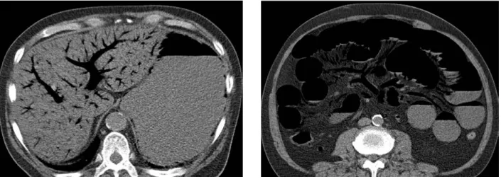
FIG.2. A 76-YEAR-OLD MALE WHO PRESENTED ABDOMINAL PAIN.
TABLE 1. CT MEASUREMENTS AND COMPARISON IN PATIENT GROUPS
Benign group
n=24 Life-threaten-ing group n=18
p value
Bowel distension 5 15 .000
Decreased bowel wall enhancement 1* 14 .000
Bowel wall defect 0* 2 .196
Extraluminal free air 13 11 .757
PVG 0 8 .000
MVG 8 12 .060
Ascites 7 13 .012
Note- Data are the number of patients and (%), PVG=portal venous gas, MVG=mesenteric venous gas. *The total number of patients is 22 because two patients with nonenhanced abdomen CT scan in benign group were excluded.
TABLE 2. PATIENT OUTCOME
Benign group
Conservative treatment, n=24
Life-threatening group
Death, n=1
Indicated to surgical intervention, n=13
Undergo emergent surgery, n=9
Refused to undergo surgery, n=4
Endoscopic examination proven duodenal ulcer and clipping, n=1
Antibiotics therapy and intensive care, n=3
medical or surgical intervention. The life-threatening group (n=18) was recommended surgical intervention at the presentation due to unstable vital signs or hos-pitalization with close observation. Of the patients in the life-threatening group, 9 underwent emergency surgery due to bowel ischemia, 1 died immediately af-ter taking the CT scan due to bowel ischemia, 4 were recommended for surgical intervention but refused, 3 underwent antibiotics therapy for ischemic bowel dis-ease and were discharged with improved status, and 1 had duodenal ulcer bleeding and improved clinical sta-tus after endoscopic bleeder clipping. The patient out-come of all enrolled patients is summarized in Table 2.
Discussion
PI is traditionally considered a surgical emer-gency with a high possibility of bowel ischemia, especially in cases associated with portomesenter-ic venous gas. However, previous studies reported that PI might occur after infection or inflammation, ulceration, surgery or trauma[20,21]. In addition, the
incidence of asymptomatic PI has been increasing in association with the development of CT scanning.
In this study, PVG had statistical significance and was only identified in the life-threatening group. However, several studies[22,23] have reported that the
PVG is not a useful indicator of bowel ischemia and is not helpful in determining the need for surgical intervention. Faberman et al.[22] analyzed 17 patients
with PMVG on CT and reported a 71% survival rate. The different result can be associated with the dif-ferent study design, as they enrolled patients with PMVG and only 9 of all 17 patients had combined PI. In our study, we enrolled a larger number of patients and all of them had PI. Additionally, all of the patients with PVG were included in the life-threatening group with statistical significance. This result supports oth-er previous largoth-er studies2,24-26, which suggested that
derwent emergent segmental resection of the colon; and the other expired immediately after taking the CT scan due to extensive small bowel ischemia. How-ever, the rest of the life-threatening group showed no significant bowel wall defect, even though about two-thirds of them showed extraluminal free air. This result may support that pneumoperitoneum with PI itself is not an ominous sign, so it is best to look for other critical signs such as bowel wall abnormality.
There were several limitations to the study. First, because this study was conducted at a tertiary re-ferral center, there is a selection bias. Second, it presents a retrospective study design, so there is a possibility of insufficient clinical information. Third, about two-thirds (66.7%) of the patients were man-aged nonoperatively, so it was not possible to con-firm the presence or absence of bowel ischemia or other pathologic findings in both groups.
CONCLUSION
It is still difficult to determine the management of patients with PI because there are various interpre-tations of the clinical significance of PI and its associ-ated CT findings.
This study revealed that the PI and concurrent PVG, bowel distension, ascites and decreased bowel wall enhancement were significantly associated with life-threatening causes of PI and unfavorable clinical outcomes. On the other hand, the presence of MVG, extraluminal free air, and bowel wall defects showed no statistical significance. Thus, it is necessary to pay attention to other ancillary CT findings when in-terpreting images of patients with PI to help charac-terize the causes of PI and determine the appropriate treatment option.
separate measurement of both CT findings.
Bowel distension and ascites were significant-ly more commonsignificant-ly identified in the life-threatening group. Concurrent bowel distension and ascites are known to be associated with high-grade obstruction and congestion. In this study, decreased bowel wall enhancement, which is a radiologic indicator of bow-el ischemia, was also significantly associated with the life-threatening group. The result is similar to that of previous larger studies25,27. Duron et al.[27] analyzed
ra-diologic findings of 150 patients diagnosed with PI on CT and compared non-operative and operative groups; dilated bowel loops and free fluid were significantly as-sociated with the operative group. In a study by Lee et al.25, that analyzed 123 patients with PI, decreased or
absent enhancement of the bowel wall on CT were as-sociated with increased mortality. Therefore, patients with PI and bowel distension or decreased bowel wall enhancement or ascites should be observed vigilantly.
Extraluminal free air, including both pneumo-peritoneum and pneumoretropneumo-peritoneum, has been considered a sign of perforated hollow viscus and weighted heavily in favor of surgical management. However, in a previous study, it was suggested that pneumoperitoneum could occur with long-stand-ing PI and rarely is associated with peritonitis28. In
this study, extraluminal free air did not significantly correlate with patient outcome, and even the benign group presented extraluminal free air in about half of the patients (54.2%). On the other hand, bowel wall defect on a CT scan, which is a direct indicator of perforated hollow viscus, was identified in only two patients among a total of 42 patients, with no statisti-cal significance. In this study, two cases with both PI and bowel wall defect resulted from transmural bow-el infarction: one patient had colon infarction and
un-RESUMO
OBJETIVO: Avaliar o desempenho diagnóstico dos achados CT em causas diferenciadoras da pneumatose intestinal (PI), incluindo causas benignas e que ameaçam a vida.
MÉTODOS: Todos os relatórios CT contendo a palavra “pneumatose” foram questionados de 10 de junho de 2006 a 31 de maio de 2015.
Um total de 42 pacientes com PI foi matriculado (idade média 63,4 anos, 23 do sexo masculino e 19 do sexo feminino) e divididos em dois grupos na base de registros médicos elétricos: grupo benigno, n = 24 e grupo com risco de vida, n = 18. Dois radiologistas anali-saram as imagens da CT e avaliaram seus achados, incluindo distensão intestinal, padrão de realce da parede intestinal, defeito da parede intestinal, gás venoso portal (PVG), gás venoso mesentérico (MVG), ar extraluminal e ascite.
RESULTADOS: Achados CT, incluindo distensão intestinal, diminuição do realce da parede intestinal. PVG e ascite foram mais comumen-te identificados em grupo com risco de vida (todos p < 0,05, respectivamencomumen-te). Todos os casos com PVG foram incluídos em grupo com risco de vida (8/18 pacientes, 44,4%). Defeito da parede do intestino, ar livre extraluminal e gás venoso mesentérico não mostraram significância estatística entre dois grupos.
CONCLUSÃO: PI e PVG concorrente, distensão intestinal, diminuição do aumento da parede do intestino ou ascites foram significati-vamente associados com causas que ameaçaram a vida e prognóstico desfavorável. Portanto, avaliar os recursos de CT auxiliares quando encontramos PI nos ajudaria a caracterizar as causas de PI e determinar a opção de tratamento apropriada.
REFERENCES
1. Heng Y, Schuffler MD, Haggitt RC, Rohrmann CA. Pneumatosis intestina-lis: a review. Am J Gastroenterol. 1995;90(10):1747-58.
2. Ho LM, Paulson EK, Thompson WM. Pneumatosis intestinalis in the adult: benign to life-threatening causes. AJR Am J Roentgenol. 2007;188(6):1604-13.
3. Knechtle SJ, Davidoff AM, Rice RP. Pneumatosis intestinalis. Surgical management and clinical outcome. Ann Surg. 1990;212(2):160-5.
4. Galandiuk S, Fazio VW. Pneumatosis cystoides intestinalis. A review of the literature. Dis Colon Rectum. 1986;29(5):358-63.
5. Read NW, Al-Janabi MN, Cann PA. Is raised breath hydrogen related to the pathogenesis of pneumatosis coli? Gut. 1984;25(8):839-45.
6. Ellis BW. Symptomatic treatment of primary pneumatosis coli with met-ronidazole. Br Med J. 1980;280(6216):763-4.
7. Yale CE, Balish E, Wu JP The bacterial etiology of pneumatosis cystoides intestinalis. Arch Surg. 1974;109(1):89-94.
8. St Peter SD, Abbas MA, Kelly KA. The spectrum of pneumatosis intestina-lis. Arch Surg. 2003;138(1):68-75.
9. Kernagis LY, Levine MS, Jacobs JE. Pneumatosis intestinalis in patients with ischemia: correlation of CT findings with viability of the bowel. AJR Am J Roentgenol. 2003;180(3):733-6.
10. Na SY, Kim KJ, Yang DH, Jung K, Ye B, Byeon JS, et al. Pneumoperitoneum in a patient with ulcerative colitis after sigmoidoscopy: is this always an indication for surgery? Inflamm Bowel Dis. 2011;17(6):E54-6.
11. Iannitti DA, Gregg SC, Mayo-Smith WW, Tomolonis RJ, Cioffi WG, Pricolo VE. Portal venous gas detected by computed tomography: is surgery im-perative? Dig Surg. 2003;20(4):306-15.
12. Lassandro F, Scaglione M, Rossi G, Grassi R, Romano L. Portomesenteric vein gas: diagnostic and prognostic value. Emerg Radiol. 2002;9(2):96-9.
13. Liebman PR, Patten MT, Manny J, Benfield JR, Hechtman HB. Hepat-ic-portal venous gas in adults: etiology, pathophysiology and clinical sig-nificance. Ann Surg. 1978;187(3):281-7.
14. Paran H, Epstein T, Gutman M, Shapiro Feinberg M, Zissin R. Mesenteric and portal vein gas: computerized tomography findings and clinical signif-icance. Dig Surg. 2003;20(2):127-32.
15. Alkhatib AA, Elkhatib FA, Alkhatib OF, Zurcher R. Pneumatosis intestinalis and gas in portal vein associated with small bowel obstruction. J Emerg Med. 2011;40(6):e125-6.
16. Hoot NR, Pfennig CL, Johnston MN, Jones I. An incidental finding? Pneu-matosis intestinalis after minor trauma. J Emerg Med. 2013;44(2):e145-7.
17. Ohtsubo K, Okai T, Yamaguchi Y, Watanabe H, Motoo Y, Matsui O, et al. Pneumatosis intestinalis and hepatic portal venous gas caused by mesen-teric ischemia in an aged person. J Gastroenterol. 2001;36(5):338-40.
18. Ong KP, Ng KH, Lim KH, Low SC, Eu KW. Pneumoperitoneum resulting from pneumatosis cystoides intestinalis: a rare complication of massive colonic dilatation. Tech Coloproctol. 2010;14(3):287-8.
19. Wright NJ, Wiggins T, Stubbs BM, Engledow A. Benign pneumatosis in-testinalis with pneumoperitoneum and typhlitis: side-effects of drug or disease induced immunosuppression. BMJ Case Rep. 2011;13;2011.
20. Feczko PJ, Mezwa DG, Farah MC, White BD. Clinical significance of pneu-matosis of the bowel wall. Radiographics. 1992;12(6):1069-78.
21. Sebastià C, Quiroga S, Espin E, Boyé R, Alvarez-Castells A, Armengol M. Portomesenteric vein gas: pathologic mechanisms, CT findings, and prog-nosis. Radiographics. 2000;20(5):1213-24.
22. Faberman RS, Mayo-Smith WW. Outcome of 17 patients with portal ve-nous gas detected by CT. AJR Am J Roentgenol. 1997;169(6):1535-8.
23. Wiesner W, Mortelé KJ, Glickman JN, Ji H, Ros PR. Pneumatosis intesti-nalis and portomesenteric venous gas in intestinal ischemia: correlation of CT findings with severity of ischemia and clinical outcome. AJR Am J Roentgenol. 2001;177(6):1319-23.
24. Griffiths DM, Gough MH. Gas in the hepatic portal veins. Br J Surg. 1986;73(3):172-6.
25. Lee HS, Cho YW, Kim KJ, Lee JS, Lee SS, Yang SK. A simple score for pre-dicting mortality in patients with pneumatosis intestinalis. Eur J Radiol. 2014;83(4):639-45.
26. Smerud MJ, Johnson CD, Stephens DH. Diagnosis of bowel infarction: a comparison of plain films and CT scans in 23 cases. AJR Am J Roentgenol. 1990;154(1):99-103.
27. Duron VP, Rutigliano S, Machan JT, Dupuy DE, Mazzaglia PJ. Computed tomographic diagnosis of pneumatosis intestinalis: clinical measures pre-dictive of the need for surgical intervention. Arch Surg. 2011;146(5):506-10.