Five-Component Self-Assembly of Cucurbituril-Based
Hetero-pseudorotaxanes
C#tia Parente Carvalho
+,
[a]Zoe Dom&nguez
+,
[a]Cristina Dom&nguez,
[a]Hamdy S.
El-Sheshtawy,
[b]Jos8 Paulo Da Silva,
[c]Jesffls F. Arteaga,
[a]and Uwe Pischel*
[a]1. Introduction
Self-assembly has received growing attention during recent years as a synthetic method for the construction of well-defined supramolecular architectures.[1–5]This interest is strong-ly driven by the desire to implement nature’s recipes in artifi-cial biomimetic systems and to obtain novel functional materi-als. The opportunities offered by the principles of self-sorting in combination with structurally and electronically pre-programmed building blocks have been demonstrated for the most diverse of systems.[4]Some of these systems make use of metal–ligand interactions,[6] whereas others embrace all-organic self-assembly.[5,7–10]
Cucurbiturils (CBs) are organic macrocycles that are com-posed of n glycoluril units linked by methylene bridges, where n=5, 6, 7, 8, 10, and 14.[10–13]Recently, they have been in the focus of many research programs owing to their fascinating host–guest chemistry. Organic compounds with positively
charged groups have been identified as preferred guests with association constants of up to 1015–1017m@1in water, matching or even surpassing those of nature’s strongest supramolecular assembly—the biotin–avidin pair.[14,15] The nanotechnological potential of CBs is currently being realized and spans from an-alytical applications, dye chemistry, materials chemistry, and supramolecular catalysis to topics inspired by the life sciences, such as biomolecule binding, drug delivery, and control of bio-logical functions.[10,13,16–32]One central aspect of the chemistry of CBs is their role as prime components in self-sorting.[33–37] The variation of the cavity size of CB homologues and conse-quently the differentiation of the strength and dynamics of guest binding can trigger thermodynamically or kinetically driven selection processes.
Importantly, the extended set of available affinity data for CB6, CB7, and CB8 binding to structurally diverse guests[10,21,34,38] provides a solid foundation for the molecular design of complex supramolecular structures containing CB macrocycles as key elements. This might ultimately lead to complex, yet well-defined systems that exhibit biomimetic fea-tures.[10,28,33,35–37,39–41]In this study, we were especially interested in the use of CBs as wheel components of pseudorotaxanes. Previous reports (see Scheme 1) have often focused on two-component assemblies.[42–44] In an extension of these studies, multicomponent pseudorotaxanes were obtained through the complexation of CBs by a pre-programmed unimolecular axle.[36,37, 39,40,45] However, the CB-templated assembly of ho-mo[5]pseudorotaxanes by means of the formation of a 1:1:1 ternary complex that holds the axle components together, has only been scarcely reported (Scheme 1).[36,46]
In the present work, we made use of heteroditopic ligands that discriminate between CB homologues (CB6, CB7, and CB8) based on well-differentiated binding constants. The reasoned molecular design enabled the high-precision self-sorting of five components. This resulted, to the best of our knowledge, in unprecedented all-CB hetero[5]pseudorotaxanes with a self-as-sembled axle and different CB homologues as wheels [5]Pseudorotaxanes can be obtained by self-sorting using
het-eroditopic guests and various cucurbituril homologues as hosts. The assembly and chemically induced disassembly of the pseudorotaxanes can be monitored by measuring the fluo-rescence of the anthracene guest in solution. Mass spectral
evi-dence for the supramolecular assemblies is obtained in the gas phase. The disassembly in the gas phase can be achieved by collision-induced dissociation leading to the corresponding [2]-and [3]pseudorotaxanes.
[a] Dr. C. P. Carvalho,+Z. Dom&nguez,+C. Dom&nguez, Dr. J. F. Arteaga,
Dr. U. Pischel
CIQSO—Center for Research in Sustainable Chemistry and Department of Chemistry, University of Huelva Campus de El Carmen, 21071 Huelva (Spain) E-mail: [email protected]
[b] Dr. H. S. El-Sheshtawy
Chemistry Department, Faculty of Science Kafrelsheikh University
33516 Kafr ElSheikh (Egypt) [c] Dr. J. P. D. Silva
Meditbio—Faculty of Sciences and Technology University of Algarve
Campus de Gambelas, 8005-139 Faro (Portugal) [++] These authors contributed equally to this work
Supporting Information and the ORCID identification number(s) for the author(s) of this article can be found under http://dx.doi.org/10.1002/ open.201600173.
T 2017 The Authors. Published by Wiley-VCH Verlag GmbH & Co. KGaA. This is an open access article under the terms of the Creative Commons Attribution-NonCommercial-NoDerivs License, which permits use and distribution in any medium, provided the original work is properly cited, the use is non-commercial and no modifications or adaptations are made.
(Scheme 1). Likewise, using only CB8 yielded all-CB8 homo[5]-pseudorotaxanes. Furthermore, the molecular design included the possibility to monitor the supramolecular processes by fluorescence spectroscopy and electrospray ionization mass spectrometry (ESI-MS).
2. Results and Discussion
2.1. Molecular DesignOur approach makes use of the previously[47] observed strong 2:1 complexation of the model compound 1 (Scheme 2a) by CB8 (K21=4.2V1012m@2; corresponding to an apparent 1:1 binding constant of 2.0V106m@1, although it is likely that the first anthracene is bound more weakly than the second). The 1:1 complex with CB7 is weaker by one order of magnitude (K11=3.0V105m@1).[47]In this study, we designed and prepared the derivatives 2 and 3, combining the same anthracene imide chromophore as in 1 with an additional motif for binding ho-mologous CB macrocycles (Scheme 2a–c and the Experimental Section). On the one hand, the aminoadamantane unit in 2 is well known to form extraordinarily strong 1:1 complexes with CB7 and somewhat weaker complexes with CB8 (K=4.2 V 1012m@1 with CB7 versus 8.2V108m@1 with CB8).[34] On the other hand, the spermidine tail in 3 should enable efficient complexation of CB6 (K=4.1 V108m@1).[38] Hence, we expected
to gain access to hetero[5]pseudorotaxanes by self-sorting for-mation based on the differentiated complexation of 2 by CB7 and CB8 and of 3 by CB6 and CB8, featuring a central ternary 2:1 anthracene–CB8 complex (Scheme 1, bottom). Likewise, for both guests the formation of homo[5]pseudorotaxanes is pre-dicted for the stoichiometric presence of only CB8. It was an-ticipated that the well-differentiated binding affinities would guarantee high precision with respect to the final assemblies, underlining a unique quality of CB host–guest chemistry.
Conveniently, the chromophoric and fluorescent nature of the anthracene-binding motif provides a handle for monitoring the axle assembly by UV/Vis absorption and fluorescence spec-troscopies. In aqueous solution (pH 6–7) compounds 2 and 3 feature a typical long-wavelength UV/Vis absorption band with a maximum at 413 nm and significant fluorescence (2: lfluo, max=496 nm, Ffluo= 0.52, tfluo= 6.75 ns; 3: lfluo, max= 497 nm, Ffluo=0.53, tfluo=6.91 ns; see the red spectra in Figure 1 for 2).[47]As a general observation, the inclusion of the anthracene in CB8 is signaled by a bathochromic shift of the long-wavelength UV/Vis absorption band (from 413 to 433 nm) and a pronounced fluorescence quenching (&90%), accompa-nied by a redshift of the emission by approximately 30–35 nm (Figure 1).[48]
Scheme 1. Examples of all-CB pseudorotaxanes and representation of the target structures in this study. [a] Ref. [39]. [b] Ref. [40]. [c] Ref. [36]. [d] Ref. [46].
Scheme 2. a) Structures of the previously prepared anthracene-2,3-imide de-rivative 1 (Ref. [47]), compounds 2 and 3 in their fully protonated form, and the CBs used in this work. b) Synthesis of 2. Note that compound 2 was ob-tained as a non-protonated amine after column chromatography under basic conditions (see the Experimental Section). c) Synthesis of 3.
2.2. Formation of [5]Pseudorotaxanes with Compound 2 and CB7/CB8 in Solution
In a first set of experiments, an aqueous solution (pH 6) of compound 2 was titrated with CB8 (Figure 2; black data points). The complexation proceeded in two well-defined phases. The addition of the first CB8 equivalent caused only a relatively small fluorescence quenching (&24 %), suggesting that mainly the aminoadamantane unit of 2 is bound along with a likely minor complexation of the anthracene unit (ap-proximately two orders of magnitude higher binding constant with the adamantane than the apparent constant with the an-thracene; see above). In a second phase, involving the addition of another 0.5 equivalents of CB8, the anthracene unit became visibly involved as indicated by the characteristic bathochromic shift of the absorption spectrum and a pronounced fluores-cence quenching (& 90% at the titration endpoint; see Figure 1). This part of the titration corresponds to the dimeriza-tion of two 1:1 complexes (formed in the first stage), involving the CB8 complexation of two anthracene units. Hence, in total
two molecules of 2 and three CB8 macrocycles interact, yield-ing the homo[5]pseudorotaxane [(2)2(CB8)3]4+ (Scheme 1, bottom).[49]
To take advantage of the binding characteristics of 2 toward CB7 and CB8, a similar experiment as described before, but with one equivalent CB7 present in the initial solution, was performed. The CB7 macrocycle binds four orders of magni-tude stronger to the aminoadamantane unit than CB8 does (see above). Therefore, it was expected that the addition of CB8 would immediately induce the dimerization by complexa-tion of two anthracene units with CB8. This was indeed the case, as experimentally corroborated by the changes in the op-tical signature, that is, fluorescence quenching and redshifted emission spectrum, and the sharp leveling off of the titration curve at 0.5 equivalents of CB8 (see Figure 2, red points). The supramolecular assembly corresponds to a hetero[5]pseudoro-taxane [(2)2CB8(CB7)2]4+ with a central CB8 and two terminal CB7 units (Scheme 1, bottom).
Also, the reverse titration, that is, addition of CB7 to a solu-tion of 2 containing 0.5 equivalents of CB8, gave rise to a char-acteristic bathochromic UV/Vis absorption shift, indicative of the inclusion of the anthracene unit into CB8 (Figure 3). The
ti-tration curve shows a typical S shape. Initially, the CB7 binds readily to noncomplexed 2 because only 0.5 equivalents CB8 are present. Upon further titration, CB7 competes with CB8 for the aminoadamantane unit of 2 and the released CB8 associ-ates to the available anthracene unit, yielding again the above-described hetero[5]pseudorotaxane. As expected for an equi-librium situation, the order of addition of the pseudorotaxane components was irrelevant for the outcome; exactly the same final spectral signature was obtained whether the order was 2–CB7–CB8 or CB8–CB7–2 (first–second–third). In general, the high uniformity of all complexation processes is underpinned by well-defined spectral changes and the occurrence of several isosbestic points in the UV/Vis absorption spectra.
Figure 1. Titration of 2 (10 mm) with CB8 (0–19.7 mm) in water (pH 6); moni-tored by a) UV/Vis absorption and b) fluorescence (lexc=419 nm). The initial
and final spectra of the titration are colored red and blue, respectively. The initial spectra are normalized to 1 at the maximum of the longest-wave-length band.
Figure 2. Fluorescence titration curves of 2 (10 mm) with CB8 (up to 19.7 mm) in water (pH 6, lexc=419 nm, lobs=490 nm) in the absence (black) and in
the presence of CB7 (15 mm, red).
Figure 3. a) UV/Vis absorption titration of 2 (10 mm) with CB7 (0–27.8 mm) in the presence of CB8 (5 mm) in aqueous solution (pH 6). The red spectrum corresponds to the starting point, and the blue spectrum marks the end-point of the titration. b) The corresponding titration curve, monitored at lobs=435 nm.
By exploiting the reversible nature of host–guest complexa-tion, it was of interest to demonstrate the disassembly of the pseudorotaxanes by means of the addition of a competitive guest for CB8. For this purpose, the sterically demanding ami-noadamantane derivative memantine was used, which is known to form strong complexes with CB8 (K= 4.3V1011m@1) but it binds much less efficiently to the smaller CB7 (K=2.5 V104m@1).[34] Titration of the [5]pseudorotaxanes [(2)2CB8(CB7)2]4+ and [(2)2(CB8)3]4+ with this competitor guest yielded the recovery of the initial UV/Vis absorption and fluo-rescence properties of the non-complexed anthracene chromo-phore (see the Supporting Information). These experiments showed that memantine can be used to disassemble the cen-tral 2:1 ternary CB8 complex, which is conveniently signaled by fluorescence enhancement.
2.3. Formation of [5]Pseudorotaxanes with Compound 2 in the Gas Phase
Solution titrations and monitoring of the changes of optical spectral features can provide information about the binding stoichiometry, and in combination with calculation of binding constants, allow for a reasoned discussion of the nature of the formed assemblies. However, it was highly desirable to draw on a complementary technique to corroborate our conclusions further. In the light of the limited CB8 solubility, which compli-cated the NMR spectroscopy experiments, ESI-MS was the ana-lytical technique of choice for this work. The use of ESI-MS ena-bled an insightful analysis of the multicomponent mixtures in the micromolar concentration range (Figure 4).[36,37,50]
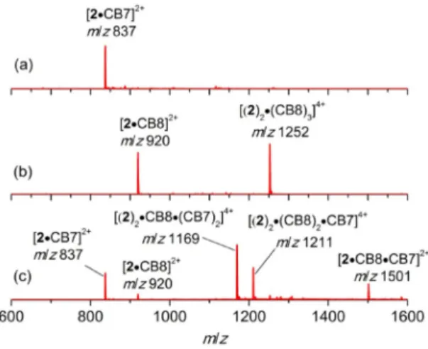
The ESI-MS spectrum of a 1:1 mixture of 2 and CB7 features one signal at m/z 837 (Figure 4a). The corresponding species was assigned as [2(CB7)]2+. The fragmentation (MS2) of this ion yielded signals that can be attributed 1) to a CB7 complex con-taining an adamantyl-substituted fragment and 2) to an un-bound anthracene-2,3-imide fragment. This finding provided support for the preferential binding of CB7 to the adamantane unit (see the MS2 spectrum in the Supporting Information).
The mass spectrum of a 2/CB8 mixture (2:3) yielded clear evi-dence for the homo[5]pseudorotaxane (Figure 4b) discussed above. Specifically, two signals at m/z 920 and 1252 were de-tected and were assigned to the 1:1 complex [2(CB8)]2+ and the homo[5]pseudorotaxane [(2)2(CB8)3]4+, respectively. Frag-mentation (MS2) of the latter ion gave the corresponding 1:1 complex [2(CB8)]2+ and the [3]pseudorotaxane [2(CB8)
2]2+, in-dicating preferential disassembly of the central 2:1 ternary CB8 complex. This is in accordance with the relative stabilities of the involved complexes (see above).
The mixture of 2, CB7 and CB8 ([2]/[CB7]/[CB8] =1:1:0.5) gave rise to a prevailing signal at m/z 1169, corresponding to the hetero[5]pseudorotaxane [(2)2CB8(CB7)2]4+, and a signal at m/z 1211, assigned to [(2)2(CB8)2CB7]4+ with one CB7 and one CB8 at the terminal positions (Figure 4c). As illustrated in Figure 5 (see the Supporting Information for the MS2 spectra),
fragmentation of the m/z 1169 ion provided as the main sig-nals those of [2(CB7)]2+ and [2(CB8)(CB7)]2+, again pointing to the disassembly of the central 2:1 ternary anthracene–CB8 complex (see the discussion above). Fragmentation of the m/z 1211 ion led to a more complex situation, derived from the unsymmetrical arrangement of different CB macrocycles at the terminal adamantane moieties. Specifically, [2(CB7)]2 + (m/z 837), [2(CB8)]2 +(m/z 920), [2(CB8)(CB7)]2+(m/z 1501), and [2(CB8)2]2+ (m/z 1585) were detected (see Figure 5 and the Supporting Information). Hence, the mass evidence, MS2 frag-mentation experiments, and the solution titrations provide clear support for the formation of [5]pseudorotaxanes contain-ing the heteroditopic buildcontain-ing block 2.
Figure 4. ESI-MS spectra of 2 (10 mm); a) with CB7 (10 mm), b) with CB8 (15 mm), c) with CB8 (5 mm) and CB7 (10 mm).
Figure 5. Principal MS2fragmentation products for the m/z 1169 and 1211
ions. The models correspond to MM2-optimized structures of the complexes. The corresponding MS2spectra are shown in the Supporting Information.
2.4. Formation of a Hetero[5]pseudorotaxane with Compound 3 and CB6/CB8
Derivative 3 was designed to investigate the molecular diversi-ty of the supramolecular five-component assembly of heter-o[5]pseudorotaxanes, using CB6 and CB8 in this case. As out-lined above, the spermidine tail of the anthracene-2,3-imide derivative 3 should provide an efficient binding motif for CB6, functioning as terminal macrocycles of the targeted hetero[5]-pseudorotaxane. As for 2, the axle was expected to organize by the formation of a ternary complex between two anthra-cene moieties and a CB8 macrocycle.
First, compound 3 was titrated with CB8, with monitoring of the changes in the UV/Vis absorption and fluorescence spectra (Figure 6). Again, the typical spectral changes as noted for the
case of guest 2 (see above) were observed.[48]Much to our ini-tial surprise, and in contrast to the results that were obtained for 2, no formation of a 2:1 ternary complex involving the an-thracene unit could be concluded. Instead, the titration curve leveled off at approximately 1 equivalent CB8, indicating a 1:1 stoichiometry of the complex with the anthracene unit (Figure 6; black data points).
However, in the presence of 1 equivalent of CB6 in the initial solution, the titration reached its endpoint with approximately 0.5 equivalents of CB8 (Figure 6, red data points). Akin to the situation observed for 2 (using CB7), this result hints at the for-mation of a hetero[5]pseudorotaxane, in which the two CB6 macrocycles occupy the terminal spermidine chains of two molecules of 3 that are joined by a central CB8 in a 2:1 ternary complex (see the general representation in Scheme 1, bottom). Interestingly, it is the CB6 complexation that somewhat
allevi-ates the coulombic repulsion between the highly charged spermidine tails, which promotes a positive cooperativity effect on the self-assembly of the desired hetero[5]pseudo-rotaxane.
The gas-phase ESI-MS studies (Figure 7) confirmed the for-mation of the pseudorotaxanes. In the presence of CB8, the [3]pseudorotaxane with the fully protonated axle 3 was ob-served, [3(CB8)2]3+at m/z 1031. However, most of 3 is integrat-ed in [2]pseudorotaxanes with varying charge status, that is, [3(CB8)]3+ (m/z 588) and [(3-H)CB8]2+ (m/z 881). Contrary to the observations made for 2, the homo[5]pseudorotaxane composed of two molecules of 3 and three of CB8 was not ob-served, which is consistent with the solution studies.
In the presence of both CB6 and CB8 ([3]/[CB6]/[CB8] = 1:1:0.5), the predicted hetero[5]pseudorotaxane [(3-H)2 -CB8(CB6)2]4+ was most abundant, as indicated by a dominant signal at m/z 1048. Other less abundant species were the [2]-and [3]pseudorotaxanes [(3-H)CB6]2+ and [3(CB8)(CB6)]3+ cor-responding to signals at m/z 715 and 920, respectively. The
MS2 fragmentation of the hetero[5]pseudorotaxane showed
the collision-induced dissociation into the corresponding frag-ments: [(3-H)CB6]2+and [(3-H)CB8(CB6)]2+ (see the Supporting Information). These observations are akin to the gas-phase chemistry of the [5]pseudorotaxanes based on compound 2, confirming the preferential dissociation of the central 2:1 ter-nary CB8 complex.
3. Conclusions
Heteroditopic fluorescent guests with high and well-differenti-ated binding constants for cucurbiturils (CB6, CB7, CB8) as well as variable complex stoichiometries (2:1 vs. 1:1) can be used for the controlled self-assembly of all-CB homo- and hetero[5]-pseudorotaxanes. This study underpins the unique strength of cucurbiturils as components in programmed self-sorting pro-cesses that lead to complex supramolecular structures that
Figure 6. Fluorescence titration curves for the addition of CB8 (up to 9.2 mm) to 3 (5 mm) in the absence (black, lobs=497 nm) and presence of CB6 (red,
11 mm, lobs= 490 nm) in water (pH 7). Inset: the development of the
absorp-tion spectra (a) and the fluorescence spectra (b) for the titraabsorp-tion in the ab-sence of CB6; initial and final spectra are colored red and blue, respectively. The initial spectra are normalized to 1 at the maximum of the longest-wave-length band.
Figure 7. ESI-MS spectra of a) 3 (20 mm) with 1.5 equiv. CB8 (30 mm), and b) 3 (10 mm) in presence of 0.5 equiv. CB8 (5 mm) and 1 equiv. CB6 (10 mm); all in water (pH 7). The models of the detected [3]- and [5]pseudorotaxanes are also shown.
might have applications as biomimetic materials. Furthermore, the tailored formation of the pseudorotaxanes can be moni-tored by optical spectroscopy, providing another functional facet to the molecular design of the axle building blocks.
Experimental Section
Materials
All reagents and solvents for synthesis were commercially available (Sigma–Aldrich) in high purities and used as received. Water was of Milli-Q quality. CB7 was synthesized according to a previously pub-lished procedure,[51] whereas CB6 and CB8 were purchased from
Sigma–Aldrich. N1,N5,N10-Tri-Boc-spermine was prepared according
to a literature procedure.[52]
Synthesis of Compound 2
1-Bromoadamantane (2.00 g, 9.30 mmol) and 3,3’-diamino-N-meth-yldipropylamine (6.75 g, 46.5 mmol) were mixed in a sealed tube. This mixture was heated at 1908C for 20 h. The mixture was al-lowed to cool to room temperature, then HCl (2m, 60 mL) and di-ethyl ether (60 mL) were added. The aqueous phase was separated and 50% NaOH solution (60 mL) was added. Finally, the product was extracted with diethyl ether (3V40 mL) and the combined or-ganic phases were dried with anhydrous Na2SO4. Removal of the
solvent gave N1-(adamantan-1-yl)-N3-(3-aminopropyl)-N3
-methylpro-pane-1,3-diamine as an oil (1.71 g, 66% yield). This material was used in the next step without further purification.
2,3-Anthracenedicarboxylic anhydride (60 mg, 0.24 mmol), N1
-(ada-mantan-1-yl)-N3-(3-aminopropyl)-N3-methyl-propane-1,3-diamine
(67.5 mg, 0.24 mmol), and triethylamine (51 mL, 0.37 mmol) were placed together with ethanol (5 mL) in a sealed tube. This mixture was heated at 808C for 72 h. Afterwards, all volatiles were removed and the residue was subjected to column chromatography on silica gel (CH2Cl2/CH3OH/NH4OH, 50:10:1). Compound 2, in its
non-protonated form, was isolated as a solid (35 mg, 28 % yield). N1-(Adamantan-1-yl)-N3-(3-aminopropyl)-N3
-methylpropane-1,3-dia-mine:1H NMR (400 MHz, CDCl 3): d=2.62 (t, J=7.2 Hz, 2H), 2.49 (t, J=7.2 Hz, 2H), 2.26 (t, J=7.2 Hz, 4H), 2.09 (s, 3H), 1.95 (brs, 3H), 1.60–1.44 (m, 16H), 1.01 ppm (brs, 3H);13C NMR (100 MHz, CDCl 3): d=56.3, 55.5, 50.2, 42.7, 42.3, 40.7, 39.0, 36.7, 31.2, 29.5, 28.6 ppm; HRMS (ESI): m/z: calcd for C17H34N3: 280.2747 [M++H]+; found:
280.2746. Compound 2: 1H NMR (400 MHz, CDCl 3): d=8.52 (s, 2H), 8.39 (s, 2H), 8.04–7.98 (m, 2H), 7.60–7.54 (m, 2H), 3.77 (t, J=7.2 Hz, 2H), 2.62 (t, J=7.2 Hz, 2H), 2.43 (t, J=7.2 Hz, 2H), 2.38 (t, J=7.2 Hz, 2H), 2.20 (s, 3H), 2.03 (brs, 3H), 1.94–1.82 (m, 2H), 1.69–1.52 ppm (m, 14H); the NH shows as a broad signal at approximately 2.6 ppm;13C NMR (100 MHz, CDCl
3): d=167.9, 133.2, 131.9, 130.0,
128.5, 127.5, 126.6, 125.7, 56.4, 55.3, 51.0, 42.3, 42.0, 39.2, 36.7, 36.6, 29.6, 27.9, 26.3 ppm; HRMS (ESI): m/z: calcd for C33H40N3O2:
510.3115 [M++H]+; found: 510.3111.
Synthesis of Compound 3
N1,N5,N10-Tri-Boc-spermine (140 mg, 0.28 mmol), triethylamine
(39 mL, 0.28 mmol), and ethanol (5 mL) were placed in a sealed tube and the solution was stirred for 15 min. Then, 2,3-anthracene-dicarboxylic anhydride (69.1 mg, 0.28 mmol) was added slowly and the resulting mixture was heated to 808C for 6 days. The mixture
was allowed to cool to room temperature, then the volatiles were removed and the residue was subjected to column chromatogra-phy on silica gel (CH2Cl2/MeOH/NH4OH, 50:10:1). The product was
obtained as a solid (148 mg, 72 % yield) and used directly in the next step without further characterization. A portion of this materi-al (99 mg, 0.14 mmol) was treated with HCl (3m) in ethyl acetate (1 mL) in a round-bottom flask. After the reaction was stirred for 90 min at room temperature the volatiles were evaporated to give compound 3, which was assumed to be the trihydrochloride salt (50 mg, 68% yield).1H NMR (400 MHz, D 2O): d=7.56 (s, 2H), 7.52– 7.45 (m, 2H), 7.32–7.26 (m, 2H), 7.20 (s, 2H), 3.42 (t, J=7.2 Hz, 2H), 3.18–3.00 (m, 10H), 2.13–2.02 (m, 2H), 2.00–1.89 (m, 2H), 1.85– 1.70 ppm (m, 4H); 13C NMR (100 MHz, CD 3OD): d=170.3, 133.7, 131.8, 131.2, 129.4, 128.6, 127.4, 125.3, 48.0, 47.9, 46.2, 45.5, 37.5, 35.9, 25.8, 24.8, 23.8 ppm (2V); HRMS (ESI): m/z: calcd for C26H33N4O2: 433.2598 [M@2H]+; found: 433.2595.
Photophysical Measurements and Titrations
All measurements were performed with air-equilibrated water solu-tions at room temperature, using quartz cuvettes (1 cm optical pathlength). Compound 2 was pre-solubilized in DMSO and the final aqueous solutions (pH 6) contained 1 vol% of the organic co-solvent. Compound 3 was sufficiently soluble in water (pH 7). The UV/Vis absorption spectra were measured with a UV-1603 spectro-photometer (Shimadzu). Steady-state fluorescence spectra (uncor-rected) were measured on a Cary Eclipse fluorimeter (Varian). The fluorescence quantum yield was measured for corrected emission spectra, with quinine sulfate in 0.05m H2SO4as a reference (Ffluo=
0.55).[53,54]The fluorescence lifetimes were determined by
time-cor-related single-photon counting (Edinburgh Instruments FLS 920). Titration experiments were performed by adding aliquots of CB stock solutions. These were accompanied by the same concentra-tion of the guest compound as present in the titrated soluconcentra-tion, thereby avoiding dilution effects in the course of the experiment. Isosbestic points of the UV/Vis absorption titration were selected as excitation wavelengths for fluorescence titrations. If required, the pH was adjusted by the addition of dilute aqueous HCl or NaOH and kept constant during the titrations. The concentration of the CB8 stock solution was determined by titration with N,N-di-methylaminophenyltropyilium perchlorate.[55]CB7 was assumed to
have 14 wt% water content (determined by1H NMR spectroscopy
in the presence of malonic acid as an internal standard). The water content of CB6 was indicated by the supplier to be 25 wt%.
Electrospray Ionization Mass Spectrometry
The electrospray ionization (ESI) mass spectra were obtained using a Bruker Esquire HCT ultra ion-trap mass spectrometer, equipped with an ESI source (Agilent). The solutions of the compounds were infused into the ESI source at a rate of 4 mLmin@1with the aid of
a syringe pump (KdScientific, model 781100, Holliston, MA, USA). Typical spray and ion optics conditions were as follows: capillary voltage, 3.0 kV; nebulizer gas pressure, 30 psi; drying gas tempera-ture, 3008C; drying gas flow, 6 L min@1; capillary exit voltage, 179 V;
skimmer voltage, 30 V. The charge status of the detected ions was determined from the isotope spacing patterns.
Molecular Modeling of the Pseudorotaxanes
Modeling was performed using the ChemOffice software package (version 8). The structures of the free CB macrocycles (CB7 and
CB8) correspond to their crystal structures[56,57]and the guests 2 or
3 were connected to the macrocycle with the Avogadro software (version 1.1.1). The structures were optimized by molecular me-chanics minimization (MM2 force field), using the ChemBio3D Ultra software (version 8).
Acknowledgements
We acknowledge the financial support of the Ministerio de Econ-om&a y Competitividad, Madrid, Spain (grants CTQ2011-28390 and CTQ2014-54729-C2-1-P for U.P. and PhD fellowship BES-2015–074458 for Z.D.), the Junta de Andaluc&a (grant P12-FQM-2140 for U.P.), and the Fundażo para a CiÞncia e a Tecnologia, Lisbon, Portugal (grant SFRH/BD/81628/2011 for C.P.C.). H.S.E.-S. is grateful for a postdoctoral stipend from the Egyptian Ministry of Higher Education, Cairo.
Conflict of Interest
The authors declare no conflict of interest.
Keywords: fluorescence · host–guest systems · macrocycles · mass spectrometry · rotaxanes
[1] A. Wu, L. Isaacs, J. Am. Chem. Soc. 2003, 125, 4831.
[2] D. Ajami, J.-L. Hou, T. J. Dale, E. Barrett, J. Rebek, Jr., Proc. Natl. Acad. Sci. USA 2009, 106, 10430.
[3] W. Jiang, C. A. Schalley, Proc. Natl. Acad. Sci. USA 2009, 106, 10425. [4] M. M. Safont-Sempere, G. Fern#ndez, F. Werthner, Chem. Rev. 2011, 111,
5784.
[5] Z. He, W. Jiang, C. A. Schalley, Chem. Soc. Rev. 2015, 44, 779.
[6] R. Joseph, A. Nkrumah, R. J. Clark, E. Masson, J. Am. Chem. Soc. 2014, 136, 6602.
[7] E. S. Barrett, T. J. Dale, J. Rebek, Jr., J. Am. Chem. Soc. 2008, 130, 2344. [8] Y. Rudzevich, V. Rudzevich, F. Klautzsch, C. A. Schalley, V. Bçhmer,
Angew. Chem. Int. Ed. 2009, 48, 3867; Angew. Chem. 2009, 121, 3925. [9] M. Chas, G. Gil-Ram&rez, E. C. Escudero-Ad#n, J. Bene tBuchholz, P.
Bal-lester, Org. Lett. 2010, 12, 1740.
[10] S. J. Barrow, S. Kasera, M. J. Rowland, J. del Barrio, O. A. Scherman, Chem. Rev. 2015, 115, 12320.
[11] J. Lagona, P. Mukhopadhyay, S. Chakrabarti, L. Isaacs, Angew. Chem. Int. Ed. 2005, 44, 4844; Angew. Chem. 2005, 117, 4922.
[12] E. Masson, X. Ling, R. Joseph, L. Kyeremeh-Mensah, X. Lu, RSC Adv. 2012, 2, 1213.
[13] K. I. Assaf, W. M. Nau, Chem. Soc. Rev. 2015, 44, 394.
[14] M. V. Rekharsky, T. Mori, C. Yang, Y. H. Ko, N. Selvapalam, H. Kim, D. So-bransingh, A. E. Kaifer, S. Liu, L. Isaacs, W. Chen, S. Moghaddam, M. K. Gilson, K. Kim, Y. Inoue, Proc. Natl. Acad. Sci. USA 2007, 104, 20737. [15] L. Cao, M. Sˇekutor, P. Y. Zavalij, K. Mlinaric´-Majerski, R. Glaser, L. Isaacs,
Angew. Chem. Int. Ed. 2014, 53, 988; Angew. Chem. 2014, 126, 1006. [16] N. J. Wheate, A. I. Day, R. J. Blanch, A. P. Arnold, C. Cullinane, J. G. Collins,
Chem. Commun. 2004, 1424.
[17] A. Hennig, H. Bakirci, W. M. Nau, Nat. Methods 2007, 4, 629.
[18] A. R. Kennedy, A. J. Florence, F. J. McInnes, N. J. Wheate, Dalton Trans. 2009, 7695.
[19] V. D. Uzunova, C. Cullinane, K. Brix, W. M. Nau, A. I. Day, Org. Biomol. Chem. 2010, 8, 2037.
[20] S. Ghosh, L. Isaacs, J. Am. Chem. Soc. 2010, 132, 4445. [21] R. N. Dsouza, U. Pischel, W. M. Nau, Chem. Rev. 2011, 111, 7941. [22] J. M. Chinai, A. B. Taylor, L. M. Ryno, N. D. Hargreaves, C. A. Morris, P. J.
Hart, A. R. Urbach, J. Am. Chem. Soc. 2011, 133, 8810.
[23] F. Tian, D. Jiao, F. Biedermann, O. A. Scherman, Nat. Commun. 2012, 3, 1207.
[24] D. Ma, G. Hettiarachchi, D. Nguyen, B. Zhang, J. B. Wittenberg, P. Y. Za-valij, V. Briken, L. Isaacs, Nat. Chem. 2012, 4, 503.
[25] B. C. Pemberton, R. Raghunathan, S. Volla, J. Sivaguru, Chem. Eur. J. 2012, 18, 12178.
[26] Y. Ahn, Y. Jang, N. Selvapalam, G. Yun, K. Kim, Angew. Chem. Int. Ed. 2013, 52, 3140; Angew. Chem. 2013, 125, 3222.
[27] L. A. Logsdon, A. R. Urbach, J. Am. Chem. Soc. 2013, 135, 11414. [28] Z. Huang, L. Yang, Y. Liu, Z. Wang, O. A. Scherman, X. Zhang, Angew.
Chem. Int. Ed. 2014, 53, 5351; Angew. Chem. 2014, 126, 5455.
[29] J. P. Da Silva, R. Choudhury, M. Porel, U. Pischel, S. Jockusch, P. C. Hub-bard, V. Ramamurthy, A. V. M. Can#rio, ACS Chem. Biol. 2014, 9, 1432. [30] F. Biedermann, W. M. Nau, Angew. Chem. Int. Ed. 2014, 53, 5694; Angew.
Chem. 2014, 126, 5802.
[31] N. Bas&lio, U. Pischel, Chem. Eur. J. 2016, 22, 15208.
[32] J. V#zquez, M. A. Romero, R. N. Dsouza, U. Pischel, Chem. Commun. 2016, 52, 6245.
[33] P. Mukhopadhyay, A. Wu, L. Isaacs, J. Org. Chem. 2004, 69, 6157. [34] S. Liu, C. Ruspic, P. Mukhopadhyay, S. Chakrabarti, P. Y. Zavalij, L. Isaacs,
J. Am. Chem. Soc. 2005, 127, 15959.
[35] P. Mukhopadhyay, P. Y. Zavalij, L. Isaacs, J. Am. Chem. Soc. 2006, 128, 14093.
[36] W. Jiang, Q. Wang, I. Linder, F. Klautzsch, C. A. Schalley, Chem. Eur. J. 2011, 17, 2344.
[37] L. Cera, C. A. Schalley, Chem. Sci. 2014, 5, 2560.
[38] M. V. Rekharsky, Y. H. Ko, N. Selvapalam, K. Kim, Y. Inoue, Supramol. Chem. 2007, 19, 39.
[39] G. Celtek, M. Artar, O. A. Scherman, D. Tuncel, Chem. Eur. J. 2009, 15, 10360.
[40] E. Masson, X. Lu, X. Ling, D. L. Patchell, Org. Lett. 2009, 11, 3798. [41] Q. Zhang, H. Tian, Angew. Chem. Int. Ed. 2014, 53, 10582; Angew. Chem.
2014, 126, 10754.
[42] V. Sindelar, K. Moon, A. E. Kaifer, Org. Lett. 2004, 6, 2665.
[43] V. Sindelar, S. Silvi, S. E. Parker, D. Sobransingh, A. E. Kaifer, Adv. Funct. Mater. 2007, 17, 694.
[44] V. Ramalingam, A. R. Urbach, Org. Lett. 2011, 13, 4898.
[45] P. Brann#, M. Rouchal, Z. Pruckov#, L. Dastychov#, R. Lenobel, T. Posp&sˇil, K. Mal#cˇ, R. V&cha, Chem. Eur. J. 2015, 21, 11712.
[46] Z.-J. Zhang, H.-Y. Zhang, L. Chen, Y. Liu, J. Org. Chem. 2011, 76, 8270. [47] C. Parente Carvalho, Z. Dom&nguez, J. P. Da Silva, U. Pischel, Chem.
Commun. 2015, 51, 2698.
[48] The fluorescence quenching is presumed to be due to the combination of environmental effects exerted by the CB8 cavity and p–p interac-tions between the guests in the case of ternary complex formation. [49] Interestingly, the different pseudorotaxanes conferred a protective
sur-rounding against chemical degradation of the guests 2 and 3. Over the course of 5 h, less than 4% decomposition (monitored by UV/Vis ab-sorption spectroscopy) was observed in all cases. This is in contrast with the moderate stability of free 2 and 3, presumably provoked by a hydrolytic ring-opening of the imide (19 % and 28% decomposition after 5 h for 2 and 3, respectively).
[50] W. Jiang, A. Sch-fer, P. C. Mohr, C. A. Schalley, J. Am. Chem. Soc. 2010, 132, 2309.
[51] C. M#rquez, F. Huang, W. M. Nau, IEEE Trans. Nanobiosci. 2004, 3, 39. [52] I. S. Blagbrough, A. J. Geall, Tetrahedron Lett. 1998, 39, 439. [53] W. H. Melhuish, J. Phys. Chem. 1960, 64, 762.
[54] W. H. Melhuish, J. Phys. Chem. 1961, 65, 229.
[55] J. V#zquez, P. Remjn, R. N. Dsouza, A. I. Lazar, J. F. Arteaga, W. M. Nau, U. Pischel, Chem. Eur. J. 2014, 20, 9897.
[56] A. L. Koner, C. M#rquez, M. H. Dickman, W. M. Nau, Angew. Chem. Int. Ed. 2011, 50, 545; Angew. Chem. 2011, 123, 567.
[57] M. V. S. N. Maddipatla, M. Pattabiraman, A. Natarajan, K. Srivastav, J. T. Mague, V. Ramamurthy, Org. Biomol. Chem. 2012, 10, 9219.
Received: December 27, 2016 Published online on March 10, 2017