Universidade de Lisboa
Faculdade de Medicina da Universidade de Lisboa
Brain Metastases: immune characterisation and the
impact over prognosis
Catarina Pinheiro Barracha Pinto de Abreu
Orientadora:
Doutora Maria Rita Dionísio, HSM-CHULN
Co-Orientadora: Profª. Doutora Sandra Casimiro, iMM, FMUL
Dissertação especialmente elaborada para obtenção do grau de Mestre
em Oncobiologia
A impressão desta dissertação foi aprovada pelo Conselho
Científico da Faculdade de Medicina de Lisboa em reunião de 16 de
Abril de 2019.
“When you change the way you look at things, the things you look at change.”
Max Planck
AMDG Para a minha Família.
AGRADECIMENTOS
Aqui termina uma etapa importante da minha vida e com isso chega também a hora de agradecer.
Em primeiro lugar, agradeço às minhas orientadoras pela oportunidade de desenvolver este projecto em que aprendi tanto. À Professora Maria Rita agradeço a motivação, a confiança e o entusiasmo desde o primeiro momento. À Professora Sandra a dedicação, o acompanhamento e a exigência. Em momentos decisivos, as suas palavras foram cruciais. Agradeço também ao Professor Luís Costa por me ter recebido no seu grupo de investigação e pelo conhecimento científico que com tanto gosto partilha. Por tudo o que me ensinaram, muito obrigada.
A todos os elementos do laboratório do Professor Luís Costa, Marta, Patrícia, Raquel, Inês, Daniela e António, agradeço a forma como me receberam, as discussões científicas sempre tão enriquecedoras e o facto de estarem sempre disponíveis para me ajudar, esclarecer e ouvir. Agradeço também à Irina que, no pouco tempo em comum no laboratório, esteve igualmente pronta para me ajudar. Por fim, quero agradecer à Patrícia Alves que, desde o dia em que cheguei ao laboratório, se tornou a minha conselheira. Obrigada por toda a ajuda, pelo tempo que dedicaste a ouvir-me, pela motivação e confiança que conseguias sempre transmitir-me e pelas peripécias e gargalhadas. Obrigada por seres minha amiga. Esta jornada sem ti não tinha tido a mesma graça.
Agradeço a todas as pessoas que me ajudaram a concretizar este projecto. Ao Dr. António Polónia, que foi incansável. Obrigada pela disponibilidade em receber-me, pelo entusiasmo com que ouvia/lia as minhas perguntas e respondia, mesmo quando eu já achava que eram questões a mais. Obrigada pela ajuda incessante desde o primeiro contacto. Agradeço também ao Pedro Pereira, na altura do Laboratório de Neuropatologia do Hospital de Santa Maria, por tudo o que me ensinou, pela enorme disponibilidade e rapidez no esclarecimento de dúvidas. Agradeço à Sara Marques pela ajuda e pela simpatia com que sempre me respondia. À Tânia Santos e à Ana Januário agradeço por todo o apoio, simpatia e tempo despendido para que fosse possível actualizar a base de dados, bem como pelas vezes em que necessitei de utilizar o microscópio, invadindo o seu local de trabalho. Aos elementos do Laboratório de Histologia e Patologia Comparada, onde passei tantas horas, agradeço a simpatia e tudo o que me ensinaram.
Agradeço ainda aos doentes, pessoas sem o contributo das quais o desenvolvimento de projectos científicos como este seria impossível.
Às amigas que levo do Mestrado em Oncobiologia, Ana, Patrícia, Neuza, Sofia (e também a Patrícia Alves), quero agradecer os momentos divertidos, as conversas científicas e todo o apoio que me deram ao longo destes 2 anos.
Obrigada aos meus amigos e à minha CVX, que sempre me acompanham: Carolina, Rita, Beatriz; Marta, Pats, Mary, Rach, Nastia, Pipa, Inês, Cris, Ivo e Lampreia; Catarina, Inês e Teresa.
Obrigada Tia Gi, Tio Francisco e Tio Bri, Meca, Bé, Diogo e Nuno e Tia Filipa. Finalmente, muito obrigada Pais e Mano e toda a minha querida Família pelo amor.
Muito obrigada a todos, Catarina
i RESUMO
O cancro é uma das principais causas de morte em todo o mundo. Estima-se que em 2018 existam mais de 18.1 milhões de casos de cancro e 0.5 milhões de novos casos em todo o mundo. As metástases cerebrais são o tumor intracraniano com maior incidência na idade adulta, ocorrendo mesmo com mais frequência que os tumores primários do cérebro. Estima-se que cerca de 20% dos doentes por cancro desenvolverão metástases cerebrais, com significativo aumento de morbilidade e mortalidade associada a cancro. A principal causa de morte por cancro está relacionada com a recaída à distância e, embora o sistema nervoso central não seja o local primário de recaída mais frequente, no caso do tumor da mama – HER2-amplificado e triplo negativo – existe tropismo para o cérebro, sendo frequente o sistema nervoso central o local primário de recaída. Historicamente, o prognóstico da doença metastática cerebral é considerado mau e as abordagens clássicas de radioterapia, nomeadamente radiocirurgia e radioterapia à totalidade do cérebro (Whole-Brain Radiotherapy, WBRT), são tratamento paliativo. Contudo, as alternativas terapêuticas que têm surgido nas últimas décadas têm vindo a alterar este paradigma. Actualmente, os pacientes vivem mais tempo após o diagnóstico e tratamento de metástases cerebrais, o que levanta novas preocupações, nomeadamente quanto à toxicidade neurocognitiva associada à terapêutica mencionada, e constitui um desafio na prática clínica. Mais ainda, a barreira hematoencefálica impede a livre passagem de diversas moléculas e de agentes quimioterápicos. No entanto, sofre disrupção ao ser lesada aquando do desenvolvimento de metástases cerebrais, o que torna muito relevante a investigação de terapêuticas eficazes que a consigam penetrar.
Em cancro do pulmão, carcinoma de células renais, melanoma e cancro da mama, a incidência de metástases cerebrais revela-se elevada, correspondendo a cerca de 16-20%, 6-10%, 6-8% e 5-6%, respectivamente. A prevalência é maior nos cancros do pulmão e da mama, mas o maior risco de desenvolvimento de lesões secundárias no sistema nervoso central é registado em melanoma.
O cancro da mama é tradicionalmente considerado não-imunogénico, no entanto, diversos estudos têm vindo a sugerir que o sistema imunitário, incluindo os linfócitos que infiltram o tumor (tumour-infiltrating lymphocytes, TILs), tem um papel na interacção hospedeiro-tumor. Os TILs, leucócitos que migram para o tumor
ii através da corrente sanguínea, são genericamente considerados indicadores de bom prognóstico e preditivos de resposta, nomeadamente em pacientes com cancro da mama tratados em neoadjuvância. Existem diferentes subpopulações de TILs com funções distintas no microambiente tumoral, incluindo os TILs CD4+ e CD8+, biomarcadores de bom prognóstico, particularmente em carcinoma ductal invasivo da mama.
Deste modo, o nosso principal objectivo foi aferir o valor prognóstico de TILs e das subpopulações TILs CD4+ e CD8+ no estroma tumoral de metástases cerebrais de cancro da mama, no contexto clínico. Os resultados obtidos em cancro da mama foram comparados com os resultados obtidos num grupo de doentes com cancros do pulmão, do rim, do cólon e melanoma, tumores considerados imunogénicos. Como objectivos secundários, propusemo-nos avaliar a presença de astrócitos reactivos, células caracterizadas por fenótipo hipertrófico e aumento de densidade e rearranjo de GFAP (proteína específica dos filamentos intermédios do citoesqueleto de elementos gliais) e que têm sido associadas a metastização cerebral, bem como verificar uma possível associação com sobrevida global; e avaliar a expressão de PD-L1, uma proteína envolvida na resposta imunitária, em metástases cerebrais. Finalmente pretendíamos verificar se o subtipo molecular se alterava entre tumor primário da mama e metástase, já que muitas vezes se observa a perda ou ganho de expressão de receptores hormonais ou mesmo HER2 no tecido metastático, com consequências clínicas importantes sobretudo ao nível da terapêutica.
A coorte utilizada para este estudo incluía 56 doentes – 34 (60.7%) do sexo feminino e 22 (39.3%) do sexo masculino – diagnosticados com metástases
cerebrais entre 2009 e 2013 e seguidos no Serviço de Oncologia Médica do HSM –
CHULN. As metástases cerebrais provinham de cancro da mama (n=25, 44.7%), cancro do pulmão (n=15, 26.8%), cancro colo-rectal (n=6, 10.7%), cancro do rim (n=6, 10.7%) e melanoma (n=4, 7.1%). Todos os doentes foram submetidos a radioterapia holocraniana após ressecção cirúrgica. A idade mediana à data do diagnóstico era de 58.50 anos e variava entre 28 e 90 anos. Por forma a estudar esta coorte, procedemos à análise da coloração hematoxilina e eosina e imunohistoquímica dos marcadores acima mencionados na sub-coorte de doentes de cancro da mama e, no caso de TILs, CD4 e CD8, comparámos com os resultados dos doentes com metástases cerebrais de outras origens.
iii A análise de sobrevivência mostrou que existe uma associação entre níveis elevados de TILs CD8+ no estroma do tumor e uma maior sobrevida no grupo de doentes que inclui todos os tipos de tumor primário (Log-rank (Mantel Cox) test: HR 2.127, 95% CI 1.002-4.518; p=0.0495). O mesmo se observou no grupo que exclui os casos com origem em cancro da mama (Log-rank (Mantel Cox) test: HR 0.4290, 95% CI 0.1370-0.8486; p=0.0296). Observou-se ainda uma tendência para uma maior sobrevida, quando os níveis de TILs são elevados. No entanto, em nenhum
dos grupos – metástases com origem em cancro da mama, incluindo todos os tipos
de tumor primário ou excluindo cancro da mama – se obteve significância estatística. Neste estudo, as amostras com TILs CD4+ ou expressão de PD-L1 foram raras. No entanto, num estudo in vitro num painel de linhas celulares tumorais, observou-se uma sobre-expressão de PD-L1 (PD-L1) nas linhas com tropismo para o cérebro e na linha com tropismo para o osso, quando comparadas com as restantes e com células epiteliais mamárias normais. A ligação de PD-L1 ao receptor PD-1 liberta um sinal inibitório que reduz a proliferação de células T com especificidade para antigénio e, simultaneamente, diminui a apoptose em células T regulatórias. No contexto tumoral, é um marcador preditivo de resposta à imunoterapia, permitindo identificar os subgrupos de doentes que beneficiarão mais da terapêutica com agentes anti-PD-1 ou anti-PD-L1, já que a sua sobre-expressão no tumor foi associada a maior agressividade do mesmo, nomeadamente em carcinoma de células renais. Finalmente, verificou-se uma alteração de subtipo molecular em 52.6% dos casos, incluindo perda ou ganho de receptores hormonais, e perda de expressão de HER2 num caso. No contexto dos objectivos secundários deste estudo, não foi possível verificar a associação com a densidade linfocitária devido ao tamanho reduzido da amostra.
Em suma, os resultados obtidos neste projecto mostram uma correlação entre elevada densidade de TILs CD8+ e maior sobrevida global, quando analisando todos os tipos de tumor primário em conjunto ou excepto cancro da mama e uma tendência para maior sobrevida global, nos casos de cancro da mama. Assim, a população linfocitária que infiltra o tumor tem relevância no contexto clínico e o estudo das suas várias subpopulações será sempre mais útil e informativo do que o da população total apenas, uma vez que o valor prognóstico varia entre subpopulações (e também é diferente comparando subpopulações e TILs totais). Os resultados apontam também para a relevância de estudar o subtipo molecular da
iv metástase cerebral, visto que pode ter impacto na progressão da doença, já que algumas linhas terapêuticas não eficazes aquando do diagnóstico do tumor primário podem revelar-se eficazes em contexto metastático, como a alteração do status do receptor de estrogénio para positivo que se registou neste estudo. Todas as análises referentes à densidade de TILs deverão ser replicadas numa coorte alargada e mais
subpopulações, bem como outras populações imunitárias – como células T
reguladoras ou linfócitos T gama-delta – deverão ser acrescentadas ao painel. A amostra de doentes com metástases cerebrais com origem em cancro da mama também deverá ser aumentada e o número de casos deverá ser semelhante entre subtipos, de forma a aprofundar o estudo da alteração de status de cada marcador e de subtipo molecular.
PALAVRAS-CHAVE
Metástases cerebrais, Microambiente tumoral, Infiltrado linfocitário (TILs), Infiltrado linfocitário CD8+ (TILs CD8+), Subtipo molecular.
v
ABSTRACT
Brain metastases are more frequent than primary tumours of the brain and breast cancer is one of the tumour types with more brain metastization propensity. Approximately 15% of women with newly diagnosed metastatic breast cancer will develop secondary lesions in the brain. In HER2-amplified and TNBC the brain can be first site of metastasis. Tumour-infiltrating lymphocytes, TILs, promising prognostic biomarkers in breast cancer, were described in brain metastases, although the brain has been considered an immune-privileged site for metastases.
Therefore, our main goal was to study the prognostic value of total, CD4+ and CD8+ stromal TILs in patients with breast cancer brain metastases, using brain metastases from immunogenic solid tumours, such as lung cancer, kidney cancer, colon cancer and melanoma, as comparators. As secondary objectives, we aimed to evaluate the presence of reactive astrocytes; and to assess the expression of PD-L1, as it is a predictive marker of benefit from immunotherapy. In parallel, we compared breast cancer and respective brain metastases patient tissue samples to assess a possible molecular subtype switch.
A positive and significant correlation between CD8+ TILs in the stroma and overall survival was found when analysing all tumours together. CD4 and PD-L1 staining were rare events in this study. However, PD-L1 was particularly up-regulated in brain tropic TNBC clones, amongst a panel of different breast cancer cell lines. Reactive astrocytes were not observed. Alterations in molecular subtype between primary tumours and secondary lesions were observed (10/19, 52.6%).
Overall, this project gives some input on T-cell infiltrates and their relevance in brain metastases from various types of tumours. It reinforces the importance of studying TILs subsets pointed out by several studies as crucial to understand their different associations with prognosis and progression in brain metastases and other tumour types. Immune microenvironment research is at the forefront of cancer research and this project raises awareness on the importance of studying immune profiles even when the immune system is thought not to have impact on the tumour and its secondary lesions, like in breast cancer.
KEYWORDS
Brain Metastases, Immune microenvironment, Tumour-infiltrating lymphocytes (TILs), CD8+ TILs, Molecular subtype.
1 TABLE OF CONTENTS RESUMO... I ABSTRACT ... V LIST OF FIGURES ... 2 LIST OF TABLES ... 3
LIST OF SYMBOLS AND ABBREVIATIONS... 4
1 INTRODUCTION ... 7
1.1 BRAIN METASTASES ... 7
1.1.1 Breast Cancer Brain Metastases ... 8
1.2 BRAIN MICROENVIRONMENT ... 9
1.2.1 Blood-Brain Barrier ... 10
1.2.2 Astrocytes ... 11
1.2.3 Tumour-Infiltrating Lymphocytes (TILs) ... 11
1.2.4 Programmed Cell Death-Protein 1 (PD-1)/Programmed Cell Death-Ligand 1 (PD-L1) Axis ... 12
2 OBJECTIVES ... 15
3 MATERIALS AND METHODS ... 16
3.1 CLINICAL SAMPLES ... 16
3.2 HAEMATOXYLIN AND EOSIN STAINING (H&E) ... 17
3.3 IMMUNOHISTOCHEMICAL STAINING (IHC) ... 18
3.4 HISTOLOGICAL EVALUATION AND IHCSCORING ... 20
3.5 STATISTICAL ANALYSIS ... 20
3.6 CELL CULTURE ... 21
3.7 RNAEXTRACTION, CDNASYNTHESIS AND REVERSE TRANSCRIPTION -QUANTITATIVE POLYMERASE CHAIN REACTION (RT-QPCR) ... 21
4 RESULTS AND DISCUSSION ... 23
4.1 PROGNOSTIC ROLE OF STROMAL TILS AND CD8+TILS IN BRMETS ... 23
4.2 DETECTION OF REACTIVE ASTROCYTES IN BRMETS ... 38
4.3 CHARACTERISATION OF PD-L1EXPRESSION IN BRMETS ... 38
4.3.1 Expression of PD-1, PD-L1 and PD-L2 in Cancer Cell Lines ... 40
4.4 ER,PR AND HER2STATUS IN PRIMARY BREAST TUMOURS AND MATCHED BRMETS ... 43
5 CONCLUSIONS AND FUTURE PERSPECTIVES ... 46
6 REFERENCES ... 47
2 LIST OF FIGURES
Figure 1 | Representation of the brain metastatic microenvironment.. ... 10
Figure 2 | Schematic representation of the PD-1/PD-L1 axis.. ... 13
Figure 3 | Flowchart of sample distribution per analysis. ... 17
Figure 4 | Representative tissue sections with high or low total or CD8+ TILs in tissue samples from patients with BrMets from BC (A) and non-BC tumours (B).. ... 26
Figure 5 | Total TILs in BrMets according to the origin of the primary tumour (A) and comparing metastases from BC with metastases from non-breast tumours (B).. ...………….2827 Figure 6 | CD8+ TILs in brain metastases according to the origin of the primary tumour (A) and comparing metastases from BC with metastases from non-breast tumours (B).. ... 29
Figure 7 | Representative images of tissue sections with the lowest (left) and the highest (right) CD4+/CD8+ TILs ratios (bold in the table).. ... 30
Figure 8 | Overall survival according to the origin of the primary tumour and comparing breast with non-breast metastatic disease (n=56)... 31
Figure 9 | 12-months-overall survival according to TILs percentage in BrMets from BC (n=24), all tumours (n=55), and non-BC (n=31).. ... 32
Figure 10 | 12 months-overall survival according to CD8+ TILs percentage in BrMets from BC (n=24), all tumours (n=55), and non-breast cancer (n=31).. ... 33
Figure 11 | Representative images of tissue sections of BC and normal brain stained for GFAP.. ... 38
Figure 12 | Representative images of tissue sections immunostained for PD-L1.. ... 39
Figure 13 | PD-1, PD-L1 and PD-L2 expression in a panel of cancer cell lines.. ... 41
Figure 14 | Representative images of BrMets tissue sections immunostained for ER, PR, HER2 and Ki67, corresponding to different molecular subtypes of BC.. ... 43
Figure 15 | Representative schema of the molecular status switch between BC primary tumours and paired BrMets. ... 44
Figure A1 | Representative images of H&E tissue sections with scores found in this study. ... 54
Figure A2 | Representative images of tissue sections immunostained for CD4 with scores found in this study.. ... 54
Figure A3 | Representative images of tissue sections immunostained for CD8 with scores found in this study.. ... 54
Figure A4 | 12 months-overall survival according to the origin of the primary tumour (top) and comparing breast with non-breast metastatic disease (bottom) (n=55).. ... 55
3 LIST OF TABLES
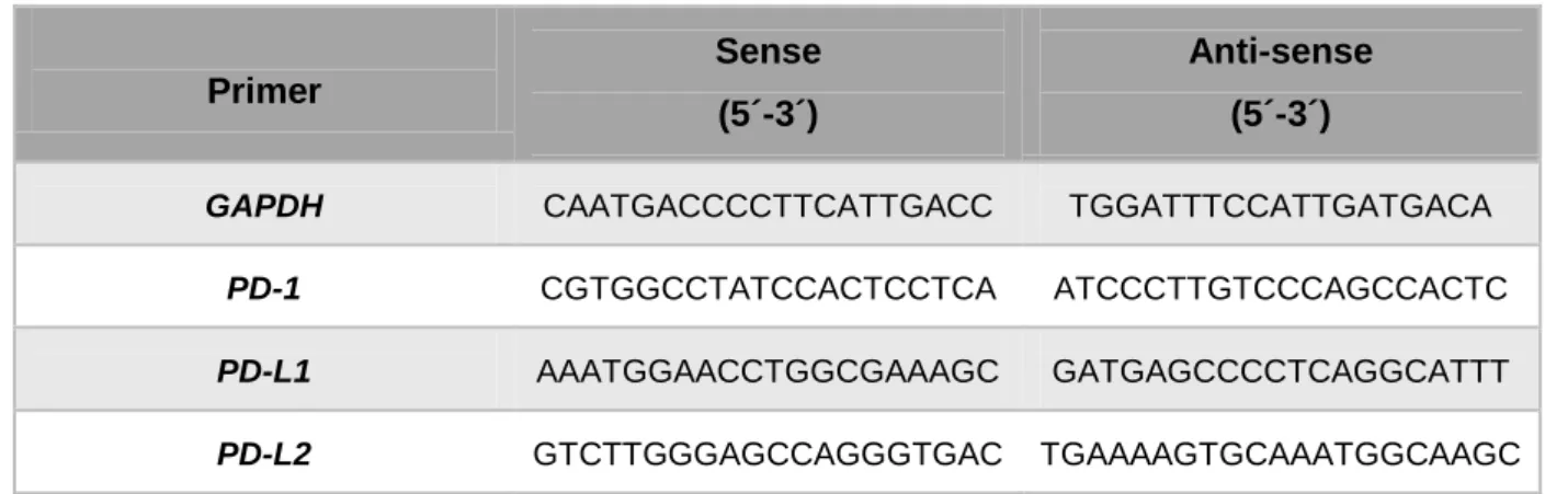
Table 1 | Antibodies and respective conditions used for the IHC staining in the present study. ... 19 Table 2 | Specific primer sense and anti-sense sequences used for gene amplification. ... 22 Table 3 | Demographic and clinicopathological characteristics of the 56 patients with BrMets originated from breast, lung, colon, kidney cancer and melanoma included in this study. ... 24 Table 4 | Demographic and clinicopathological characteristics of the 25 patients with BCBrMets included in
this study. ... 25 Table 5 | Association between clinicopathological characteristics of patients with BrMets originated from
breast, lung, colon, kidney and melanoma and total and CD8+ stromal TILs. ... 35 Table 6 | Association between clinicopathological characteristics of patients with BCBrMets and total and
4 LIST OF SYMBOLS AND ABBREVIATIONS
ºC Degree Celsius 2-ΔΔCt Fold difference μg Microgram μg/mL Microgram/millilitre μL Microlitre μm Micrometre Units/mL Units/millilitre v/v Volume/volume percent % Percentage B7-H1 B7-Homolog 1 BBB Blood-Brain Barrier BC Breast Cancer
BCBrMets Breast Cancer Brain Metastases
BrMets Brain Metastases
BT-474 B-Type-474 (breast cancer cell line)
CCLE Cancer Cell Line Encyclopedia
CD274 Cluster of Differentiation 274
CD279 Cluster of Differentiation 279
CD4 Cluster of Differentiation 4
CD8 Cluster of Differentiation 8
cDNA complementary DNA
CNS Central Nervous System
CO2 Carbon dioxide
COX2 Cycloxygenase-2
Ct Cycle threshold
CUP Cancer of Unknown Primary
DAB 3,3'-diaminobenzidine
DFS Disease-Free Survival
DMEM Dulbecco’s Modified Eagle’s Medium
EGFP Enhanced Green Fluorescence Protein
ER Oestrogen Receptor
FBS Fetal Bovine Serum
FFPE Formalin-Fixed, Paraffin-Embedded
GAPDH Glyceraldehyde-3-Phosphate Dehydrogenase
GCO Global Cancer Observatory
5
h Hour
H&E Haematoxylin and Eosin staining
HER2 Human Epidermal Growth factor Receptor type-2
HER2+ HER2-amplified
HR Hormone Receptor
H-score Histo-score
HSM – CHULN Hospital de Santa Maria – Centro Hospitalar Universitário Lisboa Norte
IARC International Agency for Research on Cancer
IHC Immunohistochemistry
INSERM Institut National de la Santé et de la Recherche Médicale
IPATIMUP Instituto de Patologia e Imunologia Molecular da Universidade do Porto
IQR Interquartile Range
MCF10A Michigan Cancer Foundation-10A (mammary epithelial cell line)
MCF7 Michigan Cancer Foundation-7 (breast cancer cell line)
MDA-MB-231 M.D. Anderson – Metastatic Breast 231 (breast cancer cell line) MDA-MB-231-BO2 M.D. Anderson – Metastatic Breast 231 – Bone tropic (breast
cancer cell line)
MDA-MB-231-BR M.D. Anderson – Metastatic Breast 231 – Brain tropic (breast cancer cell line)
MDA-MB-231-BR HER2+ M.D. Anderson – Metastatic Breast 231 – Brain tropic HER2-amplified (breast cancer cell line)
MDA-MB-231-BR HER2- Brain tropic M.D. Anderson – Metastatic Breast 231 – Brain tropic HER2-negative (breast cancer cell line)
MDA-MB-361 M.D. Anderson – Metastatic Breast 361 – Brain tropic (breast cancer cell line)
MDA-MB-435S M.D. Anderson – Metastatic Breast 435 spindle shaped variant (breast cancer cell line)
min Minute(s)
NA Not Applicable
NSCLC Non-Small Cell Lung Cancer
ON Overnight
OS Overall Survival
PBS-T Phosphate Buffered Saline with 0.05% Tween 20
PR Progesterone Receptor
6
PD-L1 Programmed Cell Death-Ligand 1
PD-L2 Programmed Cell Death-Ligand 2
PC-3 Prostate Cancer-3 (Prostate cancer cell line)
RNA Ribonucleic Acid
RT Room Temperature
RT-qPCR Reverse Transcription - Quantitative Polymerase Chain Reaction
sec Seconds
ST6GALNAC5 α2, 6-sialyltransferase
Th Helper T cell
TILs Tumour-Infiltrating Lymphocytes
TMAs Tissue Microarrays
TNBC Triple Negative Breast Cancer
Treg Regulatory T cell
WBRT Whole-Brain Radiation Therapy
7
1 INTRODUCTION
Cancer is one of the main causes of death worldwide and the number of new cases is on the rise globally, which represents a major impact on society. The World Health Organization (WHO) estimates a number of incident cases of 18.1 million in 2018 for all cancers, all ages and both sexes, with 0.5 million new cases across the world this year. According to WHO, incidence will increase to 29.5 million in 2040. These data were obtained from the Global Cancer Observatory (GCO) web-based platform (https://gco.iarc.fr/tomorrow/home), consulted in 10 November 2018.
Cancer cells can spread from the primary cancer locally, regionally and to distant sites. The formation of new tumours in parts of the body other than the site where they first formed is called metastases. Although metastases can form in most any part of the body, different types of cancer are more likely to spread to certain areas than others. Brain metastases (BrMets), or secondary brain tumours, are thought to have an incidence higher than 9% to 17% based on various studies, and the tumour types that more frequently metastasize to the brain are breast cancer (BC), lung cancer, melanoma, colon cancer and kidney cancer1. Metastatic cancer to the brain has a poor prognosis, with an average survival of less than 6 months2,3.
1.1 Brain Metastases
BrMets are one of the most frequent and devastating neurological complications related to systemic cancer, thus representing a significant cause of morbidity and mortality1,4,5. They are the commonest intracranial tumours, surpassing primary brain tumours. Despite that, there are no available data from the International Agency for Research on Cancer (IARC) neither other large-scale studies examining the incidence and prevalence of BrMets6. In Portugal these epidemiological data are also lacking. Most BrMets originate from lung cancer (36-64%), BC (15-25%) and melanoma (5-20%), which together account for 67% to 80% of all cancers7. Also, in up to 15% of patients with presentation of BrMets, these will correspond to a diagnosis of cancer of unknown primary (CUP)5.
BrMets have traditionally been managed with whole-brain radiation therapy (WBRT) or stereotactic surgery, and are typically associated with a poor prognosis3. Still, early detection and improvements in the treatment of primary tumour and systemic disease over the past decades resulted in the increase of survival of
8 patients with BrMets8,9. However, the frequency of the diagnosis of BrMets appears to be increasing also, occurring in 20% to 40% of patients with cancer in the United States of America1,10. The increased rate of diagnosis and lack of effective therapies are of significant concern and turn BrMets in a burgeoning clinical challenge. In view of these facts and in order to improve patients’ outcomes, it is clear that there is a need to better understand the brain’s milieu in the context of metastatic disease. 1.1.1 Breast Cancer Brain Metastases
BC is the most prevalent cancer in women worldwide, and approximately 15% of women with newly diagnosed metastatic BC will develop BrMets. However, this percentage is referring only to clinically apparent cases, since autopsy studies reveal that the incidence may be higher11.
BC is a heterogeneous disease divided into different molecular subtypes with clinical implications, according to the status of molecular markers such as oestrogen receptor (ER), progesterone receptor (PR), human epidermal growth factor receptor type-2 (HER2) and Ki67, a proliferation marker12. It is classified as Luminal A (ER+, HER2-, PR high, Ki67 low), Luminal B HER2- (ER+, HER2-, PR low or Ki67 high), Luminal B HER2+ (ER+, HER2+, any PR, any Ki67), HER2-Amplified (HER2+) (HER2+, ER and PR absent) or Triple Negative Breast Cancer (TNBC) (ER and PR absent, HER2-).
Patients with TNBC and HER2+ BC are at highest risk for central nervous system (CNS) recurrence, and incidences of breast cancer brain metastases (BCBrMets) as high as 30% to 40% have been reported for these molecular subtypes13,14,15,16. Retrospective studies have shown that BC subtype impacts survival in patients with BCBrMets, being TNBC the subtype with the poorest prognosis17,18. Median survival from the diagnosis of BCBrMets historically ranges between 2 and 16 months, depending on the involvement of the CNS, the extent of extracranial metastatic disease, performance status and administration of local or systemic therapy19,20. Differences in disease progression in patients with BCBrMets, which translate into different outcomes, highlight the need for a tailored approach in the care of these patients.
Despite the report of controversial results in published studies, the small size of many of the cohorts used and the little known about the following topic, compelling evidence has shown that there are molecular changes occurring in extracranial BC
9 metastases that could make a difference to treatment21,22. The selection of HER2+ clones in 24% to 48% of the cases, of ER negative clones in 7% to 13% of the cases or of PR negative ones into positive in the metastasis are alterations that could enable the prescription of treatments in the metastatic setting that were not an option for the primary tumour23,24,25.
As previously mentioned, there is a need to know the brain microenvironment and to unravel the crosstalk between this particular metastatic niche and tumour cells. In the context of BC and other brain tropic tumours, this could provide new and more efficient therapeutic strategies in the future.
1.2 Brain Microenvironment
Organotropism or the propensity of tumour types to disseminate to specific organs, such as the brain, has always been a hot topic in cancer research26. It is consensual that tumour cells will grow in congenial environments and that cancer cells acquire specialised functions to overtake specific organs, but is still not well understood how the metastatic niche is pre-conditioned by the tumour cells in the micro- to macrometastases progression. However, it is known that for the brain metastatic process there is the formation of perivascular niche, at least at early stages, which has been associated with stem-like and resistant cellular phenotype
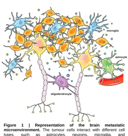
per se in brain tumours27,28. The blood-brain barrier (BBB) components are the most likely candidates to provide this supportive niche for cancer cells29. The composition of tumour microenvironment depends on the tumour site, meaning that the brain microenvironment is different from that of extracranial sites30. It consists of various specialised cell types, such as brain endothelial cells, astrocytes and glial cells that may influence tumour growth (Figure 1). Finally, the presence of immune cells has been described in metastatic solid tumours to the brain, such as BC, lung cancer and melanoma, although their prognostic significance remains unclear31.
Overall, it seems that the dissemination of cancer cells to the brain results from a symbiotic relationship between the tumour cells and the host, thus it is critical to elucidate the underlying molecular features of the metastatic cascade to further study and develop more effective therapy modalities that may interfere in this relationship, such as immunotherapy5,7. As the tumour microenvironment may deeply impact on the efficacy of cancer immunotherapies, the histologic assessment of
10 tumours and their immune contexture is becoming decisive. Quantifying and characterising the immune infiltrate, and assessing the presence of prognostically relevant cell subsets, such as CD8+ T cells, as well as determining the existence of actionable immunotherapy targets, as programmed cell death-ligand 1 (PD-L1), represent a new era in the clinical management of BrMets, namely in melanoma.
1.2.1 Blood-Brain Barrier
The brain has been considered an immune-privileged site for metastases, a ‘sanctuary’ where cancer cells can lay dormant in the CNS, behind the BBB10,32
. The BBB has a protective role of the brain and is formed by three main types of cells – endothelial cells, astrocytes, and pericytes – that limit the invasion of the brain parenchyma by circulating molecules, immune cells and antibodies33,34.
However, any disseminated tumour may be able to subvert the BBB and reach the brain, mainly the brain parenchyma1. Gene expression analysis revealed that specific genes, like α2, 6-sialyltransferase
(
ST6GALNAC5), mediate cancer cell passage through it35,36. The expression of ST6GALNAC5 is normally restricted to thebrain, but it was found to enhance BC cells adhesion to brain endothelial cells. Along Figure 1 | Representation of the brain metastatic
microenvironment. The tumour cells interact with different cell
types, such as astrocytes, neurons, microglia, and
11 with cycloxygenase-2 (COX2) and other genes, ST6GALNAC5 mediates BC cells infiltration through the BBB.
Furthermore, debate continues as to what extent therapeutic resistance is related to inadequate delivery of drugs to the brain versus intrinsic tumour resistance and/or stromal protective effects37.
1.2.2 Astrocytes
Astrocytes are non-proliferative cells in the normal adult brain that control homeostatic functions in health and disease. They can be activated upon injury and be involved in gliosis, characterised by proliferation or hypertrophy of several types of glial cells or, in its most extreme form, glial scar formation38. They then assume a reactive hypertrophic phenotype, characterised by the upregulation and rearrangement of the glia-specific cytoskeletal intermediate filament protein glial fibrillary acidic protein (GFAP)39,40. Hypertrophic activated astrocytes have been associated with BrMets as BrMets can induce the strong local activation of astrocytes41. In fact, accumulation of microglia and activated astrocytes around and within lesions has been shown, with astrocytes having direct contact with cancer cells42,43.
As the most abundant cell type in the CNS, astrocytes account for the majority of the interactions that cancer cells will be exposed to during brain metastatic process, both in early and late stages, and are emerging as essential regulators of BrMets progression as they have a secretory nature and can function as oncogenic signals for the tumour cells38,41,44. However, it has been reported that they show different roles, depending on the stage of the disease44. Overall, initially astrocytes act as an innate host defense system preventing disease progression, while in late stages they can favour it, providing protection against tumour cell death45,46.
1.2.3 Tumour-Infiltrating Lymphocytes (TILs)
Supporting evidence of the role played by immune infiltrating cells in brain tumours is increasing, although the CNS being allegedly considered an immune-privileged site. Despite brain’s apparently limited capacity for inflammatory response, BrMets contain tumour-infiltrating lymphocytes (TILs), drivers of the ‘selective pressure’ on CNS tumours, in the tumour stroma and in an intratumoral location47
12 TILs, which include T and B cells at a density that varies according to tumour type and stage of the disease, can be a clinically significant prognostic biomarker in various types of tumours48. Their presence in tumours is generally a good prognostic sign and despite their role in BrMets being still not clear with conflicting data from different primary tumour sites, reviews of the literature concluded that TILs density in CNS metastases was strongly associated with improved overall survival (OS)49,50,51.
BC has not been traditionally considered an immunogenic cancer type. Nevertheless, increasing evidences suggest that an effective immune response may greatly impact on the clinical behaviour of this malignancy. Also in BC, TILs are associated with favourable prognosis, especially in early TNBC and HER2+ BC phenotypes – the ones with more brain tropism –, and may positively influence the response to systemic therapies52,53. Studies with the same cutoff for TILs positivity suggested survival benefit in TNBC patients in their presence54,55.
It is extremely difficult to characterise and quantify all TILs subpopulations, and the prognostic value of a specific subset may well not represent the total impact of TILs on survival, as TILs subsets have their own roles in the immune microenvironment and in metastatic BC. For example, CD4+ T cells include helper T (Th) and regulatory T (Treg) cells, whereas CD8+ lymphocytes are the main immune effector cells. The prognostic value of CD8+ T cells varies according to tumour type and, therefore, more prospective studies are warranted to confirm it. However, a meta-analysis with 25 studies concluded that high density of CD8+ T cells is an indicator of good prognosis in BC patients56. Furthermore, an association between higher amounts of CD8+ lymphocytes within the invasive margin and significantly longer disease-free survival (DFS) was reported in BCBrMets57. The study of the CD4+/CD8+ TILs ratio may be an important parameter in cancer patients. Although it varies between different types of cancer, an increase has been observed in patients with several cancers, such as BC58,59.
1.2.4 Programmed Cell Death-Protein 1 (PD-1)/Programmed Cell Death-Ligand 1 (PD-L1) Axis
The effectiveness of immune-modulating agents within the CNS could be limited. However, there is growing evidence that immune therapies may be effective,
13 namely in patients with BrMets from melanoma, providing durable clinical responses60.In primary CNS tumours, a study showed promising preclinical data with immune-modulating antibodies61. Several lines of evidence suggest that T cells within the tumour microenvironment are the drivers of response to immune-modulation therapies47,62.
Programmed cell death-protein 1 (PD-1), also known as CD279 (cluster of differentiation 279), helps regulating the autoimmune response by down-regulating the immune system and suppressing T-cell inflammatory activity. However, this can also suppress antitumour immunity. PD-1 is normally expressed on the surface of immune cells, but recent studies revealed a widespread tumour-intrinsic expression of PD-1 in cancer, such as melanoma and lung cancer63,64.
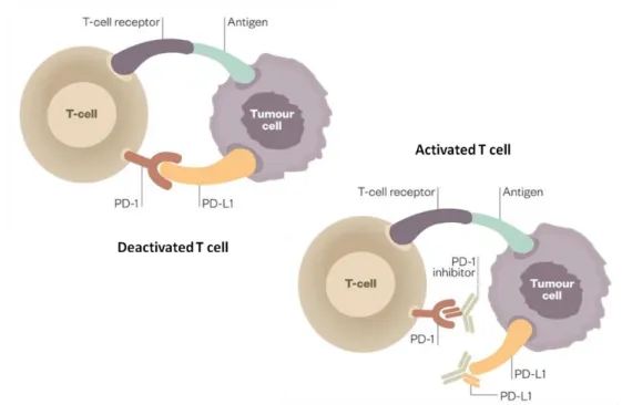
PD-L1, also known as CD274 (cluster of differentiation 274) or B7-H1 (B7-Homolog 1), similar to programmed cell death-ligand 2 (PD-L2 or PDCD1LG2), is a PD-1 ligand and an immune regulatory transmembrane protein expressed by tumour cells65. Therefore, targeting the PD-1/PD-L1 axis, by blocking it, is associated with tumour regression in several malignancies (Figure 2).
It has been described that a high level of PD-L1 expression in tumour cells correlates with poor prognosis in several cancers, including breast, lung and renal
Figure 2 | Schematic representation of the PD-1/PD-L1 axis. T cells are deactivated upon
PD-1 and PD-L1 interaction and activated when inhibitors block this interaction. Adapted from 103.
14 cancers, and melanoma66. However, other studies have suggested a positive or inexistent correlation between PD-L1 expression and survival67,68,69. Specifically in BC, PD-L1 was shown to be most frequently expressed in basal-like tumours, though its expression was rare70. Thus, its prognostic value is controversial. Finally, a recent study defined PD-L1 and PD-L2 expression as a common occurrence in BCBrMets, irrespective of primary tumour or BCBrMets phenotypes71. The role played by PD-L2 is still not clear66.
15
2 OBJECTIVES
The primary objective of this project was to retrospectively investigate if the immune compartment of BCBrMets, characterised by the presence of stromal TILs (total, CD4+ and CD8+), was associated with OS as endpoint, using a randomised retrospective cohort of patients with BrMets from other tumours as comparator.
As secondary objectives we proposed to: a) characterise the presence of activated astrocytes (using GFAP as a surrogate marker) and the expression of PD-L1 in BCBrMets, and to explore a possible association with the patients’ outcome; b) assess the molecular alterations between primary breast tumours and paired BrMets, namely in ER, PR, HER2 and Ki67 expression.
16
3 MATERIALS AND METHODS
3.1 Clinical Samples
In this study we used a retrospective cohort of 56 formalin-fixed, paraffin-embedded (FFPE) samples from BrMets tissue. The use of tissue samples was approved by the institutional review board of Hospital de Santa Maria – Centro
Hospitalar Universitário Lisboa Norte (HSM – CHULN) (approval number 556/14). All
tissue samples used were initially obtained for pathological diagnosis and are part of the general sample archive of the Neuropathology Laboratory, HSM – CHULN. The requirement for written informed consent was waived due to the retrospective nature of the study. During the analysis, the observers were fully blinded for patients’ personal data, as samples were identified by their registration number and, in a later phase, by their tissue microarrays (TMAs) positioning.
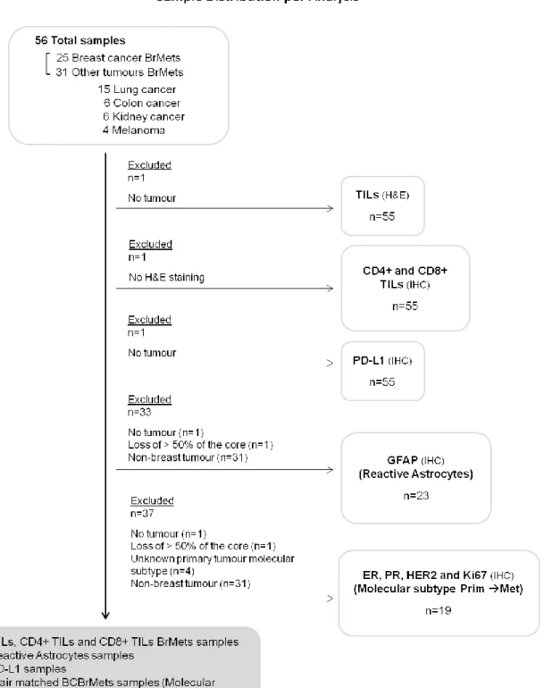
The cohort included 56 patients diagnosed with BrMets between 2009 and 2013 and followed at Department of Medical Oncology of HSM – CHULN; 34 (60.7%) women and 22 (39.3%) men, with a median age at BrMets diagnosis of 58.50 years (range 28-90 years), and with BrMets arising from BC (n=25, 44.7%), lung cancer (n=15, 26.8%), colon cancer (n=6, 10.7%), kidney cancer (n=6, 10.7%) and melanoma (n=4, 7.1%). The time between first recurrence and metastization to the brain varied between 0 – 5 BC patients had the brain as primary site of recurrence – and 24 months. The median survival after BrMets was 7.5 months. All of the patients received holocranial radiotherapy after surgical resection. Sample distribution per analysis is represented in Figure 3.
17
Figure 3 | Flowchart of sample distribution per analysis.
3.2 Haematoxylin and Eosin Staining (H&E)
For Haematoxylin and Eosin staining (H&E), 5 m sections of TMAs were depparafinized in xylene, rehydrated in decreasing concentrations of ethanol (100%, 95%, 70%); each for 5 minutes (min) and placed in distilled water. Tissue sections
18 were stained with Harris Haematoxylin for 3 min, washed in running tap water for 5 min, and dipped in 70% ethanol. After staining with alcoholic eosin, tissue sections were dehydrated in increasing concentrations of ethanol, cleared in xylene for 10 min and mounted with a solvent-based mounting media, Quick-D (Klinipath).
3.3 Immunohistochemical Staining (IHC)
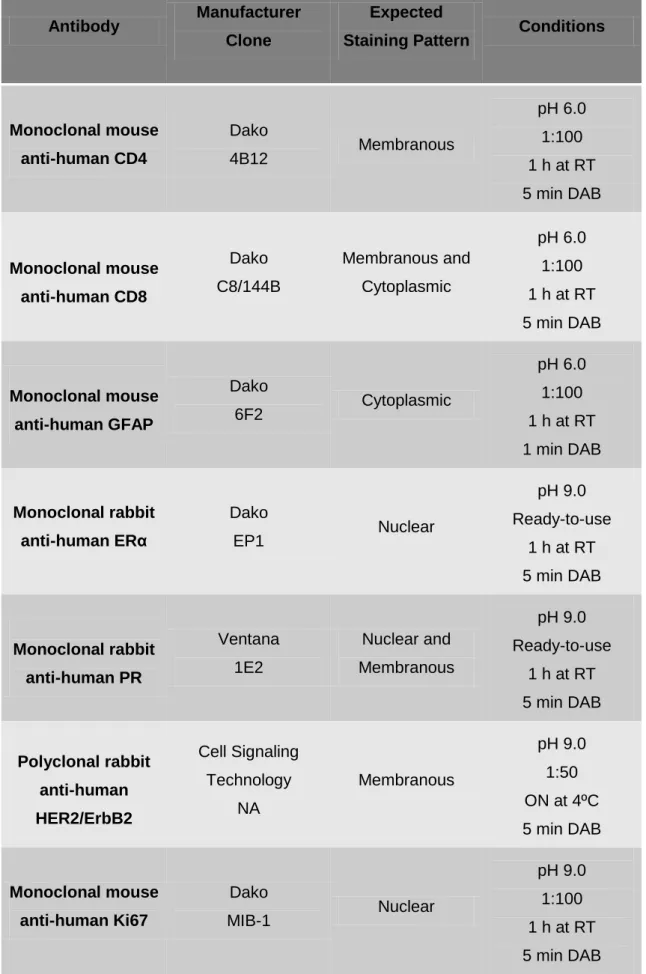
For immunohistochemistry (IHC), 5 m sections of TMAs were processed using the PT Module Thermo Scientific for Tissue Specimens (Dako), with Antigen Retrieval pH 6.0 or pH 9.0 solution (Dako) at 94ºC for 20 min. Sections were washed with Phosphate Buffered Saline (Sigma) with 0.05% Tween 20 (VWR, PROLABO) (PBS-T) for 5 min at room temperature (RT). The activity of endogenous peroxidase was blocked with Blocked Endogenous Peroxidase Solution (Dako) for 10 min at RT, followed by three 5 min rinses in wash buffer. Total protein blockade was performed using Protein Block Solution (Dako) for 20 min at RT, followed by incubation with the following primary antibodies, diluted in Antibody Diluent (Dako): anti-CD4, anti-CD8, anti-GFAP, anti-HER2, anti-ERα, anti-PR and anti-Ki67 (for additional details and working conditions see Table 1). Tissue sections were rinsed in wash buffer two times, 10 min each, and incubated with the visualisation system Dako REAL EnVision Detection System, Peroxidase/DAB+, Rabbit/Mouse (Dako) for 1 hour (h) at RT. After incubation, slides were rinsed in wash buffer as described above, incubated with 3,3'-diaminobenzidine (DAB+ Chromogen, Dako REAL), and rinsed in PBS-T and Elix water, each for 5 min. Slides were counterstained with Harris Hematoxylin for 10 seconds (sec), washed in running water for 5 min, dehydrated, cleared and mounted as described for H&E staining.
PD-L1 staining was performed at Instituto de Patologia e Imunologia Molecular da Universidade do Porto (IPATIMUP), using the Ventana BenchMark XT Staining System, with a 36 min incubation time for the monoclonal mouse anti-human PD-L1 (1:70) (22C3, Dako) and the OptiView DAB IHC Detection Kit (Ventana Medical Systems). Membranous and cytoplasmic staining pattern was expected.
The absence of primary antibody was used as negative control for all the markers.
19 Table 1 | Antibodies and respective conditions used for the IHC staining in the present study.
Antibody Manufacturer Clone
Expected
Staining Pattern Conditions
Monoclonal mouse anti-human CD4 Dako 4B12 Membranous pH 6.0 1:100 1 h at RT 5 min DAB Monoclonal mouse anti-human CD8 Dako C8/144B Membranous and Cytoplasmic pH 6.0 1:100 1 h at RT 5 min DAB Monoclonal mouse anti-human GFAP Dako 6F2 Cytoplasmic pH 6.0 1:100 1 h at RT 1 min DAB Monoclonal rabbit anti-human ERα Dako EP1 Nuclear pH 9.0 Ready-to-use 1 h at RT 5 min DAB Monoclonal rabbit anti-human PR Ventana 1E2 Nuclear and Membranous pH 9.0 Ready-to-use 1 h at RT 5 min DAB Polyclonal rabbit anti-human HER2/ErbB2 Cell Signaling Technology NA Membranous pH 9.0 1:50 ON at 4ºC 5 min DAB Monoclonal mouse anti-human Ki67 Dako MIB-1 Nuclear pH 9.0 1:100 1 h at RT 5 min DAB
20 3.4 Histological Evaluation and IHC Scoring
All H&E and IHC slides were analysed by a Pathologist, with each case being represented by 3 cores. The evaluation and scoring of every biomarker resulted from the mean of the 3 corresponding cores per case. TILs density was presented as the percentage of total TILs in the tumour stromal compartment using visual assessment of H&E-stained TMAs. Samples were classified as: absent (0% TILs), slight (30% TILs), moderate (30%> TILs 60%), and marked (>60% TILs)72. CD4+ TILs and CD8+ TILs were classified as described for TILs after IHC staining and percentages were normalised to total stromal TILs.
PD-L1 expression was classified as positive (membranous and cytoplasmic staining in ≥1% of tumour cells or stromal TILs) or negative (membranous and cytoplasmic staining in <1% of tumour cells or stromal TILs)73.
GFAP was used as a marker of reactive astrocytes. Astrocytes in tumour sections were classified qualitatively as not altered (normal GFAP cytoplasmic staining intensity) or altered (abnormal GFAP cytoplasmic staining intensity).
Hormone receptors (ER and PR), HER2, and Ki67 were classified according to the guidelines of the Template for Reporting Results of Biomarker Testing of
Specimens from Patients with Carcinoma of the Breast, from the College of American
Pathologists (2014). ER and PR were considered positive if ≥1% positive cells. For HER2, staining intensity was classified from 0 to 3: (0) absence of staining, (1) weak, (2) moderated and (3) strong staining, and samples were classified as negative (0 or 1+), equivocal (2+) or positive (3+). Ki67 was classified as low (<10% positive cells), borderline (20%-30% positive cells) or high (>30% positive cells).
3.5 Statistical Analysis
For statistical analysis, patients were dichotomised into low and high, according to TILs and CD8+ TILs percentage cutoff, selected using 12-months OS as endpoint and Cutoff Finder (http://molpath.charite.de/cutoff/)74.
Demographic and clinicopathologic data of patients were described using frequencies for categorical variables, and central tendency and range for continuous variables. Univariate association of these characteristics and TILs and CD8+ TILs percentage was done through Kruskal-Wallis test, Fisher’s exact test, χ2 test and Mann-Whitney test as applicable. OS was estimated using Kaplan-Meier curves and
21 differences were determined using the log-rank (Mantel-Cox) test. A significance level of P-value <0.05 was set for all statistical analyses.
Statistical analysis was carried out using the software GraphPad Prism 6 for Windows (GraphPad Software).
3.6 Cell Culture
All cell lines were cultured in supplemented Dulbecco’s Modified Eagle’s Medium (DMEM) (Gibco) with 10% (v/v) fetal bovine serum (FBS) (Gibco) and 1% (v/v) Penicillin Streptomycin (Pen Strep, 10,000 Units/mL Penicillin, 10,000 μg/mL Streptomycin) (Gibco). Cells were kept at 37ºC with 5% CO2 in a humidified atmosphere, and medium was changed every two or three days.
The BC brain tropic cell lines MDA-MB-231-BR HER2+ and MDA-MB-231-BR HER2- were kindly provided by Patricia S. Steeg and David Lyden Lab, at Cornell University. MCF-10A, MB-231, MB-435S, MCF7, ZR-75, SK-BR-3, MDA-MB-361 and PC-3 cell lines were purchased from ATCC. MDA-MB-231-BO2, T-47D and BT-474 cells were provided by Phyllippe Clézardin Lab, Institut National de la
Santé et de la Recherche Médicale (INSERM).
3.7 RNA Extraction, cDNA Synthesis and Reverse Transcription - Quantitative Polymerase Chain Reaction (RT-qPCR)
Ribonucleic Acid (RNA) extraction (NZY Total RNA Isolation kit, NZYtech) was performed according to the manufacturer’s protocol. Total RNA was quantified by spectrophotometry, using NanoDrop ND-1000 (Thermo Fisher Scientific) and 1 μg of total RNA was used to synthesize complementary DNA (cDNA), with Oligo(dT)18 primer and the NZY M-MuLV First-Strand cDNA Synthesis Kit (NZYTech), according to the manufacturer’s instructions.
Genes of interest were amplified by semi-quantitative real time PCR in the ViiA 7 Real-Time PCR System (Applied Biosystems), using specific primers (Invitrogen, Table 2) in a 10 μL reaction volume with SYBR Green PCR Master Mix (Applied Biosystems).
Target gene expression was normalised against the housekeeping gene
22 Primer Sense (5´-3´) Anti-sense (5´-3´)
GAPDH CAATGACCCCTTCATTGACC TGGATTTCCATTGATGACA
PD-1 CGTGGCCTATCCACTCCTCA ATCCCTTGTCCCAGCCACTC
PD-L1 AAATGGAACCTGGCGAAAGC GATGAGCCCCTCAGGCATTT
PD-L2 GTCTTGGGAGCCAGGGTGAC TGAAAAGTGCAAATGGCAAGC
23
4 RESULTS AND DISCUSSION
4.1 Prognostic Role of Stromal TILs and CD8+ TILs in BrMets
Histologic assessment of the immune context of tumours has been showing prognostic relevance75. Thus, the determination of the amount of immune infiltrate as well as its composition, in particular of prognostically relevant immune cell subtypes such as CD8+ T cells, for example, is becoming central.
As stated before, TILs and CD8+ TILs have been shown to have prognostic value across a broad range of tumour types and to be positively correlated with OS, suggesting that TILs might reduce metastatic spread48,50,51,54. Therefore, as a primary objective we aimed to quantify total, CD4+ and CD8+ stromal TILs in BCBrMets, and to assess their prognostic role in the context of brain metastatic disease.
Although we focused on BCBrMets, we used a retrospective cohort of FFPE TMAs, which included not only samples from BCBrMets but also from BrMets from other types of tumours. This allowed us to use these non-breast cancer samples as comparators in our analysis, enriching the results of this project. Demographic and clinicopathological characteristics of the 56 patients with BrMets, included in this study, 25 of which with BCBrMets, are presented in Tables 3 and 4, respectively. For the analysis of total TILs and CD8+ TILs on H&E and IHC tissues, respectively, we excluded 1 case due to absence of tumour in the TMAs cores used for H&E staining.
24 Table 3 | Demographic and clinicopathological
characteristics of the 56 patients with BrMets originated from breast, lung, colon, kidney cancer and melanoma included in this study.
Total (N=56)
Age at diagnosis of BrMets (years)
Median 58.50 Range 28-90 IQR 15.75 Gender Female 34 (60.7%) Male 22 (39.3%) Primary tumour Breast cancer 25 (44.6%) Lung cancer 15 (26.8%) Colon cancer 6 (10.7%) Kidney cancer 6 (10.7%) Melanoma 4 (7.2%)
Time between BrMets and death (months)
Median 7.5 Mean 11.07 Range 1-72 Survival status Deceased 54 (96.4%) Alive 2 (3.6%)
25
Total (N=25) Age at diagnosis of BrMets (years)
Median 57 Range 28-86 IQR 17 Gender Female 25 (100%) Male 0 (0%) TNM stage IA 2 (8%) IB 1 (4%) IIA 7 (28%) IIB 4 (16%) IIIA 4 (16%) IIIB 3 (12%) IIIC 1 (4%) IV 0 (0%) Unknown 3 (12%) Histology Ductal carcinoma 18 (72%) Lobular carcinoma 1 (4%) Unknown 6 (24%) ER statusⱡ Positive 7 (28%) Negative 16 (64%) NA 2 (8%) PR statusⱡ Positive 11 (44%) Negative 12 (48%) NA 2 (8%) HER2 statusⱡ Positive 10 (40%) Negative 9 (36%) Equivocal 4 (16%) NA 2 (8%) Ki67 statusⱡ High 8 (32%) Low 5 (20%) Borderline 10 (40%) NA 2 (8%)
Molecular subtype* Primary tumour Metastasis
Luminal 9 (36%) 7 (28%) Luminal B HER2+ 3 (12%) 5 (20%) HER2+ 7 (28%) 6 (24%) TNBC 2 (8%) 5 (20%) Unknown 4 (16%) 0 (0%) NA 0 (0%) 2 (8%)
Molecular subtype discordance between primary tumour and metastasis
Unknown 4/25 (16%)
NA 2/25 (8%)
Adjuvant therapy 25 (100%)
BrMets , Brain Metastases; IQR , Interquartile Range; ER , Oestrogen Receptor; PR , Progesterone Receptor;
HER2 , Human Epidermal Growth Factor Receptor type-2; TNBC , Triple Negative Breast Cancer; NA , Not Applicable, meaning absence of tumour or loss of > 50% of the cores.
ⱡSatuses refer to the metastasis; *Molecular subtype classification is according to the 2015 St Gallen Consensus Conference, recommended by the ESMO Clinical Practice Guidelines; Luminal comprises Luminal A and Luminal B HER2- subtypes.
10/19 (52.6%)
Table 4 | Demographic and clinicopathological characteristics of the 25 patients with BCBrMets included in this study.
26 TMAs H&E slides were used to observe tissue morphologic characteristics and also to quantify total TILs (Figure A1). Next, samples were dichotomised into low or high TILs, using the cutoffs 1%, 3% and 0.5% for BC cases, non-BC cases and all tumour types, respectively, as these were the best cutoff points found by Cutoff Finder after testing other cutoffs (median; positive (>0) vs negative (0)). These cutoffs were not close to the 50% cutoff point reported as the best for dichotomisation of lymphocyte-predominant BC, due to overall low TILs density in our TMAs samples. Moreover, 50% was chosen to analyse only lymphocyte-predominant BC, a subset of TNBC and we had only five TNBC samples in our fifty six tissue samples cohort53.
CD4+ and CD8+ TILs were assessed by IHC and normalised to total TILs (Figures A2 and A3, respectively). Next, samples were dichotomised into low or high TILs, using the cutoffs 75%, 45% and 45% for BC cases, non-BC cases and all tumour types, respectively, like in TILs. Representative images of H&E and IHC staining are displayed in Figure 4.
Figure 4 | Representative tissue sections with high or low total or CD8+ TILs in tissue samples from patients with BrMets from BC (A) and non-BC tumours (B). Example of TILs on H&E sections is indicated by a white
circle; Example of CD8+ TILs in IHC sections is indicated by a black circle. Magnification X200.
27 T I L s P e r c e n ta g e Bre as t (n =2 4) No n-b rea st (n= 31 ) 0 5 1 0 1 5 2 0 2 5 3 0 3 5 p = 0 . 2 3 3 4 P e r c e n ta g e Bre as t (n =2 4) Lu ng (n =1 5) Co lon (n =6 ) Kid ne y ( n= 6) Me lan om a ( n= 4) 0 5 1 0 1 5 2 0 2 5 3 0 3 5 T I L s p = 0 . 1 2 4 1
Across all samples, total TILs ranged between 0% and 30%, corresponding to absent or slight TILs, respectively (Figure 5). Median TILs ranged between 1.33% and 8.33%, depending on tumour type.
Figure 5 | Total TILs in BrMets according to the origin of the primary tumour (A) and comparing metastases from BC with metastases from non-breast tumours (B). Corresponding mean percentage,
median percentage and range (%) are registered in the table (C). P-value was calculated with Kruskal-Wallis (A) or Mann-Whitney test (B), and significance was set as p<0.05.
Figure 4 | Representative tissue sections with high or low total or CD8+ TILs in tissue samples from patients with BrMets from BC (A) and non-BC tumours (B). Low TILs and CD8+ TILs on the left, high TILs and CD8+
TILs on the right. Example of TILs on H&E sections is indicated by a white circle; Example of CD8+ TILs in IHC sections is indicated by a black circle. Magnification X200. (Continued)
28 Figure 5 | Total TILs in BrMets according to the origin of the primary tumour (A) and
comparing metastases from BC with metastases from non-breast tumours (B).
Corresponding mean percentage, median percentage and range (%) are registered in the table (C). P-value was calculated with Kruskal-Wallis (A) or Mann-Whitney test (B), and significance was set as p<0.05. (Continued)
Regarding specific subsets of TILs, we observed the presence of both CD4+ and CD8+ TILs in BrMets, although CD4 staining was a rare event in this study, with only four positive cases. The scarce positive data prevented us from further investigating their relevance in this cohort. However, we must take into account that CD4+ TILs absence was consistently observed and that this may be of particular relevance and deserve further studies in larger cohorts. Moreover, the use of TMAs is a limitation of this study, inasmuch as TMAs surely limit the representativeness of the tissue heterogeneity.
Concerning CD8+ T cells, 23/55 cases (41.8%) scored 100%. Given the low percentages of total TILs, this implies that even in those cases of abundant CD8+ TILs, they correspond to a small part of the immune cell population. It would be important to quantify the other immune cell subpopulations in those BrMets system to further characterise the microenvironment of BrMets.
Distribution of CD8+ TILs across the different types of tumours is depicted in Figure 6. As in total TILs, we did not observe a difference between tumour types.
29 C D 8 + T I L s P e r c e n ta g e Bre as t (n =2 4) No n-b rea st (n= 31 ) 0 5 0 1 0 0 p = 0 . 8 7 0 4 P e r c e n ta g e Bre as t (n =2 4) Lu ng (n =1 5) Co lon (n =6 ) Kid ne y ( n= 6) Me lan om a ( n= 4) 0 5 0 1 0 0 C D 8 + T I L s p = 0 . 9 1 9 7
Figure 6 | CD8+ TILs in BrMets according to the origin of the primary tumour (A) and comparing metastases from BC with metastases from non-breast tumours (B). Corresponding mean
percentage, median percentage and range (%) are registered in the table (C). P-value was calculated with Kruskal-Wallis (A) or Mann-Whitney test (B), and significance was set as p<0.05.
Since it has been suggested that the ratio CD4+/CD8+ TILs may impact the prognosis in melanoma, BC and squamous cell carcinoma of the cervix, we analysed the CD4+/CD8+ TILs ratio in the four cases with CD4+ TILs, two from melanoma, one from BC and one from lung cancer, even though it did not allow us any statistical inference (Figure 7)58,59,76.
30 Melanoma
Figure 7 | Representative images of tissue sections with the lowest (left) and the highest (right) CD4+/CD8+ TILs ratios (bold in the table). TILs are
pointed out by black arrows. Ratios of the cases with positive CD4 staining. Magnification X200.
TILs subsets have their own roles in BC progression and CD8+ TILs, the main effective cells in the immune response, have been related to better DFS48,56,77.
31 T im e a f t e r m e t a s t iz a t io n ( m o n t h s ) P e r c e n t s u r v iv a l 0 5 0 1 0 0 1 5 0 0 5 0 1 0 0 B r e a s t ( n = 2 4 ) N o n - b r e a s t ( n = 3 1 ) p = 0 . 0 2 9 2* H R = 0 . 5 8 0 3 ( 0 . 2 9 9 5 - 0 . 8 8 3 9 ) T im e a f t e r m e t a s t iz a t io n ( m o n t h s ) P e r c e n t s u r v iv a l 0 5 1 0 1 5 2 0 2 5 3 0 0 5 0 1 0 0 9 0 1 2 0 1 5 0 p = 0 . 0 2 7 5* L u n g ( n = 1 5 ) C o lo n ( n = 6 ) K id n e y ( n = 6 ) M e la n o m a ( n = 4 ) B r e a s t ( n = 2 4 ) T im e a f t e r m e t a s t iz a t io n ( m o n t h s ) P e r c e n t s u r v iv a l 0 5 1 0 1 5 0 5 0 1 0 0 B r e a s t ( n = 2 4 ) p = 0 . 0 8 3 7 L u n g ( n = 1 5 ) C o lo n ( n = 6 ) K id n e y ( n = 6 ) M e la n o m a ( n = 4 )
So next we aimed to assess the prognostic role of total TILs and CD8+ TILs in BrMets, by quantifying its association with OS.
Samples were dichotomised into low and high TILs and CD8+ TILs according to best cutoff value when analysing OS as endpoint. The exception was the sub-analysis of TILs in BCBrMets, where we could not use Cutoff Finder, and selected the median as dichotomisation cutoff, since it was the method tested associated with the best P-value.
We started by assessing the contribution of the primary tumour type to survival rates and 12-months survival rates (Figures 8 and A4, respectively). The OS curves were different according to the tumour type (p=0.0275), and better for BC patients, in comparison to non-breast patients (p=0.0292, median survival 11 vs. 7 months).
Figure 8 | Overall survival according to the origin of the primary tumour and comparing breast with non-breast metastatic disease (n=56). P-value was calculated using log-rank (Mantel-Cox) test, and significance
32 T im e a f t e r m e t a s t iz a t io n ( m o n t h s ) P e r c e n t s u r v iv a l 0 5 1 0 1 5 0 5 0 1 0 0 T IL s h ig h ( n = 4 2 ) T IL s lo w ( n = 1 3 ) p = 0 . 5 3 6 0 H R = 0 . 7 8 7 0 ( 0 . 3 2 3 8 - 1 . 7 6 7 ) A l l T u m o u r s T im e a f t e r m e t a s t iz a t io n ( m o n t h s ) P e r c e n t s u r v iv a l 0 5 1 0 1 5 0 5 0 1 0 0 T IL s h ig h ( n = 1 2 ) T IL s lo w ( n = 1 9 ) p = 0 . 5 5 9 0 H R = 0 . 7 8 1 2 ( 0 . 3 2 9 6 - 1 . 7 9 0 ) N o n - b r e a s t T im e a f t e r m e t a s t iz a t io n ( m o n t h s ) P e r c e n t s u r v iv a l 0 5 1 0 1 5 0 5 0 1 0 0 T IL s lo w ( n = 1 8 ) T I L s h ig h ( n = 6 ) p = 0 . 4 9 0 8 H R = 0 . 6 6 5 7 ( 0 . 1 6 8 6 - 2 . 3 0 2 ) B r e a s t
Next we assessed the impact of total TILs and CD8+ TILs on OS (Figures 9 and 10, respectively).
Figure 9 | 12-months-overall survival according to TILs percentage in BrMets from BC (n=24), all tumours (n=55), and non-BC (n=31). Cutoff values for dichotomisation (%) 1, 0.5, 3, respectively. P-value was calculated using log-rank (Mantel Cox) test, and significance was set as p<0.05.
33 T im e a f t e r m e t a s t iz a t io n ( m o n t h s ) P e r c e n t s u r v iv a l 0 5 1 0 1 5 0 5 0 1 0 0 C D 8 + T I L s h ig h ( n = 1 9 ) C D 8 + T IL s lo w ( n = 1 2 ) p = 0 . 0 2 9 6 * H R = 0 . 4 2 9 0 ( 0 . 1 3 7 0 - 0 . 8 4 8 6 ) N o n - b r e a s t T im e a f t e r m e t a s t iz a t io n ( m o n t h s ) P e r c e n t s u r v iv a l 0 5 1 0 1 5 0 5 0 1 0 0 C D 8 + T I L s h ig h ( n = 1 2 ) C D 8 + T IL s lo w ( n = 1 2 ) p = 0 . 3 9 7 1 H R = 0 . 6 2 1 1 ( 0 . 1 9 4 0 - 1 . 8 7 6 ) B r e a s t T im e a f t e r m e t a s t iz a t io n ( m o n t h s ) P e r c e n t s u r v iv a l 0 5 1 0 1 5 0 5 0 1 0 0 C D 8 + T IL s lo w ( n = 2 1 ) C D 8 + T I L s h ig h ( n = 3 4 ) p = 0 . 0 4 9 5 H R = 2 . 1 2 7 ( 1 . 0 0 2 - 4 . 5 1 8 ) * A l l T u m o u r s
Figure 10 | 12 months-overall survival according to CD8+ TILs percentage in BrMets from BC (n=24), all tumours (n=55), and non-BC (n=31). Cutoff values for dichotomisation (%) 75, 45, 45, respectively. P-value was calculated using log-rank (Mantel Cox) test, and significance was set as p<0.05.