Universidade do Algarve Faculdade de Ciências e Tecnologia
Reproductive biology of the species
Leucoraja naevus from Portuguese
continental waters
Catarina Maia
Master thesis submitted for the partial fulfillment of the title of Master of Marine Biology
Universidade do Algarve Faculdade de Ciências e Tecnologia
Reproductive biology of the species
Leucoraja naevus from Portuguese
continental waters
Catarina Maia
Master thesis submitted for the partial fulfillment of the title of Master of Marine Biology Internal supervisor: Prof. Dr. Karim Erzini
External supervisor: Profa. Dra. Ivone Figueiredo
I would like to thank everyone who helped me in IPIMAR and University:
First I would like to thank Dr. Ivone Figueiredo and Dr. Karim Erzini for the opportunity to perform this work and the availability and encouragement shown over the same;
I would also like to express my immense gratitude to Dr. Barbara Serra-Pereira for the help, encouragement and support (tireless!!!!) that greatly facilitated my work;
My sincere thanks to José do Lago and Neide Lagarto for their help in sampling and friendship;
As Teresa, Ana Rita and Inês, Miguel and Nuno, who not only gave me the motivation but also by the availability and friendship shown.
I also thank to all IPIMAR workers, including Carmo and Cristrina for their help and suggestions in histology;
Tanks to PNAB that partially supported my work;
My eternal gratitude to my parents and Francisco who were always by my side and supported me unconditionally.
Skate populations tend to be highly vulnerable to exploitation as a result of the main life history characteristics (slow growth, late maturity and low fecundity). The cuckoo ray, Leucoraja naevus, one of the skate species landed in Portugal, is mainly a by-catch species from the artisanal mixed-fishery, and for this reason it is very difficult to acquire information about its basic biology. The two main goals of the present work were to estimate the length at first maturity, fecundity and reproductive cycle of the species Leucoraja naevus and to compare the microscopic structure and chemical nature of the secretions produced by the oviducal gland of the species Leucoraja naevus with other skate species taking into account their reproductive strategy.
Although the species has been studied in other NE Atlantic geographic areas, namely Celtic Sea, Irish Sea and North Sea, this study is the first to provide information on the reproductive biology of Leucoraja naevus in Southern European waters. Length at first maturity was estimated at 57 cm and 55 cm TL for males and females, respectively. Active females were observed in every months of the year suggesting that reproduction may occur year round, with a reproductive peak during winter months between January and April. Females release follicles in batches of 5. However, it was not possible to determine the number of reproductive episodes realized by a female during a 1-year period.
Leucoraja naevus, Raja brachyura, Raja miraletus, Raja undulata, Raja microocellata and Raja montagui, have similar microscopic structure divided in four zones: club, papillary, baffle and terminal. However, differences were found between Leucoraja naevus and the remaining species in the chemical nature of the secretions produced in the papillary zone, that seems to have a higher content of sulphated acid mucins, and in the terminal zone that, like in Raja microocellata, lacks serous gland tubules. The differences found are related with the reproductive strategy displayed by each species, namely with the capsule and spawning habitats characteristics. Sperm was found in all species as laterally aligned bundles in the deep recessed tubules of the baffle zone, adjacent to the muscle layer, suggesting short-term sperm storage.
These results provide essential information on the reproductive biology not only about the species Leucoraja naevus but also about other by-catch species inhabiting
management plan.
Keywords: Cuckoo ray, fecundity, maturity, reproductive season, oviducal gland, reproductive strategy, Portugal, Rajidae
Devido a determinadas características do seu ciclo de vida, nomeadamente crescimento lento, maturação tardia e baixa fecundidade, as raias são organismos extremamente vulneráveis à exploração. A raia de São Pedro ou raia de dois olhos, Leucoraja naevus, uma das espécies desembarcadas nos portos portugueses, constitui um importante by-catch da pesca artesanal e por esta razão é muito difícil obter informação sobre a sua biologia. Os dois principais objectivos deste trabalho foram estimar o comprimento de primeira maturação, a fecundidade e a sazonalidade reprodutiva da espécie Leucoraja naevus e a comparação da estrutura microscópica e natureza química das secreções produzidas pela glândula oviductal da espécie Leucoraja naevus com outras espécies tendo em atenção a sua estratégia reprodutiva.
Apesar de existirem já vários estudos sobre a espécie em outras áreas geográficas do NE Atlântico, nomeadamente no Mar Céltico, Mar da Irlanda e no Mar do Norte, o presente estudo constitui a primeira abordagem acerca da biologia reprodutiva da espécie em águas sul europeias. O comprimento de primeira maturação foi estimado em 57 cm e 55 cm de comprimento total para machos e fêmeas, respectivamente. Foram observadas fêmeas activas em todos os meses do ano, sugerindo que a reprodução ocorre durante todo o ano, com um pico de reprodução entre os meses de Janeiro e Abril. As fêmeas libertam folículos em batches de 5 sem, no entanto, ter sido possível determinar a fecundidade potencial da espécie nem o número de episódios reprodutivos que uma fêmea é capaz de realizar durante um ano.
A glândula oviductal é uma estrutura discreta e especializada presente em peixes cartilagineos e tem como função, entre outras, a produção da cápsula. Todas as espécies aqui abordadas, nomeadamente Leucoraja naevus, Raja brachyura, Raja miraletus, Raja undulata, Raja microocellata e Raja montagui, têm a mesma anatomia e estrutura microscópica, partilhando a mesma zonação interna: zona club, papillary, baffle e terminal. Contudo, foram encontradas algumas diferenças entre a espécie Leucoraja naevus e as restantes espécies na natureza química das secreções da zona papillar, que parece ter um maior conteúdo de mucinas ácidas sulfatadas, e na zona terminal que, à semelhança da espécie Raja microocellata, não possui túbulos serosos e consequentemente não forma pêlos no exterior da cápsula. As diferenças encontradas estão relacionadas com a estratégia reprodutiva levada a cabo por cada espécie,
onde se realiza a desova. Foi encontrado esperma em todas as espécies aqui estudadas alinhado lateralmente em túbulos situados na porção mais caudal da zona baffle perto do tecido muscular, sugerindo um armazenamento de esperma a curto prazo.
Os presentes resultados fornecem informação essencial sobre a biologia reprodutiva não só da espécie Leucoraja naevus mas também sobre outras espécies by-catch da costa continental portuguesa, constituindo assim uma importante ferramenta para a aplicação de planos de gestão apropriados.
Palavras-chave: Raia de S. Pedro, fecundidade, maturação, época reprodutiva, glândula oviductal, estratégia reprodutiva, Portugal, Rajidae
I. General introduction...1
II. General methodology...3
III. Reproductive biology of the species Leucoraja naevus from Portuguese continental waters...6
IV. Comparison of the oviducal gland of Leucoraja naevus with other skate species from mainland Portugal...22
V. Final considerations...41
VI. References...43
1
I. General introduction
Cartilaginous fishes (Class Chondrichthyes) comprise about 1200 species and represent one of the oldest living classes of vertebrates (Ebert and Compagno 2007). The skates, order Rajiformes, are the most diverse order among Chondrichthyes, comprising at least 27 genera and more than 245 species (Ebert and Compagno 2007).
Skates inhabit a variety of substrates at depths from shallow inshore areas, as open coasts, bays and estuaries, to depths of 3000 m (Ruocco et al. 2006) in temperate to tropical waters (Ebert and Compagno 2007). Like others cartilaginous fishes, skates have sophisticated and complex reproductive cycles with life-history characteristics, like density dependent reproductive potential, slow growth rates, late maturity and low fecundity, that make them highly vulnerable to exploitation (Dulvy et al. 2000).
Despite the increasing number of valid species described over the last century (Ebert and Compagno 2007), the last 20 years has seen a rapid and increasing decline in skates populations, mainly due to directed fisheries and by-catch (Andreson 1990). There is already some legislation in respect to elasmobranch fisheries, such as the forbiddance of landing of a few species, the application of Total Allowable Catches (TAC’s) and discrimination of landings for a number of species (Clarke 2009). However, despite their increasing economic importance around the world, the majority of countries still lack proper elasmobranch management plans, with elasmobranchs generally receiving less attention than exploited bony fishes, often even being landed as aggregated species (Stevens et al. 2000, Coelho and Erzini 2006), making it difficult to obtain accurate data on the real landed quantities by species.
Due to all these factors it is very important to collect accurate and complete data on the biology and ecology of the exploited species. One of the typical approaches for elasmobranch fisheries management are the life-history or demographic and mortality type models, that includes information on age, growth, maturity and fecundity (Clarke 2009). All this information is very important for assessing age structure, growth and reproduction patterns, for understanding the response of the populations to fisheries exploitation (Anderson 1990) and therefore, to apply better management programmes (Pauly and Morgan 1987, Conrath and Musik 2002).
In Portugal, skates constitute an important by-catch of the mixed fishery, namely from the artisanal (61%) and trawl (31%) sectors (Figueiredo et al. 2007), representing
2
55% (around 1313 ton) of the elasmobranch total landings in 2009 (Serra-Pereira et al. 2009a). The most important landed species are Raja brachyura, Raja clavata, Raja montagui and Leucoraja naevus.
The present work is divided in two parts: I) reproductive biology of the species Leucoraja naevus, where the main goal was to collect information on the reproductive biology, with the assessment of parameters such as length at maturity, reproductive potential/fecundity and reproductive season, and II) Comparison of oviducal glands of reproducing females of the species Leucoraja naevus with other skate species regarding their reproductive strategies, where the main goal was to provide information on the reproductive strategy and spawning habitat preferences of each species.
I.2 Leucoraja naevus (Müller & Heule, 1841): species characterization
The coocko ray (Fig. 1A), Leucoraja naevus, is distributed in Eastern Atlantic waters from the North Sea to north-west Africa and the Mediterranean, being found in shallow inshore waters down to depths of 250 m (Walker 1998) (Fig. 1B). This species has a light grey/brown dorsal surface covered by small spines and can be easily identified by the presence of yellow black eyespots in each pectoral fin. The ventral surface is smooth and white. Around the inner margin of the eyes there are 9 to 13 thorns and a large triangle of thorns on the shoulders. On each side of the midline of the tail there are two rows of thorns which, in adults, continue onto the back. This species has a short snout with slightly concave pectoral fins (Stehmann and Bürkel 1984).
The maximum total length (TL) observed for the species was 73 cm in the North Sea (Walker 1998). It reproduces year round in the NE Atlantic and is considered one of the most fecund skate species, being able to lay around 90 eggs per year (Walker 1998, Du Buit 1976).
3 Figura 1: (A) Leucoraja naevus. (B) Distribution map of Leucoraja naevus (source: fishbase.org).
II. General Methodology
The specimens observed in the present study were obtained from i) fishing hauls performed during IPIMAR research surveys and ii) from commercial artisanal fleets along the Portuguese coast (Fig.1). In both cases, samples were collected under the scope of National Data Collection Program (PNAB, DCR) during the period between 2003 and 2010. A total of 856 specimens of Leucoraja naevus, 21 Raja montagui, five Raja brachyuran, ten Raja miraletus, eight Raja undulata and one specimen of Raja microocellata were sampled.
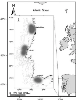
4 Figure 1: Map of the NE Atlantic coast with details of the continental coast of Portugal. The main landing ports (Matosinhos, Peniche and Portimão) for skates and corresponding fishing areas (shaded grey) are identified.
For each specimen the following measures (Fig. 2) were taken: i) total length (TL in mm) as the distance from the tip of the rostrum to the end of the tail, ii) disc width (DW in mm) as the distance between the extremities of the pectoral fins, iii) disc length (DL in mm) as the distance from level of tip of snout to level of rear lobe of the disk. and iv) total weight (TW in g). For males, inner clasper length (CL in cm) was measured as the distance from the tip of the clasper to the centre of the cloaca.
5
Before dissection of specimens, sex was macroscopically determined by the presence (male) or absence (females) of claspers. In males, the external maturity stage was assessed by visual inspection according to the following scale: stage 1 (immature): flexible claspers shorter than the pelvic fins; stage 2 (maturing): flexible claspers longer than the pelvic fins; stage 3 (mature): claspers completely developed and firm and; stage 4 (active): claspers completely developed and firm, generally with a dilated tip and a reddish colour in the interior and, possibly, with sperm.
For all specimens internal maturity stage was assessed by visual inspection of the reproductive organs according to the maturity scale for elasmobranchs (Stehmann 2002) (Table 1). The weight of liver and stomach, as well the gutted weight were recorded.
Table 1: Internal maturity scale for males and females adapted from Stehmann (2002).
Male Female
Stage 1 (immature):
Gonads with a homogeneous content and a light colour. Smooth and narrow sperm ducts, hardly distinguishable.
Stage 2 (maturing):
Gonads with developing lobules and sperm ducts beginning to coil.
Stage 3 (mature):
Completely developed gonads with a reddish colour and lobules occupying the total surface. Developed sperm ducts coiled throughout their length and with sperm inside.
Stage 4 (active):
Completely developed gonads with a reddish colour and lobules occupying the total surface. Developed sperm ducts coiled throughout their length and with sperm flowing inside.
Stage 1 (immature):
Homogeneous and light coloured gonad without differentiate ovarian follicles. Narrow uterus with a filamentous aspect. Absent oviducal gland.
Stage 2 (maturing):
Ovaries with transparent walls and ovarian follicles in the beginning of differentiation with several sizes and some showing a yellow colour. Oviducal glands are being formed. Uterus is wider than the previous stage.
Stage 3 (mature):
Totally developed and firm ovaries containing large follicles colouring orange. Oviducal gland and uterus fully developed.
Stage 4 (active):
Capsules in formation and presence of follicles in the oviducal gland.
Stage 5 (advanced):
Presence of a completely formed capsule containing a follicle in the anterior uterus.
Stage 6 (extrusion):
Completely formed hard capsule with fibres situated in the posterior uterus ready to be extruded.
6
III. Reproductive biology of the species Leucoraja naevus from Portuguese continental waters
1. Introduction
In Portuguese continental waters, skates constitute an important by-catch of the mixed fishery, namely from artisanal and trawl sectors (Figueiredo et al. 2007), representing 55% (around 1313.5 ton) of the elasmobranchs total landings in 2009 (Serra-Pereira et al. 2009). The cuckoo ray, Leucoraja naevus, is one of the most frequently landed species at the Portuguese ports (Machado et al. 2004) with about 5% (around 66 ton) of the total elasmobranch landings in 2009 (Serra-Pereira et al. 2010). Despite their increasing landings and significant income for fisherman in recent years, skates tend to receive less attention than some exploited bony fishes, probably because of their low economic value. Information on species-specific landing data is scarce and species from this group are landed in aggregated categories that include several species (Coelho and Erzini 2006, Figueiredo et al. 2007).
Skates are K- strategist species with a relatively long life cycle and characterized by slow growth rate, low fecundity and late sexual maturity. Their low reproductive potential make them particularly vulnerable to exploitation (Stevens et al. 2000). A good understanding on their reproductive biology is crucial to perceive how their populations react to exploitation and through this to propose adequate conservation measures (Clarke 2009). Important aspects related to reproductive biology include determination of age and length at maturity, fecundity, reproductive seasonality and delimitation of spawning areas (Conrath and Musik 2002).
The species L. naevus is distributed along the Eastern Atlantic from the North Sea to the Mediterranean and north-west Africa, from shallow inshore waters down to depths of 250 m (Kyne et al. 2008, Walker 1998). Like other Chondrichthyan fishes, L. naevus displays a complex and sophisticated reproductive mode (Quiroz et al. 2009). This species is oviparous with internal fertilization (Frisk and Miller. 2009) and release eggs protected by a capsule on the seabed (Quiroz et al. 2009). Clark (1922) reported, in laboratory conditions with temperatures differing very little from the normal habitat
7
where egg capsules are laid, that full formed juveniles hatch after 8 months of development. Walker (1998) and Du Buit (1976) stated that this species reproduces year round and is able to lay around 90 eggs per year. Length at first maturity (L50) was
estimated for several geographic regions in the NE Atlantic. In the North Sea L50 is
attained at about 55 cm of total length (TL) for both males and females (Walker 1998), while in the Irish Sea was estimated as 56 cm TL for females and 57 cm TL for males (Gallagher 2005) and in the Celtic Sea this species attain sexual maturity at 59 cm TL for both sexes.
In Portuguese continental waters there is no information on the reproductive biology of the species L. naevus. The main purpose of this study was to provide information on the reproductive biology of this species in continental Portuguese waters, in particular length at maturity, fecundity and seasonality patterns of the reproductive cycle.
2. Material and methods
Sampling
The specimens of Leucoraja naevus observed in the present study came from from i) fishing trawl tows performed during IPIMAR research surveys and ii) landings of the artisanal fleets operating along the Portuguese coast. In both cases, samples were collected under the framework of National Data Collection Program (PNAB, DCR) from 2003 to 2010. A total of 856 specimens (501 females and 355 males) were sampled (Annex A, Fig. I).
Total length (TL in cm) was measured as the distance from the tip of snout to the end of the tail, and disc width (DW in cm) as the distance between the extremities of the pectoral fins. The total weight (Tw in g), gutted weight (EVw in g), gonad weight (Gw in g) and liver weight (Lw in g) were recorded.
Sex and maturity were assessed by visual inspection of reproductive organs according to the maturity scale for elasmobranchs proposed by Stehmann (2002). Maturity data were further grouped in three maturity stages, immature (stage 1),
8
maturing (stage 2) and mature (stages 3, 4, 5 and 6) for females and immature (stage 1), maturing (stage 2) and mature (stages 3 and 4) for males. The active specimens, here referred as “reproducing”, correspond to females assigned to maturity stages 4, 5 and 6 and males assigned to stage 4.
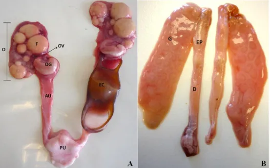
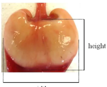
Different measures of reproductive structures were taken from both females and males. In females, mature follicles (here considered as follicles with a yellowish colour), were counted and its diameter measured (in mm) in the two ovaries. Anterior and posterior uterus width (in cm) and weight (in g), width, height and length (in cm) of oviducal glands were measured (Fig. 4A). In males, clasper length (in cm), as the distance from the centre of the cloaca to the posterior tip of the clasper, and epididymis and sperm ducts width (in cm) were measured (Fig 4B). Egg capsule length, width, thickness, anterior horn length, anterior horn width, posterior horn length, inter-posterior horn width were measured (in cm) and weighed (in g) (Fig 5).
Figure 4: Reproductive system of Leucoraja naevus. (A) Female reproductive system: Ov – oviduct; OG – oviducal gland; O – ovary; F – follicles; EC- egg capsule: AU – anterior uterus; AP – posterior uterus. (B) Male reproductive system: G – gonad; EP – epididymis; D – sperm ducts.
9 Figure 5: Capsule from Leucoraja naevus.
Data analysis
The exploratory data analysis included plotting the percentage of reproducing specimens in the sampling, both for males and females, by month.
The gonodosomatic index (GSI) was calculated for each specimen as:
and the hepatosomatic index (HIS) as:
Eviscerated weight (EVw) was used to avoid any influence of the gonad and stomach on the weights. For reproducing specimens, boxplots of the gonodosomatic index and of the hepatosomatic index by month were constructed. The reduced number of individuals sampled did not allow the analysis of interannual variations (Annex A, Fig. II and III).
The χ2
test was applied to test the null hypothesis of no differences in the proportion of reproducing individuals between months. Analysis of variance (ANOVA) was adjusted to compare mean GSI and HSI values at different months of the year.
10
The measurements taken from the gonads, oviducal glands and uteri in females and from gonads, claspers and sperm ducts in males were analysed by TL and by maturity stage.
Maturity ogives were fitted to the reproductive data. To avoid underestimation of the L50 the data were restricted to the last and first quarter of the year (which
correspond to the peak of reproduction). Maturity ogives were determined for each sex through the fitting of a Generalized Linear Model (GLMs; McCullagh and Nelder 1989) in which a binominal error distribution and a logit link were considered:
Where p is the proportion of fish in a mature condition; l is the length class; is the error term and and are parameters. The significance of the model was tested with an ANOVA test.
Percentiles of 25%, 50% and 75% were estimated as:
The errors of each estimate were also determined.
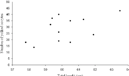
The potential fecundity of females (here considered as the number of follicles released in a year) was investigated as the number of yolked follicles (diameter ≥ 4 mm) in ovaries of maturity stage 3 females with GSI value greater than the 75% percentile (GSI>3.18).
The median number of follicles released by female in each reproductive episode - batch fecundity, was estimated as the number of yolked follicles with diameter ≥ 20 mm (follicles in a ovulation phase (Du Buit 1976) in reproducing females with a GSI above the percentile 75% (GSI>3.49).
11
3. Results
Reproductive season
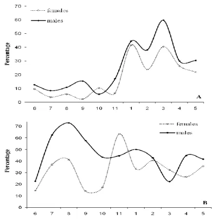
Females with egg capsules were found throughout the year, indicating that this species reproduces year round. However, the percentage of reproducing individuals increased during the last quarter of the year reaching relatively high values between January and April. In the remaining quarters of the year the percentage of reproducing individuals remained constant at low level (Fig. 6A). The test χ2 supported the existence of a reproduction peak (χ2: p<0.05). The temporal evolution of the percentage of maturity stage 3 individuals showed a seasonal pattern with a peak displaced to earlier months relatively to that of reproducing individuals (Fig. 6B). The high percentages of maturity stage 3 individuals occurred during the third quarter (χ2: p<0.05).
Figure 6: Percentage of reproducing (A) and of mature individuals (maturity stage 3) (B) males and females by month over a 1-year period. Solid line represents the monthly variation of reproducing males and dotted line represents the monthly variation of reproducing females.
12
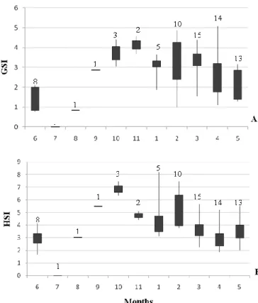
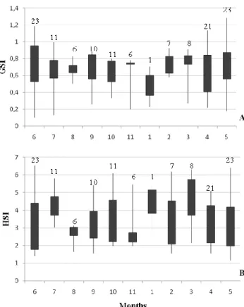
GSI of reproducing females along the year presented higher values between October and March (Fig. 7A). However, the variability of GSI was quite high along all month of the year. The analysis of variance of GSI values along the year showed significant differences between months (ANOVA: F=3.6, p<0.05).
The pattern of HSI values is somehow similar to that of GSI (Fig. 7B). However the analysis of variance of HIS values did not show significant differences between months (ANOVA: F=6.5, p<0.05).
At small TL the liver weight increased slowly with TL. However, near L50 the
rate of increase is higher (Fig 8). The maximum value of liver weight was about 165 g corresponding to a female with 66.5 cm TL (Fig. 8).
Figure 7: Seasonal variation of the gonodosomatic index (GSI) (A) and hepatosomatic index (HSI) (B) over a 1-year period for reproducing females. Range and interquartile range in GSI and HSI values is indicated. Numbers above the boxplot represent the number of individuals considered.
13 Figure 8: Relationship between liver weight and total length for females. Black dots represent mature individuals, white dots represent maturing individuals and crosses represent immature individuals.
GSI and HSI values of males were lower and more stable along the months than those of females (Fig. 9A and B). In males no clear trend on GSI and HSI was observed. The analysis of variance did not show significant differences between months (ANOVA: F=1.4, p>0.05; F=1.1, p>0.05, respectively).
At small TL the liver weight increased slowly with TL. However, near L50 the
rate of increase is higher (Fig 10). The maximum liver weight observed in males was about 112 g corresponding to a male with 67.7 cm TL (Fig. 10).
14 Figure 9: Seasonal variation of the gonodosomatic index (GSI) (A) and hepatosomatic index (HSI) (B) over a 1-year period for mature males. Range and interquartile range in GSI and HSI values is indicated. Numbers above the boxplot represent the number of individuals considered.
Figure 10: Relationship between liver weight and total length for males. Black dots represent mature individuals, white dots represent maturing individuals and crosses represent immature individuals.
Maturity
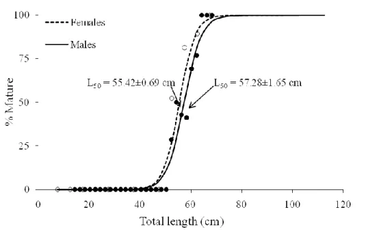
The confidence intervals of length at maturityof malesand females represented a high overlap, suggesting that both sexes mature at about the same TL.
15
In females the estimate of L50 was 55±0.69 cm TL (r2=0.28; p-value<0.05) (Fig.
11) and the interquartile maturity range was estimated as 52 and 59 cm TL (Table 2). The smallest mature female measured 47.3 cm TL and the largest 71.8 cm TL. L50
corresponded to 77% of the maximum observed TL (71.8 cm), appearing to mature very close to maximum TL. The uterus posterior width increased slowly with size at small lengths and the rate increased for lengths higher than L50 (Fig. 12A).
Males L50 was estimated as 57±1.65 cm TL (r2=0.26; p-value<0.05) (Fig. 11)
and the interquartile maturity range was estimated between 54 and 61 cm TL (Table 2). The smallest mature male measured 50.5 cm TL and the largest 68.2 cm TL. The estimate of L50 corresponded to 80% of the maximum observed TL (71.8 cm), and
similarly to females appearing to mature very close to maximum TL. In small males (for TL < 55 cm) claspers length grow slowly with TL, then during maturation claspers length increase at faster rate with TL (Fig. 12B).
Figure 11: Maturity ogives for males (solid line) and for females (dotted line). Dark points represent the proportion of mature males by TL (5cm class interval) and white points represent the proportion of matures females by TL (5cm class interval). L50 of males anf females are indicated by arrows.
Table 2: Estimates of L25, L50 and L75 with associated error for both sexes.
Sex N L25 L50 L75
Males 101 53.58±2.36 57.28±1.65 60.99±1.05
16 Figure 12: Relationship between uterus posterior width and total length for females(A) and relationship between clasper length and total length for males (B). Black dots represent mature individuals, white dots represent maturing individuals and crosses represent immature individuals. Arrows on X-axis denote 50%maturity.
Fecundity
In maturity stage 3 females with GSI>3.18 (this threshold correspond to 75% percentile of GSI values in maturity stage 3 females), the number of yolked follicles at different TL was quite variable, ranging from 14 to 43 (Fig. 13).
Batch fecundity for reproducing females with a GSI>3.49 and follicles with diameter ≥ 20 mm, was estimated to be a median of 5 follicles per batch (Table 3).
17 Figure 13: Number of yolked follicles (diameter ≥4 mm) versus total length of maturity stage 3 females with GSI values higher than 3.18.
Table 3: Estimates of 25%, 50% and 75% percentiles of the number of follicles per batch for reproducing females with a GSI value superior to 3.49.
Percentile 25% 50% 75%
Number of oocytes per batch 3 5 6
Egg capsules
There were measured 56 egg capsules and the maximum number of egg capsules found in a female was 3, one in each side of the anterior uterus and one in the posterior uterus. Egg capsules were convex on both sides, smooth and semi-transparent without hairs and lateral keels, and had a ovulated shape, measuring 52±6.0 cm in length excluding horns, 34±2.7 cm in width and 12±3.9 cm of thickness (Fig. 14). Posterior and anterior horns were different in shape and size. Porterior horns measured 40±19.4 cm in length and tapered towards the tips and anterior horns measured 21±3.5 cm in length. The transparency of the egg capsule and the absence of external hairs unabled to observe the vitello and the developing embryo inside the capsule.
18 Figure 14: Egg capsule of Leucoraja naevus
4. Discussion
This study constitute the first approach to understanding the reproductive biology of L. naevus in continental portuguese waters and is therefore, a first tool to monitor the effects of fisheries on the population. However, previous studies of the biology of the species were realized for other NE Atlantic geographic areas, namely in the Celtic Sea (Du Buit 1976), North Sea (Walker 1998) and in Irish Sea (Gallagher et al. 2005).
Reproductive season
L. naevus might reproduce year round. Howerver a peak season was detected during winter months. This conclusion was based on the proportion of reproducing males and females, GSI and HSI. In the Celtic Sea it was found that 56% of the sampled females, contained follicles with a diameter >20mm in February, which also supports the existence of a peak of spawning (Du Buit 1976). In the North Sea, females carriyng eggs were only reported in summer months (Walker 1998). Differences on the reproductive seasons between geographic areas are relatively common for many species since reproduction can be highly influenced by environmental (Frisk and Miller 2009).
There was a temporal co-occurrence of reproducing individuals, with males and females being active at the same time. So in this species there is a synchrony in the reproductive activity between sexes.
19
In females, GSI and HIS values were, in general, higher and less stable than in males. The high variations within months could indicate the presence of females in different phases of the reproductive season (Oddone and Vooren 2005).
A higher increase in the liver weigth for males and females at the onset of maturity can be raleted to reproductive activity. While in females liver reserves can be used in the production of gametes, gestation and production of egg capsules that are deposited in a short interval of time (Oddone and Velasco 2006), in males liver reserves could have a different function and being more related with migration or feeding success as in the species Rhizoprionodon terraenovae (Colonello et al. 2007), not suffering so much variation throughout the year.
Length at first maturity
L. naevus did not display sexual dimorphism in length-at-first-maturity. This was also observed in other small skates species suach as L. wallacei and L. melitensis (Ebert 2005). Sexual dimorphism in length-at-first-maturity is a common feature in elasmobranchs, with males commonly reaching maturity at smaller sizes and earlier in the life cycle (Ebert 2005). However, there are species, like Psammobatis extenta, that present the opposite pattern, with females reaching maturity earlier and at smaller sizes than males, and this could be related with the fact that delayed maturity in females is more applicable to species that attain large TL (Quiroz et al. 2009).
Several authors have investigated length-at-first-maturity for L. naevus at different geographic locations and estimated different lengths. Du Buit (1976) reported that both males and females attain sexual maturity at 59 cm TL in Celtic Sea, while Walker (1998) estimated 55 cm TL for both males and females, in the North Sea and Gallagher (2005) estimated 57 cm TL for males and 56 cm TL for females in the Irish Sea. Similar differences in sizes at maturity between different geographic locations have been reported for species like Amblyraja radiata or Leucoraja erinacea. Those differences can be due either to habitat or environmental differences or to different fishing pressures (Ebert and Compagno 2007, Ebert et al. 2008). It is known that fishing pressure can affect the abundance of a species and lead to changes in life-history parameters (Walker and Hislop 1998). In the central and north-western North Sea, the increase of fishing mortality of thornback ray, R. clavata, determined faster grow rate and smaller length of first maturity (Walker and Hislop 1998).
20
In Portuguese continental waters, L. naevus attain sexual maturity at nearly 80% of the maximum observed TL (71,8cm TL). Such high percentage is in accordance with observations by Cortés (2000) for other elasmobranch. Elasmobranch fishes tend to undergo an extended juvenile phase and grow very little after maturity (Ebert 2005). The trade-off of delayed maturity could result in a longer gestation period and lower growth rates, making the species less able to withstand high fishing pressures (Frisk and Miller 2009).
Reproductive organs such as claspers and uterus tend to develop gradually during the immature stages and suffering an abrupt increase at the onset of maturity (Colonello et al. 2007, Templeman 1987). Such differences suggest that those measurements could be used as indicators to differenciate between immature and mature individuals.
Fecundity
Due to the high variability found in the number of yolked follicles, even when only maturity stage 3 females with a GSI value greater than 3.18 were considered to insure that will be the ones closest to reproduction, it was not possible to determine the potencial fecundity of the species neither the number of reproductive episodes carried out by one female during a entire year.
Follicles were found in varies developing stages in both ovaries, indicating that follicles development is asynchronous, but it was not possible to determine if it is a determinate or indeterminate spawaner due to the lack of information on resting and regenerating stages and because of the possibility of occurrence of atretic losses (Murua and Saborido-Rey 2003). Recently, Serra-Pereira et al. (in press) suggested a new terminology for assessing maturity in elasmobranch fishes where a resting and a regeneration stage are considered. In the future a more accurate collection of data in respect to these stages and for evaluating the possibility of occurrence of atresy will be important. Estimating fecundity based on ovarian fecundity could not translate the real reproductive potential, mainly because we are dealing with a species that produces follicles over a long period of time. On other hand, maintaining specimens in captivity could alter the behaviour compared to the natural environment (Kyne et al. 2008).
The median number of released follicles per batch was 5 and the maximum number os follicles with diameter ≥20mm found in the two ovaries was 10, which was
21
close to that reproted by Du Buit (1976) who found a maximum of 8 follicles for both ovaries. For this estimate only reproducing females with a GSI value greater than 3.49 were considered to ensure that only females in the beginning of the reproductive season were taken into account. The low value of batch fecundity in L. naevus contrasts with the batch fecundity observed in other skates species such as R. clavata (Serra-Pereira et al. in press). That difference on batch fecundity can be related with the fact that L. naevus is a small species and it seems to be more advantageous to release eggs in small batches due to the small space available to support larger follicles.
Other authors such as Du Buit (1976) and Walker (1998) estimated the total fecundity for this species at about 90 eggs per year. These estimates were determined by assuming a constant egg laying rate (0.5 eggs per day) during the reproductive cycle without a resting period and did not enter into consideration the number of follicles in ovaries.
Fishing pressure might have strong effects on several life history traits of cartilageneous fishes (Oddone and Vooren 2005). In some areas of the Northeast Atlantic smaller species like L. naevus and R. radiata have increased in abundance in recent years, possibly due to the fact that small and early maturing species are less susceptible to exploitation (Kyne et al. 2008) and to the additional food and habitat availability, resulting from the removal of larger species (Gallagher et al. 2005). More studies on L. naevus in Portuguese waters, namely on age, growth, nursery grounds and habitat preferences, are needed to evaluate the resilience of this species to fishing in this area.
22
IV. Comparison of the oviducal gland of Leucoraja naevus with other skate species from mainland Portugal
1. Introduction
The order Rajiformes (skates) comprises 245 species, being the most diversified group among cartilaginous fishes (Ebert and Compagno 2007). Although they constitute a diversified group, there are several common features of their life history that make them vulnerable to fishing exploitation (Figueiredo et al. 2007), mainly slow growth, late maturity, low fecundity, long gestation time and a high energetic investment in reproduction (Stevens et al. 2000).
Chondrichthyans present a great diversity of reproductive strategies (Hamlett et al. 1998) ranging from oviparity (egg-laying) to viviparity (live-bearing) (Dulvy and Reynolds 1997, Kormanik 1993), but in common all have internal fertilization (Hamlett et al. 2002). Skates, 40% of sharks and holocephelans, (Smith. et al. 2004) are oviparous species that produce a resistant protein egg capsule (Koob and Cox 1993, Kormanik 1993, Serra-Pereira et al. in press, Wourms and Demski 1993) that protects the embryo during development (Galindez and Estecondo 2008). The fertilization occurs in the oviduct (Knigth et al. 1996) and the egg passes through the oviducal gland (Galíndez and Estecondo 2008). When in the oviducal gland, the egg is first surrounded by the egg jelly (Hamlett et al. 2005) that provides hydrodynamic support to the embryo during development (Koob and Straus 1998). Then the egg enters the species specific egg capsule, passes through the uterus when completely formed, to be released to the external environmental (Hamlett et al. 2005). Once deposited in the external environmental, the embryo develops without parental care, being nourished by the yolk sac contents (Carrier et al. 2004).
The egg capsule display an important role protecting the embryo from predation, wave action, metabolic waste accumulation (Hamlett et al. 2005, Knight et al. 1996) and pathogens (Lucifora and García 2004, Kormanik 1993), due to its toughness and strength, flexibility, high permeability to small ions and low molecular weight substances and moderate extensibility (Knight et al. 1996).
23
The oviducal gland is a specialized organ situated in the anterior portion of the oviduct (Smith et al. 2004) and is responsible for the encapsulation of the fertilized egg (Hamlett and Hysell 1998).
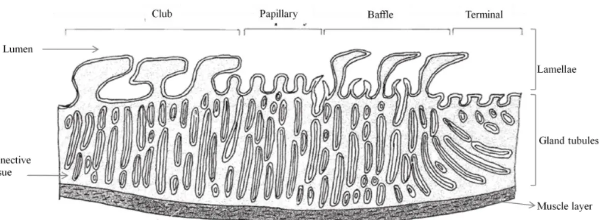
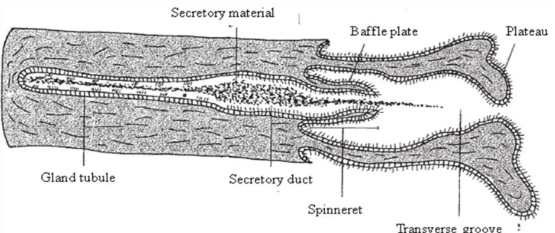
The oviducal gland performs four main functions: transport of fertilized eggs to the uterus, egg jelly production, formation of the species specific tertiary egg envelope, and, in some elasmobranchs, sperm storage (Hamlett et al. 1998, Storrie et al. 2008). Although the development and architecture of the oviducal gland is related to the reproductive mode and cycle stage (Galíndez et al. 2010), oviducal glands from all oviparous species share the same gross anatomy and microscopic organization design principles and the relevance of each zone is correlated with the species specific egg capsule (Hamlett et al. 2005). Morphologically, the oviducal gland from oviparous species is a complex structure composed of two similar dorsoventral flattened halves with upturned lateral margins (Hamlett et al. 1998). Internally, the oviducal gland can be divided, based on their distinct lamellae and position in the oviducal gland, in four histologically different zones (from the oviduct to the uterus): club, papillary, baffle and terminal (Fig. 16) (Galíndez and Estecondo 2008, Hamlett et al. 1998, Smith et al. 2004).
Figure 16: Diagram of a generic oviducal gland showing club, papillary, baffle and terminal zones (adapted from Hamlett et al. 1998).
Except for the terminal zone, that does not form discrete transverse grooves (Storrie et al. 2008), all the zones have the same basic structure, consisting of gland tubules surrounded by connective tissue and opening to the oviducal gland lumen through transverse grooves situated between the lamellae (Hamlett et al. 1998). Club and papillary zones secrete the egg jelly layers that surround the fertilized egg before
24
being introduced into the egg capsule, and can be differentiated through roughly T-shape lamellae in the club and digit-T-shaped lamellae in the papillary (Hamlett et al. 1998). The baffle zone produces the tertiary egg envelope. A crystalline liquid is produced in secretory tubules and then delivered to the lumen through the spinneret region (Fig 17) (Hamlett et al. 2005, Knigth et al. 1996) originating the proteinaceous multilaminate egg capsule (Oddone 2005, Smith et al. 2004). This zone is easily identified due to the characteristic lamellae with two types of projections: the baffle plates forming the spinneret region and the plateau projections altering with transverse grooves (Galíndez and Estecondo 2008, Hamlett et al. 1998, Smith et al. 2004). The most posterior zone, the terminal zone, is the site of formation of fine surface hairs that adorn the egg capsule in oviparous species that have a fibrous egg capsule and is also described to be the site of sperm storage in other species (Smith et al. 2004, Storrie et al. 2008). In oviparous, compound tubular glands situated deeply into the terminal zone secrete hairs that pass through ducts (Hamlett et al. 1998) and are then coated by a mucous (Serra-Pereira et al. in press, Smith et al. 2004) produced in mucous gland tubules situated near the lumen (Hamlett et al. 2005).
Figure 17: Diagram of a generic baffle unit showing a gland tubule, secretory duct, spinneret with baffle plates, the plateau projections and transverse groove (adapted from Hamlett et al. 1998).
For some chondrichthyes, several authors had already described the structure and functions of the oviducal gland. Metten (1939), Threadgold (1957) and Rusaouën (1976) described the morphology and functions of the oviducal gland of Scyliorhinus canicula and capsule formation was later addressed by Rusaouën-Innocent (1976), Feng and Knight (1992, 1994) and Knight et al. (1996). Hamlett et al. (1998) carried out a
25
comparative study of the oviducal gland from several elasmobranch species, Smith (2004) addressed the microscopic organization of the oviducal gland of the holocephalan Callorhynchus milii, Galindez et al. (2008, 2010) studied the morphology of the oviducal gland from two rajid species and Serra-Pereira et al. (in press) studied the development of the oviducal gland of Raja clavata.
The species investigated in the present work are L. naevus, R. brachyura, R. miraletus, R. undulata, R. microocellata and R. montagui, that constitute important by-catches from the artisanal mixed-fishery in Portugal (Machado et al. 2004). Except for the species L. naevus that inhabit depths down to 250 m and deposit their eggs in more offshore waters (Ellis et al. 2004), in general these species inhabit depths down to 100 m and some may migrate to inshore shallow waters during Spring/Summer to reproduce (Coelho and Erzini 2006, Serena 2005). Little is known about nursery grounds, but egg-laying substrates are generally sandy or muddy bottoms (Serena 2005). The species L. naevus and R. microocellata possess a more ovulated and smooth egg capsule without hairs and with different horns in both extremities and R. brachyura, R. monatgui, R. miraletus and R. undulata have capsules with rectangular shape, similar horns and covered by fibers.
The comprehension of reproductive biology features and reproductive strategies of skate species is an important component for a more accurate stock assessment of these fishery resources. The egg capsule characteristics are correlated with the characteristics of the spawning habitat and, therefore, can provide clues about habitat preferences of each species here addressed. The objective of the present work was to compare the microstructure and the nature of the secretions of the oviducal gland from mature females of L.naevus with the remaining species and relate it with the species specific reproductive strategy.
2. Material and methods
Sampling
L. naevus (n=50), R. Brachyura (n=5), R. Montagui (n=21), R. Microocellata (n=1), R. Miraletus (n=10) and R. undulata (n=8) females were caugh during from i)
26
fishing trawl tows performed during IPIMAR research surveys and ii) the artisanal fleets operating along the Portuguese coast. Samples were collected under the scope of National Data Collection Program (PNAB, DCR) during the period between 2003 and 2010.
Only mature females (corresponding to maturity stages 3, 4, 5 and 6) were selected for this study, being the reproductive stages assessed according to Stehmann (2002) maturity scale for elasmobranchs. The oviductal glands (OG) were extracted measured in width, height and thickness (to the nearest mm) (Fig. 18), weighed (in g) and preserved in 10% buffered formaldehyde.
Figure 18: Measures taken from the oviducal gland.
Histological procedures
The morphological structure of the oviducal gland and the presence and nature of the secretions produced were analysed using a selection of oviducal glands from mature females. Sagittal sections of about 3 mm thicker were removed from the middle of the oviducal glands. The samples were processed using an automated tissue processing machine (Leica TP1020, Germany). The protocol consisted of: 1) dehydration through a series of alcohols from 70% to absolute ethanol; 2) clearing with xylene; and 3) impregnation and embedding in paraffin wax. Embedding of the samples in paraffin wax blocks was made using the heated (58ºC) paraffin embedding system (Leica EG 1140H, Germany). The paraffin blocks were then sliced, in sagittal sections of 3 μm of thickness, using a rotary microtome (Leica RM2125RT, Germany). Hematoxylin and Eosin (H&E) staining technique was applied, staining the nucleus
27
black and the cytoplasm pink. To analyse the chemical nature of the secretions produced by the different glandular zones, a combined Alcian blue and PAS (Periodic Acid Schiff) staining technique was applied. Histological procedures used by Serra-Pereira et al. (in press) were followed. The histological slides were observed using a stereo microscope (Olympus SZX9, USA) and an optic microscope (Carl Zeiss Axioplan 2 imaging, Germany). The former was used to observe the whole gland sections and the latter was used to analyse the gland structure in more detail. The surface length was measured as the portion occupied by the lamellae of each zone, and the glandular area as the portion occupied by the gland tubules in each zone using imaging software TNPC 4.1 (coupled with the stereo microscope).
3. Results
All species share with L. naevus the same design principles, so this species was taken as a model to illustrate common features. Macroscopically, the oviducal gland is composed of two similar dorsoventral flattened halves with upturned lateral margins (Fig. 19A). Internally, it is possible to observe the four distinct zones that correspond to the club, papillary, baffle and terminal (Fig. 19 B and C). When analysed histologically, in sagittal section (Fig. 19D), the four distinct zones were easily identified, having all a basic structure of gland tubules surrounded by a connective tissue and lamellae lining the lumen.
Figure 19: Leucoraja naevus. (A) External anatomy of the OG showing the upturned lateral margins (lm). The position of oviduct (ov) is indicated. (B) Internal view of the OG showing the club (c), papillary (p), baffle (b) and terminal (t). (C) Sagittal section of the OG showing club (c), papillary (p), baffle (b) and terminal (t). the position of the oviduct (ov) is indicated. (D) Sagittal section of the OG showing the club (c), papillary (p), baffle (b) and terminal (t) zones (H&E) ×40.
28
Club is composed of lamellae truncated basally with a roughly t-shaped (Fig 20 A). The lamellae epithelium is simple columnar with sustentacular ciliated cells and secretory cells (Fig 20 B). Simple tubular glands connect to the lumen between adjacent club lamellae and are composed of two types of cells: sustentacular pyriform ciliated cells with apical nuclei and secretory cells with basal nuclei (Fig. 20C) that produce a PAS+AB+ secretory product.
Figure 20: Leucoraja naevus. (A) Club T-shaped lamellae.×200 (H&E) (B) Club lammelae epithelium with two types of cells: sustentacular cells (a) with cillia (ci) and secretory cells (s). ×200 (H&E). (C) Club gland tubule with sustentacular cells (a) and secretory cells (s). ×400 (H&E).
Papillary is composed of digit-shaped lamellae (Fig. 21A). The lamellae epithelium is columnar with cilia, composed of two types of cells: ciliated and secretory cells (Fig. 21B). Gland tubules are similar those of the club zone, with two types of cells stained PAS+AB+. The caudal most row of the papillary zone near the baffle zone, present more vacuolated gland tubules (Fig. 21C), with secretory product stained intensely AB+.
29 Figure 21: Leucoraja naevus.(A) Papillary digit-shaped lamellae. ×200. (H&E). (B) Papillary lamellae epithelium with two types of cells: ciliated cells (a) with cillia (ci) and secretory cells (s). ×400 (H&E). (B) Papillary gland tubules (p) adjacent to the baffle (b). ×400. (H&E).
The baffle zone is composed of lamellae with two types of projections: the baffle plates forming the spinneret region and the plateau projections that are apically flattened alternating with transverse grooves (Fig. 22A). Lamellae epithelium is simple columnar ciliated with two types of cells: sustentacular cells with apical nuclei and secretory cells with basal nuclei (Fig. 22B). Gland tubules are simple and unbranched most frequently containing secretory product and comprised two cell types: serous secretory cells with basal nuclei and pyriform sustentacular ciliated cells with apical nuclei (Fig. 22C). Gland tubules are confluent with the secretory duct that delivers secretory PAS-AB- product in the spinneret region and it is extruded between the baffle plates.
30 Figure 22: Leucoraja naevus. (A) Baffle plates (Bp) forming the spinneret region (sr) and the plateau projections (pp) alternating with transverse grooves (tg). ×100. (B) Ballfe lamellae epithelium with two types of cells: sustentacular cells (a) and secretory cells (s). ×400. (C) Baffle gland tubules with two types of cells: sustentacular cells (a) and secretory cells (s) showing secretory product (sp). ×400. (H&E).
The smallest and more posterior zone is the terminal zone. Terminal zone is not organized in lamellae and do not form discrete transverse grooves. Surface epithelium is simple columnar ciliated with two types of cells: sustentacular ciliated cells and secretory cells (Fig. 23).
Figure 23: Leucoraja naevus. (A) Terminal zone not organized in lamellae.×200 (H&E). (B) Terminal epithelium with two types of cells: sustentacular cells (a) and secretory cells (s).×400 (H&E).
Differences were found between L. naevus and the remaining species in the portion occupied by each zone in the gland and in the number of lamellae, in staining affinities in the papillary zone and in the gland tubules from the terminal zone.
Differences found in the portion occupied by each zone in the gland and the respective number of lamellae are identified in Table 4.
31 Tabela 4: Glandular area (GA), surface length (SL), in percentage, and number of lamellae for each zone and species. The standard deviation is indicated.
Club Papillary Baffle Terminal
Species N GA* SL Nº lamellae SL Nº lamellae GA SL Nº lamellae GA SL L.naevus 50 20±3 15±2 8-10 15±3 12-18 76±3 40±3 30-38 30±4 3±1 R.montagui 21 17±3 13±3 13-16 14±3 11-15 77±3 36±5 23-33 5±2 37±8 R.brachyura 5 13 19* - - - 80 34 36-42 6 47 R.miraletus 10 18±2 24±6 - - - 64±3 32±13 20-22 18±4 44±7 R.microocellata 1 19 27* 16 - 20 81 47 30 3 25 R.undulata 8 20±4 23±4* 14-15 - 15-19 73±4 32±11 44-45 7±2 18±9
*The measure was made considering club and papillary.
Except for the species L.naevus, in which club and papillary stained positively PAS/AB and is not possible to distinguish them (Fig. 24A), all the remaining species show two different zones. In these species, the gross portion of papillary gland tubules, the ones in contact with the club, stained positively PAS/AB and the ones near the lumen stained more intensely PAS+, being possible to distinguish from the club ones (Fig. 24B).
Figure 24: (A) Leucoraja naevus. Club and papillary staining PAS+AB+. ×40. (B) Raja montagui. Encircled area is the zone where it is possible to distinguish papillary from club, staining more intensely PAS+. ×40.
The terminal zone also presents some differences. In L.naevus and R. microocellata, terminal zone gland tubules are elongated with mucous secretory cells and sustentacular cells (Fig. 25A) stained AB+ (Fig. 25B). In the remaining species, terminal zone is composed of two types of gland tubules: the ones composed of serous
32
and mucous secretory cells that secrete a PAS-AB+ product (Fig. 25C) and the ones composed only of mucous secretory cells that secrete a AB+ product (Fig. 25D). Serous tubules are predominantly situated more deeply into the terminal zone and mucous secretory tubules are closely to the OG lumen.
Figure 25: (A) Leucoraja naevus terminal gland tubules with sustentacular cells (a) and mucous secretory cells (ms).×400 (H&E). (B) Leucoraja naevus terminal gland tubules staining PAS-AB+. ×100. (C) Raja montagui terminal gland tubules with two types of secretory cells: serous secretory cells (ss) and mucous secretory cells (ms). ×400. (D) Raja montagui terminal gland tubules composed by serous secretory cells staining PAS-AB- and mucous secretory cells staining AB+. ×200.
Presence of sperm
Sperm was found in oviducal glands from all species. It was possible to observed sperm as laterally aligned bundles in the most caudal portion of the baffle zone near the muscle tissue in all species (Fig. 26A, B, C, D,E and F). Baffle zone tubules containing sperm had the same morphology and histology of the typical baffle gland tubules with sustentacular cells and secretory cells stained PAS-AB-. No matrix was found involving the sperm. In L. naevus and R. montagui, sperm was observed in non-aggregated individual form in the oviduct, papillary, baffle and terminal zones and in
33
one specimen of R. brachyura the presence of a large quantity of sperm in baffle lamellae was observed.
Figura 26: Sperm (e) as laterally aligned bundles in the most caudal portion of the baffle zone. (A) Leucoraja naevus. (B) Raja montagui. (C) Raja miraletus. (D) Raja microocellata. (E) Raja undulata. (F) Raja brachyura. ×400 (H&E).
Egg capsule material
Except for R. brachyura and R. miraletus, a brown material was observed in the oviducal glands of all species. In L. naevus (Fig. 27A) and R. undulata (Fig. 27D) the brown material was found in the baffle zone adjacent to the terminal and in the papillary in maturity stage 3 females not carrying eggs. This material was observed in papillary, baffle, terminal and in the muscle tissue adjacent to the baffle zone, in one maturity stage 4 R. montagui (Fig. 27 B). In the species R. microocellata (Fig. 27 C) it was found in papillary and baffle zones, in a maturity stage 4 female. This material has the same color of the final stage of the egg capsule.
34 Figure 27: Brown material (bm). (A) Brown mateial in the baffle zone adjacent to teminal zone in Leucoraja naevus. (B) Brown material in the terminal zone in Raja montagui. (C) Brown mateial in the baffle zone adjacent to teminal zone in Raja microocellata. (D) Brown mateial in the baffle zone adjacent to teminal zone in Raja undulata. ×400 (H&E).
4. Discussion
The oviducal gland of the species discussed in the present study all share the same anatomy and microscopic structure, as well with most chondrichthyans (Hamlett et al. 2005). Nevertheless, differences were detected between species, namely related to the relative size of glandular area and surface length of the lamellae liny the lumen of the four distinct zones. These differences vary interspecifically according to the egg capsule characteristics. The egg capsule is species specific, and its shape is related with the three-dimensional arrangement of the secretory gland tubules in the oviducal gland (Smith et al. 2004).
Club and papillary zones
In all species here discussed club reacted positively to PAS/AB staining, indicating the presence of glycoproteins, namely neutral and sulphated acid mucins. Differences were found between species in the chemical nature of the secretions being produced in the papillary zone indicated by differential affinities to PAS/AB staining.
35
In L. naevus, the papillary zone has two distinct secretory regions; i) most papillary gland secretory material stained positively PAS/AB, indicating that the secretory nature produced is glycoprotein, namely neutral and sulphated acid mucins and ii) the ones adjacent to the baffle that stained intensely AB, indicating a high portion of sulphated acid mucins being produced. In R. montagui, R. miraletus, Raja microocellata, R. brachyura and R. undulata, it was possible to divide papillary in three different zones. Despite the gross portion of gland tubules stained positively PAS/AB, which indicates that the secretions produced are neutral and sulphated acid mucins, the gland tubules near the lumen stained intensely PAS+AB- indicating a higher portion of neutral mucins and absence of sulphated acid mucins, and the ones adjacent to the baffle reacted intensely to AB indicating that those gland tubules secrete a sulphated acid mucin. The differences in papillary PAS/AB affinity is not unexpected once that the egg jelly display different viscosities resulting from different concentrations in carbohydrates (Koob and Straus 1998). In several studies with S. canicula (Feng and Knigth 1994, Threadgold 1957, Rusauen 1976) it was concluded the presence of carbohydrates, neutral mucopolysaccharide and acidic polysaccharides. Koob and Strus (1998) identified, in R. erinacea, four types of carbohydrates and Serra-Pereira (in press) identified in R. clavata, mucins.
The production of different types of mucins by the papillary gland tubules secretory cells were observed in several studies (e.g. Galíndez et al. 2010, Nalini 1940, Serra-Pereira et al. in press), suggesting that the most caudal gland tubules, adjacent to the baffle, secrete a sulphated acid material that function as a interface between the egg jelly and the forthcoming egg capsule, serving as a binding layer between them (Galíndez et al. 2010, Hamlett et al. 1998, Knigth et al. 1996, Nalini 1040, Smith et al. 2004). The sulphated acid mucins that are present in the chemical composition of the egg jelly produced by club and papillary zones may constitute an important barrier against pathogens (Bansil and Turner 2006). Other possible function of the egg jelly may be to contribute with maternal immunoglobulins to the embryo (Cateni et al. 2003), once that oviparous species can be born without a complete formed immune system (Galíndez and Estecondo 2008). There is no indication that the egg jelly function as a carbohydrate source to the developing embryo, once that are no loss of it during the liquefaction process (Koob and Straus 1998). L. naevus has the longer gestation period, about 8 months (Clark 1922), and a semi-transparent egg capsule and therefore, possibly is more susceptible to predation and pathogens. Lucifora and García (2004)
36
conducted a study in the southwestern Atlantic where they related the elasmobranchs eggs predation rate with the chemical composition and egg nutritive value, so this little difference in the content of sulfated acid mucins could work as an important chemical defense against predation.
After more or less of one third of the development, the egg jelly is liquefied and the embryonic tail pump water into the capsule (Kook and Straus 1998). Egg jelly functions are not yet clarified, but it seems to be essential to the embryo in the early stages of development (Smith et al. 2004), namely providing hydrodynamic support during embryogenesis (Koob and Straus 1998, Smith et al. 2004) and functioning as a lubricant during encapsulation (Hamllet et al. 1998).
Baffle zone
The baffle zone of the oviducal gland of the studied species share similar structure and the fact that its secretions stained negativelly to PAS and AB. The major differences were detected in the number of lamellae, total glandular area and surface length.
The staining techniques used in the present work gave a negative result, indicating that baffle zone do not secrete a carbohydrate rich material and, therefore, suggesting a protein nature. Amongst elasmobranchs there are different types of secretions produced by the baffle zone (Knight et al. 1996, Koob and Cox 1993, Smith et al. 2004, Serra- Pereira et al. in press). In Raja erinacea there were identified six proteins containing high levels of glycine, serine, tyrosine and proline (Koob and Cox 1993) and in Chiloscyllium griseum (Krishnam 1959) and Scyliorhinus canicula, (Knigth et al. 1996) the main capsule constituent is collagen.
The species R. brachyura and R. undulata are the ones who show a baffle zone composed of more lamellae which might be related to a more robust egg case. The baffle gland tubules secrete a crystalline liquid that emerges between the baffle plates (Hamlett et al. 1998, 2005, 2002), oriented by the extraordinary structure of the spinnerets (Knight et al.1996, Smith et al. 2004) originating the several capsule lamella (Hamlett et al. 2005). The number of transverse grooves determines the number of layers that constitute the capsule wall and its thickness (Hamlett et al.1998, Serra-Pereira et al. in press). Of all the species here observed, R. miraletus is the one with less lamellae in the baffle zone and, therefore, it is expected to have a thinner and