ii
DIREITOS DE AUTOR E CONDIÇÕES DE UTILIZAÇÃO DO TRABALHO POR TERCEIROS
Este é um trabalho académico que pode ser utilizado por terceiros desde que respeitadas as regras e boas práticas internacionalmente aceites, no que concerne aos direitos de autor e direitos conexos. Assim, o presente trabalho pode ser utilizado nos termos previstos na licença abaixo indicada.
Caso o utilizador necessite de permissão para poder fazer um uso do trabalho em condições não previstas no licenciamento indicado, deverá contactar o autor, através do RepositóriUM da Universidade do Minho.
Licença concedida aos utilizadores deste trabalho
Atribuição CC BY
iii
ACKNOWLEDGMENTS
I spent the last two years studying Biotechnology and I strongly believe, from the knowledge I acquired, that Biotechnology will revolutionize our entire world.
First and foremost, I want to thank my supervisors. Over these last nine months, Dr Tatiana Aguiar provided me with the most appropriate and thoughtful guidance in every step of my research project. I had the privilege to work with a fantastic person whose valuable knowledge contributed significantly to improve my skills and guided me to look at molecular biotechnology in a more comprehensive way. I esteem her curiosity and commitment to Science and I would like to thank her for all the commitment she also dedicated to me. I also would like to thank her for introducing me the wonderful world of glycosylation. I must add a special thanks to Dr Carla Oliveira, my co-supervisor, for all the guidance, all the knowledge she transmitted me about recombinant protein production and for teaching me that organization is a very important skill.
I am very grateful to Professor Lucília Domingues for receiving me with open arms at the Molecular Biotechnology Laboratory and for introducing me to the molecular biotechnology world. I am very thankful for the great advices when they were necessary. I must add a special word of thanks to Professor Peter Philippsen. I cannot leave unmentioned the suggestions he gave me in my work. My special thanks to everyone at the Molecular Biotechnology Laboratory for such a pleasant working atmosphere and help. I am particularly grateful to Ana, Sara Baptista, Joana, Carlos, Pedro and Rui.
I also want to thank my friends Bruna Basto, Rafael Marques, Eduarda Gomes, Mário Azevedo, Gonçalo Carvalho, Marta Teixeira and Tiago Gião for the love and patience everyday throughout these 5 years in the university. For all the good moments, I am deeply grateful. I must thank my Father, my Mother, my Brother and my Husband for their unconditional love and support. I would just like to thank them for always valuing me, for all they have sacrificed for me and for giving me the opportunity to pursue my dreams. I must ultimately thank my parents for teaching me that when you dedicate all your efforts to something, one day someone will value you for what you have done.
Lastly, I would like to thank the support given by FCT and by the European Regional Development Fund under the scope of Compete2020 and Norte2020: strategic funding of UID/BIO/04469/2019, Project EcoBioInks4SmartTextiles (PTDC/CTM-TEX/30298/2017–POCI-01-0145-FEDER-030298) and BioTecNorte operation (NORTE-01-0145-FEDER-000004).
iv
STATEMENT OF INTEGRITY
I hereby declare having conducted this academic work with integrity. I confirm that I have not used plagiarism or any form of undue use of information or falsification of results along the process leading to its elaboration.
v
GLYCANZYME - Characterisation of novel enzymes for protein glycoengineering
ABSTRACT
One of the most common protein post-translational modifications occurring in eukaryotes is glycosylation, which is critical for several biological processes. There are different enzymes involved in the processing of N-glycans in eukaryotes, among them endo-β-N-acetylglucosaminidases (ENGases), whose physiological roles are still poorly understood. ENGases of the glycoside hydrolase (GH) family 85 are a class of enzymes (EC 3.2.1.96) that, in addition to hydrolytic activity against the diacetylchitobiose core of N-glycans, can also display transglycosylation activity. Given their usefulness for the analysis and glycoengineering of glycopeptides and glycoproteins, these enzymes have become a focus of biotechnological interest.
Envisioning the identification of novel GH85 ENGases with useful action, this thesis focused on the characterization of two new putative ENGases of this family, one from the filamentous fungus Ashbya gossypii (AgENGase) and another from the yeast Zygosaccharomyces rouxii (ZrENGase). Previous experimental results hinted at the existence of ENGase activity in these organisms and in their genome one homologue gene exists that should code for a putative GH85 ENGase (AFR597W in A. gossypii and ZYRO0B07216g in Z. rouxii). Bioinformatic approaches were initially used to assess the potential activity of these putative ENGases and important residues for the hydrolytic and transglycosylation activity were found to be conserved in AgENGase. Only the putative 3D model of ZrENGase presented the typical (β8/α8)-TIM-barrel structure of ENGases. To characterise these proteins, their coding genes were cloned
into an Escherichia coli expression plasmid and their recombinant production was optimized by testing different E. coli expression strains, production media and induction conditions. However, the recombinant proteins produced remained mainly in the insoluble fraction after cell lysis and further improvements in their production and purification will be required to generate enough protein for activity studies and eventual application. Nevertheless, different protocols were already optimized in the scope of this thesis for the future evaluation of the deglycosylation activity of these recombinant ENGases.
Considering the bioinformatic analysis and the results from enzymatic assays performed in crude cell extracts of wild A. gossypii, the AgENGase action could be located in the intracellular space. Envisioning the understanding of the physiological role of the putative AgENGase, a gene with no homolog in Saccharomyces cerevisiae and with unknown function, the disruption of the A. gossypii AFR597W was also performed. Thereby, AgENGase was found to be important for sporulation, as the generated afr597w mutants lost the ability to sporulate. Nevertheless, further studies will be needed to fully understand the physiological and enzymatic function of this enzyme.
vi
Glycanzyme - Caracterização de novas enzimas para glico-engenharia de proteínas
RESUMO
Uma das modificações pós-tradução de proteínas mais comuns que ocorrem em eucariotas é a glicosilação, que é crítica para vários processos biológicos. Existem diferentes enzimas envolvidas no processamento de N-glicanos em eucariotas, entre elas as endo-β-N-acetilglucosaminidases (ENGases), cujos papéis fisiológicos ainda são pouco compreendidos. ENGases da família glicosil hidrolase (GH) 85 são uma classe de enzimas (EC 3.2.1.96) que, além da atividade hidrolítica no núcleo de diacetil-quitobiose de N-glicanos, também podem exibir atividade de transglicosilação. Dada a sua utilidade para a análise e a glico-engenharia de glicopéptidos e glicoproteínas, essas enzimas apresentam-se com forte interesse biotecnológico.
Tendo em vista a identificação de novas ENGases da família GH85 de interesse, esta tese focou a caracterização de duas novas supostas ENGases dessa família, uma do fungo filamentoso Ashbya gossypii (AgENGase) e outra da levedura Zygosaccharomyces rouxii (ZrENGase). Resultados experimentais anteriores sugeriram a existência de atividade ENGase nesses organismos e, no seu genoma, encontrou-se um gene homólogo que poderá codificar para putativas ENGases da família GH85 (AFR597W em A. gossypii e ZYRO0B07216g em Z. rouxii). Abordagens bioinformáticas foram usadas inicialmente para avaliar a atividade potencial dessas ENGases e foram identificados resíduos importantes para a atividade hidrolítica e de transglicosilação conservados na putativa AgENGase. Por outro lado, apenas o suposto modelo 3D previsto para a putativa ZrENGase apresentou a estrutura típica de um barril TIM (β8/α8) das ENGases. Para caracterizar essas proteínas, os seus genes foram clonados
num plasmídeo de expressão de Escherichia coli e a sua produção recombinante foi otimizada testando diferentes estirpes de expressão de E. coli, meios de produção e condições de indução. No entanto, as proteínas recombinantes produzidas permaneceram principalmente na fração insolúvel após a lise celular e serão necessárias melhorias adicionais na sua produção e purificação para gerar quantidade de proteína suficiente para estudos de atividade e eventuais aplicações. No entanto, diferentes protocolos já foram otimizados no âmbito desta tese para avaliação futura da atividade de desglicosilação dessas ENGases recombinantes.
Considerando a análise bioinformática e os resultados de ensaios enzimáticos realizados em extratos celulares brutos de A. gossypii wild, foi possível determinar a localização da AgENGase como sendo intracelular. Visando compreender o papel fisiológico da suposta AgENGase foi também realizada a interrupção do gene AFR597W de A. gossypii. Dessa forma, a AgENGase foi considerada importante para a esporulação, pois os mutantes afr597w gerados perderam a capacidade de esporular. No entanto, serão necessários mais estudos para entender completamente a função fisiológica e enzimática dessa enzima.
Palavras-chave: Ashbya gossypii; endo-β-N-acetilglucosaminidases; produção de proteínas recombinantes
vii
TABLE OF CONTENTS
DIREITOS DE AUTOR E CONDIÇÕES DE UTILIZAÇÃO DO TRABALHO POR TERCEIROS ... ii
ACKNOWLEDGMENTS ... iii STATEMENT OF INTEGRITY ... iv ABSTRACT ... v RESUMO ... vi INTRODUCTION ... 2 1. Overview on glycosylation ... 2
1.1. Glycosylation and its biological significance ... 2
1.2. Diversity of glycans ... 3
1.3. N-glycan biosynthetic pathway ... 6
1.4. Synthetic methods for glycoprotein engineering ... 8
2. ENGases ... 11
2.1. Mechanism of action ... 13
2.2. ENGases applications ... 14
3. A. gossypii ... 16
4. Z. rouxii ... 17
5. Recombinant protein production in Escherichia coli: advantages and challenges ... 18
OBJECTIVES ... 20
MATERIALS AND METHODS ... 22
1. Bioinformatics ... 22
2. Strains and media ... 22
3. Molecular Biology ... 24
3.1. DNA constructs ... 24
3.1.1. Cloning and expression plasmid construction of putative AgENGase and ZrENGase-encoding genes 24 3.1.2. Disruption of AFR597W gene in A. gossypii ... 28
3.2. A. gossypii transformation and screening of transformants ... 28
4. Production and purification of recombinant ENGases ... 29
4.1. SDS- and Native-PAGE Analysis ... 30
5. ENGase/chitinase activity detection ... 31
5.1. Analysis of ENGase deglycosylation activity ... 31
viii
activity in A. gossypii... 32
RESULTS AND DISCUSSION ... 35
1. Bioinformatic characterization ... 35
2. Production and purification of recombinant AgENGase and ZrENGase ... 41
2.1. Expression plasmid construction ... 42
2.2. Optimization of production and purification of the recombinant proteins ... 43
3. Optimization of electrophoresis- and HPLC-based methodologies to analyse ENGase deglycosylation activity ... 53
4. Analysis of intra and extracellular ENGase/chitinase activity in A. gossypii ... 56
5. Disruption of the AFR597W gene in A. gossypii and characterization of the generated mutant ... 58
CONCLUSION ... 62
REFERENCES ... 64
ix
LIST OF FIGURES
Figure 1. Common classes of eukaryotic glycans. The common structures of a proteoglycan, a
glycosylphosphatidylinositol (GPI)-anchored glycoprotein, a glycoprotein and a glycosphingolipid are represented. Figure adapted from 1. ... 2
Figure 2. Most common glycosidic bonds. The following glycosidic bonds are represented: A) (β-N
-acetylglucosamine)-N-asparagine; B) (α-N-acetylgalactosamine)-O-serine/threonine; and C) (β-N -acetylglucosamine)-O-serine/threonine. The monosaccharides through which the glycans are bound are shown in gray. Figure taken from 9. ... 3
Figure 3. Types of N-glycans found in eukaryotes. N-glycans, linked to the Asn-X-Ser/Thr consensus
sequence on eukaryotic glycoproteins, can be classified into three fundamental types: oligomannose, complex and hybrid. Each N-glycan contains a common core, Man3GlcNAc2Asn. Figure taken out from 14. ... 5
Figure 4. Processing and maturation of an N-glycan. The mature Dol-P-P-GlcNAc is transferred to the
Asn-X-Ser/Thr consensus sequence of a protein, during protein synthesis, as the proteins are translocated to the ER. After transferring fourteen sugars to the protein, α-glycosidases I and II, present in ER, remove three glucose residues and mannosidase removes a mannose residue. These reactions are directly linked to lectin-assisted glycoprotein folding, which determines whether the glycoprotein continues to the Golgi or is degraded. If folding is correct, the glycoproteins continue maturation in this organelle. Figure adapted from 11. ... 7
Figure 5. N-glycan processing in the Golgi of different eukaryotic taxa. Figure adapted from 1. ... 8
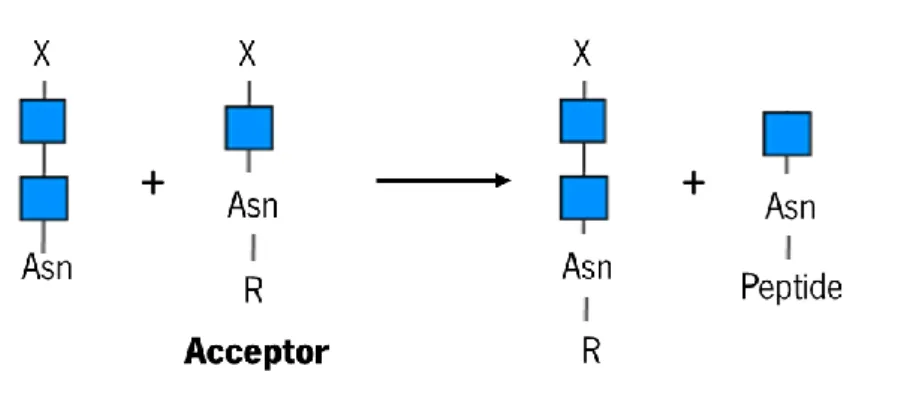
Figure 6. Scheme of transglycosylation reaction catalysed by some ENGase of GH85 family. Figure
adapted from 58. ... 12
Figure 7. Hydrolytic mechanism of action of ENGases of the GH85 family. The mechanism involves an
acid-base catalysis reaction supported by the side chain of a residue of glutamate and asparagine. The intermediate formed is an oxazoline. Figure adapted from65. ... 13
Figure 8. Life cycle of A. gossypii. The main phases of the filamentous fungus A. gossypii life cycle are illustrated: a) and b) represent the isotropic growth phase; c), d) and e) represent the polar growth phase; and f) represents the sporulation phase. Figure adapted from 93. ... 16
Figure 9. Life cycle of Z. rouxii. In most of the life cycle, Z. rouxii cells remain in a haploid state and then
reproduce by budding, like most conventional yeasts, leading to the formation of a transient heterokaryotic zygote. Afterwards, the diploid zygote undergoes meiosis and sporulation and the haploid state is finally restored. Figure adapted from108... 18
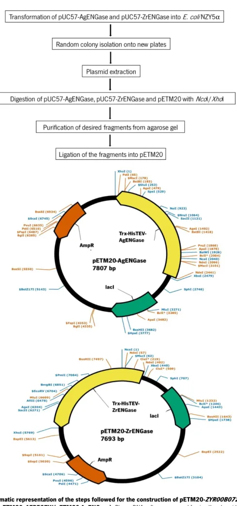
Figure 10. pETM20 vector sequence features. ... 25 Figure 11. Schematic representation of the steps followed for the construction of
pETM20-ZYR00B07216g (pETM20-ZrENGase) and pETM20-AFR597W (pETM20-AgENGase). PlasmaDNA
software was used for in silico plasmid construction and representation. ... 26
Figure 12. Multiple alignment of several ENGases of the GH85 family, namely the putative ENGases of A. gossypii and Z. rouxii, and corresponding dendogram. Multiple alignment was performed using the
Clustal Omega tool and the dendogram was built based on the multiple alignment performed and representend using the FigTree tool. Residues conserved between the homologues are shaded in black. The putative or experimentally determined residues involved in the hydrolytic (red box) and transglycosylation activity (green box) are represented. Accession numbers for the represented ENGases are described in Table 7. Data included in the poster presented at the 4th Meeting of Medicinal Biotechnology (Appendix E). ... 37
Figure 13. Multiple alignment of several ENGases of the GH85 family, namely the putative ENGases
from A. gossypii and Z. rouxii. Multiple alignment was performed using Clustal Omega tool. Residues conserved
x
residues that may be conserved in Endo-M, Endo-Om and the putative ZrENGase and AgENGase are represented in a red box. Accession numbers for the represented ENGases are described in Table 7. ... 38
Figure 14. Endo-A structure used as template for the homology-based models of AgENGase and ZrENGase. Endo-A mutant (PDB: 2VTF.1.A) structure was used as a template for the construction of the
homology-based models of AgENGase and ZrENGase. This structure was visualized in The Pymol Molecular Graphics System, Version 2.1.0 Schrödinger, LLC. ... 39
Figure 15. Prediction of the 3D structure of the putative AgENGase and ZrENGase from homology-based models. The models (A and D) were built using the SWISS-MODEL server and were visualized using The
Pymol Molecular Graphics System, Version 2.1.0 Schrödinger, LLC. A- Predicted model for the possible AgENGase 3D structure, B- Representation of the solvent accessible surface of the putative AgENGase; C- Representation of residues that may establish hydrophobic interactions around the catalytic pocket (Tyr-341 132, Tyr-134, Phe-135, Phe-207 and Phe-139), D- Predicted model for the possible 3D structure ZrENGase, E- Representation of the solvent accessible surface of the putative ZrENGase, F- Representation of residues that may establish hydrophobic interactions around the catalytic pocket (Phe-111, Tyr-317, Tyr-113, Phe-114 and Phe-183). Data included in the poster presented in the 4th Meeting of Medicinal Biotechnology (Appendix E). ... 39
Figure 16. Overlapping of the generated models for the putative AgENGase and ZrENGase. A- Model
resulting from superposition of the predicted model for the possible AgENGase with the predicted model for the possible ZrENGase; B- Overlapping of the secondary structure of the possible AgENGase (in yellow) with that of the possible ZrENGase (in red). ... 40
Figure 17. Confirmation of E. coli NZY5α clones containing AgENGase and
pETM20-ZrENGase. A- Colony PCR products were assessed by electrophoresis on a 1 % TAE agarose gel and the image
was acquired with an UV transilluminator. A1-A4 correspond to the clones tested for pETM20-AgENGase transformants (one expected amplicon with 2045 bp) and Z1-Z4 correspond to the clones tested for pETM20-ZrENGase transformants (one expected amplicon with 1931 bp). B- After plasmid extraction, digestion with restriction enzymes was performed to verify if the insert was in the correct orientation. DNA fragments were separated by electrophoresis on a 1 % TAE agarose gel and the image was acquired with an UV transilluminator. Lane 1 corresponds to pETM20-AgENGase digested with PstI (two expected fragments with 6445 and 1362 bp) and lane 2 corresponds to pETM20-ZrENGase digested with XbaI and XhoI (two expected fragments with 5329 and 2364 bp). ... 42
Figure 18. Recombinant production of AgENGase and ZrENGase in E. coli Origami 2 (DE3) and
SHuffle T7 Express in LB and AI media at 37 and 20 ℃. Insoluble (IF) and soluble (SF) protein fractions
were resolved in a 15 % acrylamide gel and stained with Coomassie Brilliant Blue. Bands with the predicted MW of the fusions TrxA-His-tag-TEV-AgENGase (92 kDa) / ZrENGase (88 kDa) are in the red boxes. Note that insoluble fraction at 37 ℃ was diluted 1:5 in order to facilitate band visualization. ... 44
Figure 19. Recombinant production of AgENGase and ZrENGase in E. coli Origami 2 (DE3) and
SHuffle T7 Express in LB and TB media at 20 ℃. Insoluble (IF) and soluble (SF) protein fractions were
resolved in a 15 % acrylamide gel and stained with Coomassie Brilliant Blue. Bands with the predicted MW of the fusions TrxA-His-tag-TEV-AgENGase (92 kDa) / ZrENGase (88 kDa) are in the red boxes. ... 45
Figure 20. Recombinant production of AgENGase and ZrENGase in E. coli BL21 (DE3) in LB, TB and
AI media at 20 ℃. Total (TF) and soluble (SF) protein fractions were resolved in a 15 % acrylamide gel and
stained with Coomassie Brilliant Blue. Bands with the predicted MW of the fusions TrxA-His-tag-TEV-AgENGase (92 kDa) / ZrENGase (88 kDa) are in the red boxes. ... 46
Figure 21. Recombinant production of AgENGase and ZrENGase in E. coli Origami 2 (DE3) in LB
medium at 18 ℃. Insoluble (TF) and soluble (SF) protein fractions were resolved in a 15 % acrylamide gel and
stained with Coomassie Brilliant Blue. Bands with the predicted MW of the fusions TrxA-His-tag-TEV-AgENGase (92 kDa) / ZrENGase (88 kDa) are in the red boxes. ... 47
xi
Figure 22. Recombinant production of AgENGase and ZrENGase in E. coli Origami 2 (DE3) in LB
medium at 16 ℃ with and without additives (1 M sorbitol, 0.8 M NaCl, 0.05 M sodium phosphate buffer pH 7.4). Insoluble (TF) and soluble (SF) protein fractions were resolved in a 15 % acrylamide gel and
stained with Coomassie Brilliant Blue. Bands with the predicted MW of the fusions TrxA-His-tag-TEV-AgENGase (92 kDa) / ZrENGase (88 kDa) are in the red boxes. ... 48
Figure 23. Lysis of a pellet collected from the production of the putative ZrENGase in E. coli Origami
2 (DE3) at 4 ℃ in LB medium with 1 M sucrose or a combination of 1 M sucrose and 0.2 M arginine.
Insoluble (TF) and soluble (SF) protein fractions were resolved in a 15 % acrylamide gel and stained with Coomassie Brilliant Blue. Bands with the predicted MW of the fusion TrxA-His-tag-TEV-ZrENGase (88 kDa) are in the red boxes. ... 49
Figure 24. Lysis of pellets collected from the production of the putative ZrENGase in E. coli Origami
2 (DE3) at 16 ℃ in LB medium and of the putative AgENGase in E. coli Origami 2 (DE3) at 4 ℃ in
LB medium in the presence of 2 M urea. Insoluble (TF) and soluble (SF) protein fractions were resolved in a
15 % acrylamide gel and stained with Coomassie Brilliant Blue. Bands with the predicted MW of the fusions TrxA-His-tag-TEV-AgENGase (92 kDa) / ZrENGase (88 kDa) in the insoluble fraction are in the red boxes and in the soluble fraction are in green boxes. ... 50
Figure 25. Recombinant production of ZrENGase in E. coli Origami 2 (DE3), BL21 (DE3) and SHuffle
T7 Express in LB medium supplemented or not with 0.2 M NaCl, 0.5 % (w/v) glycerol and 0.2 M arginine at 16 ℃. Insoluble (TF) and soluble (SF) protein fractions were resolved in a 15 % acrylamide gel and
stained with Coomassie Brilliant Blue. Bands with the predicted MW of the fusions TrxA-His-tag-TEV-AgENGase (92 kDa) / ZrENGase (88 kDa) are in the red boxes. ... 51
Figure 26. Column purification of the soluble protein fractions from productions in E. coli Origami
2 (DE3) at 20, 18, 16 and 4 ℃ of the putative ENGases. Fraction that did not bind to the column (FT),
fraction from the last washing step (LW) and fractions were proteins were eluted (E1 and E2) were resolved in a 15 % acrylamide gel and stained with. Bands with the predicted MW of the fusions TrxA-His-tag-TEV-AgENGase (92 kDa) / ZrENGase (88 kDa) are in the red boxes. Black arrow is pointing to the band that may correspond to the putative ZrENGase. ... 52
Figure 27. Gel electrophoresis detection of Endo-H ENGase deglycosylation activity using different substrates. Band shifts resulting from the reaction of the referred substrates with a model ENGase, Endo-H, were
detected in a 15 % acrylamide gel. 1- RNase B from bovine pancreas (negative control), 2- RNase B deglycosylated with Endo-H, 3- β-galactosidase from A. niger (negative control), 4- β-galactosidase deglycosylated with Endo-H, 5- invertase from S. cerevisiae (negative control), 6- invertase deglycosylated with Endo-H. The black arrows indicate the deglycosylation shift of each substrate, the red arrow is pointing to possible contaminants of the β-galactosidase substrate solution and the asterisk represents the band corresponding to Endo-H. ... 53
Figure 28. Chromatogram obtained from the analysis of the reaction of β-galactosidase with
Endo-H (A) as well as the native control (B) by Endo-HPLC. Deglycosylation of glycoproteins was monitored by HPLC
using a Jasco chromatograph equipped with an ELSD detector (SEDEX 85, SEDERE) and a Prevail Carbohydrate ES column (5 μm, 250 × 4.6 mm, Altech). A mixture of acetonitrile-water (60:40 % (v/v)), pumped at 0.9 mL/min, was used as mobile phase. The injection volume was defined as 20 μL. ... 54
Figure 29. Chromatogram obtained from the analysis of the reactions of invertase with Endo-H (A)
as well as the negative control (B) by HPLC. Deglycosylation of glycoproteins was monitoredby HPLC using
a Jasco chromatograph equipped with an ELSD detector (SEDEX 85, SEDERE) and a Prevail Carbohydrate ES column (5 μm, 250 × 4.6 mm, Altech). A mixture of acetonitrile-water (60:40 % (v/v)), pumped at 0.9 mL/min, was used as mobile phase. The injection volume was defined as 20 μL. ... 55
Figure 30. Substrates provided in the Chitinase Assay Kit used to detect β-N
-acetylglucosaminidase/chitobiosidase activity. A- 4-Methylumbelliferyl N-acetyl-β-D-glucosaminide, B-
xii
Figure 31. Specific ENGase/chitinase activity in A. gosspii crude cell extracts (intracellular) and
concentrated culture supernatants (extracellular). Specific enzymatic activities were normalized per
amount of total protein in the reaction mix. Error bars represent the standard deviation from two biological replicates. ... 57
Figure 32. Schematic representation of the strategy used for the disruption of AFR597W in A.
gossypii, and verification PCR of the generated mutants. (A) Construction of the A. gossypii mutant strain. The AFR597W gene was replaced by the loxP-GEN3-loxP cassette. (B) Verification of the integration of the lox
P-GEN3-loxP cassette in the AFR597W locus and confirmation of its excision by colony PCR with primers F3.F/F3.R and V3-Kan_FW/F3.R. ... 59
Figure 33. Colony radial growth measured in A. gossypii afr597w mutant strain and another A.
gossypii mutant strain that also has resistance to G418 (control) after 4 days of growth on selective agar-solidified AFM. The values represent the mean of two biological replicas. Error bars represent the standard
xiii
LIST OF TABLES
Table 1. Summary of the predicted catalytic residues involved in the hydrolytic and transglycosylation activity of some representative ENGases of the GH85 family. Residues that were
experimentally determined to be essential for hydrolytic or transglycosylation activity are represented in bold. ... 14
Table 2. Bacterial strains used in this work as cloning and expression hosts. ... 23
Table 3. Media used in this work for bacterial growth. ... 23
Table 4. pETM20 vector features... 25
Table 5. Primers used in this work. Overhang sequences are underlined. ... 27
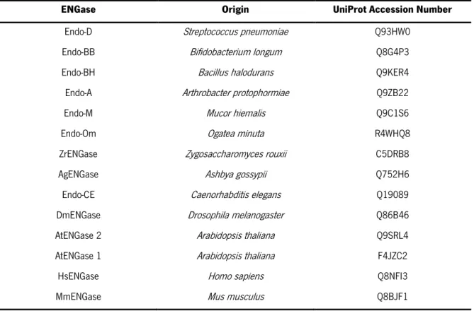
Table 6. ENGases of the GH85 family. The UniProt Accession Number and the origin of the ENGases used to perform a multiple alignment with the putative ENGases of A. gossypii and Z. roxii are represented. ... 36
Table 7. Summary of several computed features for the putative ENGases. The determination of several physical and chemical parameters (theorical pI, predicted MW and extinction coefficient) of the putative ENGases was done using ProtParam tool. Dissulfide bonds were predicted using DiANNA web server. ... 41
Table 8. Total protein concentration determined for intracellular content and concentrated culture supernatants. ... 56
xiv
LIST OF ABBREVIATIONS AND ACRONYMS
AFM Ashbya full medium
AI Auto-Induction
BSA Bovine Serum Albumin
CAZy Carbohydrate-Active enZymes
CV Column volume
Dol-P Dolicol phosphate
Dol-P-Glu Dolicol phosphate glucose
Dol-P-Man Dolicol phosphate mannose
Dol-PP-GlcNAc N-acetylglucosamine dolicol pyrophosphate
EDTA Ethylenediamine tetra acetic acid
ELSD Evaporative Light Scattering Detector
ENGases Endo-β-N-acetylglucosaminidases
ER Endoplasmic Reticulum
ERAD Endoplasmic Reticulum Associated Degradation
G418 Geneticin GAG Glycosaminoglycan Gal Galactose GDP Guanosine diphosphate GH Glycoside hydrolases GlcNAc N-acetylglucosamine
GlcNAc-P N-acetylglucosamine 1-phosphate
GPI Glycosylphosphatidylinositol
GST Glutathione S-transferase
HPLC High-Perfomance Liquid Chromatography
IgG Immunoglobulin G
IMAC Imobilized Metal Afinity Chromatography
IPTG Isopropyl β-D-1-thiogalactopyranoside
KEGG Kyoto Encyclopedia of Genes and Genomes
LB Luria-Bertani
MALDI-TOF Matrix-assisted laser desorption/ionization-time-of-flight
Man Mannose
xv
MCP-3 Monocyte Chemotactic Protein-3
MS Mass spectrometry
MW Molecular weight
NCL Native Chemical Ligation
NGT N-glycosyltransferase
NMR Nuclear magnetic resonance
NOHBY NO Homolog in Baker's Yeast
NusA N-utilizing substance A
OD Optical density
ORF Open reading frame
OST Oligosaccharide transferase
PDB Protein Data Bank
PMSF Phenylmethylsulphonyl fluoride
PNGase Peptide:N-glycosidase
QC Quality Control
QMEAN Qualitative Model Energy ANalysis
RMSD Root-mean-square deviation
RNase B Ribonuclease B
SCO Single Cell Oil
SDS Sodium dodecyl sulphate
SDS-PAGE Sodium dodecyl sulphate–polyacrylamide gel electrophoresis
SNFG Symbol Nomenclature for Glycans
SOB Super Optimal Broth
SPPS Solid-Phase Peptide Synthesis
SUMO Small ubiquitin-like modifier
TAE Tris-Acetate-EDTA
TB Terrific broth
TIM Triosephosphate isomerase
TrxA Thioredoxin A
xvi
1
2
INTRODUCTION
1. Overview on glycosylation
1.1. Glycosylation and its biological significance
Glycobiology is the science that deals with the study of the structure, biosynthesis, biology and evolution of carbohydrates (glycans) distributed in Nature in the context of the biological supports to which they are attached. Glycans are commonly covalently attached to proteins or lipids to form glycoconjugates (glycoproteins, glycolipids and proteoglycans). Considering the structural variability of the glycans, glycoconjugates are hybrid molecules that may present new functionalities and specificities1. The most
common classes of eukaryotic glycans are represented in Figure 1.
Figure 1. Common classes of eukaryotic glycans. The common structures of aproteoglycan, a glycosylphosphatidylinositol
(GPI)-anchored glycoprotein, a glycoprotein and a glycosphingolipid are represented. Figure adapted from 1.
A proteoglycan has one or more glycosaminoglycan (GAG) chains attached to a protein through a typical core region ending in a xylose residue that is linked to the hydroxyl group of a serine residue. On
3
the other hand, a glycophosphatidylinositol anchor is a glycan bridge between phosphatidylinositol and a phosphoethanolamine (amide linkage to the carboxyl terminus of a protein). A glycosphingolipid (often called a glycolipid) consists of a glycan usually attached via glucose or galactose to the terminal primary hydroxyl group of the lipid moiety ceramide2.
Glycans are involved in several important cellular communication processes, such as cell adhesion3, pathogen-host interactions4 and immune response5. Protein glycosylation, a common and
essential processing strategy in eukaryotes, differs from other post-translational covalent modifications in relation to the size and complexity of the added group and the magnitude of the cellular machinery that is required for their synthesis and modulation6. This post-translational modification can affect the intrinsic
properties of proteins, such as folding7, stability8, solubility8 and intracellular traffic9. Depending on the
type of glycosidic linkage (Figure 2), the protein glycosylation can be divided into N- or O-glycosylation.
Figure 2. Most common glycosidic bonds. The following glycosidic bonds are represented: A) (β-N-acetylglucosamine)-N-asparagine;
B) (α-N-acetylgalactosamine)-O-serine/threonine; and C) (β-N-acetylglucosamine)-O-serine/threonine. The monosaccharides through
which the glycans are bound are shown in gray. Figure taken from 9.
In N-glycosylation, the binding occurs between the N-acetylglucosamine (GlcNAc) of the N-glycan and the nitrogen atom of the side chain of an asparagine residue located in the Asn-X-Ser/Thr consensus region (where X denotes any amino acid except proline). In O-glycosylation, the first monosaccharide of the glycan (e.g., α-N-acetylgalactosamine, GlcNAc or fucose in mammals and mannose in fungi) binds to the protein through the oxygen atom of the side chain of a serine or threonine residue4,10.
1.2. Diversity of glycans
In general, glycans present a great variety of monosaccharide units connected by different types of glycosidic bonds, structured in a linear or branched way, and can be modified artificially by chemical
4
or enzymatic methods11. There is a symbolic nomenclature for glycans accepted by the scientific
community, the SNFG (Symbol Nomenclature for Glycans), which will be used in this dissertation (Appendix A)12.
Most of the reactions involved in the glycoprotein biosynthetic pathway are not complete, which leads to an incomplete addition of glycans to the proteins and subsequent incomplete processing of glycans already bound to the proteins, thereby creating structural diversity. The diversity generated from the sub-stoichiometric transfer of a glycan into a protein is called macro-heterogeneity. On the other hand, the diversity generated from the sub-stoichiometric glycan processing is called micro-heterogeneity. In general, macro-heterogeneity of glycoproteins is caused by the presence or absence of a glycan at a given site in a protein, while the micro-heterogeneity of glycoproteins is caused by the presence of different glycan structures in the same site in a protein13.
At the organism level, the structural diversity of glycans can vary between cells and between organelles due to differential expression of the processing enzymes. The structural diversity of glycans can still be caused by reactions of phosphorylation, methylation, among others13. Based on the type of
glycosidic linkage involved in their attachment to proteins, glycans can be categorized into N-glycans or O-glycans. All the N-glycans present in eukaryotes present a common core, Manα1-3 (Manα1-6) Manβ1-4GlcNAcβ1-4GlcNAcβ1-Asn-X-Ser/Thr, and are classified into three types (Figure 3), according to the modifications made to this base sequence: i) oligomannose, in which there is only extension of the glycan structure with mannose residues (Man); ii) complex, in which the extension of the core is made from ramifications initiated with the addition of GlcNAc residues; and iii) hybrid, in which the core branch extends from Manα1-6 with Man residues and from Manα1-3 with the initial addition of one or two GlcNAc residues. Complex N-glycans may have more than six GlcNAc-initiated branches (called antennas) and each one may be elongated with repeats of Galβ1-4GlcNAc (where Gal represents a residue of galactose).
5
Figure 3. Types of N-glycans found in eukaryotes. N-glycans, linked to the Asn-X-Ser/Thr consensus sequence on eukaryotic
glycoproteins, can be classified into three fundamental types: oligomannose, complex and hybrid. Each N-glycan contains a common core, Man3GlcNAc2Asn. Figure taken out from 14.
Rapid advances in high-throughput mass spectrometry (MS) techniques have allowed an increase in the number of comprehensive glycomics studies based on glycoconjugate fragmentation and its analysis. This type of approach allows evaluating: i) the degree of heterogeneity; ii) the type of glycosylation; iii) the glycosylation sites; iv) the identification of the glycan-linked protein; v) the branching of glycans; vi) the number and length of antennas in complex glycans, their composition and substitution by groups of fucose, sialic acid or other groups; and vii) the determination of complete sequences of individual glycans. Based on the information obtained from these studies it was possible to verify the evolutionary trends in N-glycan processing in eukaryotes1.
In fungi, N-glycan processing is restricted by the glycoprotein folding quality control (QC) process and is limited by the diversity of sugar residues that can be used in its extension1. The N-glycans present
in fungi are mainly composed of mannosyl residues (high-mannose type), although some fungi may also exhibit glucose, xylose, fucose, pyruvate or phosphate residues15,16. Generally, filamentous fungi produce
shorter high-mannose type N-glycans, while yeasts produce more extensive high-mannose type N-glycans, often branched to generate hypermannose outer chains17. Interestingly, the diversity of N-glycans
produced by the filamentous fungus Ashbya gossypii (syn. Eremothecium gossypii), which was characterized by MALDI-TOF (Matrix-Assisted Laser Desorption/Ionization-time-of-flight) MS, and nuclear magnetic resonance (NMR)18, resembles that of yeasts more than that of others filamentous fungi with
respect to composition and extent (N-glycans of high-mannose type in the range of Man4-18GlcNAc2,
sometimes phosphorylated)18. Complex N-glycans such as those present in mammals have not yet been
detected in fungi16. N-glycans present in plants have no branching in the core, but have a xylose residue
6
There are several classes of enzymes involved in N-glycosylation, including glycosidases (EC 3.2.1.-) and glycosyltransferases (EC 2.4.x.y). Glycosyltransferases catalyse the transfer of sugar residues (monosaccharides) from activated donor substrates to specific acceptor molecules, forming glycosidic bonds, while glycosidases catalyse the hydrolysis of the glycosidic bond between two or more sugar residues, or between a carbohydrate and a non-carbohydrate moiety in a regio and stereospecific manner3.
The major glycosidases involved in the deglycosylation of N-glycans occurring in Nature are the peptide:N-glycosidase (PNGase; EC 3.5.1.52), which hydrolyses the glycosylamine linkage between two N-acetylglucosamine residues and an asparagine (generating a free peptide and an intact oligosaccharide with the N-N’-diacetylchitobiose unit at the reducing end), and endo-β-N-acetylglucosaminidase (ENGase; EC 3.2.1.96), which cleaves the oligosaccharide linkage between the N-N’-diacetylchitobiose core of asparagine-linked glycans20.Given its relevance to the aim of this project, in section 2 a brief bibliographic
review on ENGases is provided. These enzymes are also quite useful for the artificial synthesis of glycoproteins, as discussed in section 1.4.
1.3.
N
-glycan biosynthetic pathwayIn eukaryotes, the glycoproteins produced in greater amounts and of greater relevance at the cellular level are those that present N-glycans. As such, this topic will discuss its biosynthesis.
N-glycans biosynthesis (Figure 4) in eukaryotes initiates on the cytoplasmic face of the endoplasmic reticulum (ER) membrane, with the transfer of an N-acetylglucosamine 1-phosphate (GlcNAc-P), from uridine diphosphate-N-acetylglucosamine (UDP-GlcNAc), to a lipid precursor, dolicol phosphate (Dol-P), to generate N-acetylglucosamine dolicol pyrophosphate (Dol-PP-GlcNAc). This reaction is catalysed by the enzyme N-acetylglucosamine-1-phosphotransferase. Subsequently, still in the cytoplasmic side of the ER, Man5GlcNAc2-P-P-Dol is generated from the transfer of sugar residues from
UDP-GlcNAc and GDP-Man, catalysed by specific glycosyltransferases. The Man5GlcNAc2-P-P-Dol
precursor is further translocated to the lumen of the ER, where the formation of the final sugar precursor, Dol-P-P-glycan, is performed by the addition of four Man residues from Dol-P-Man and of three glucose residues from Dol-P-Glc. Afterwards, the glycan constituted by fourteen sugars is transferred en bloc into an Asn-X-Ser/Thr consensus sequence of a protein that is being synthesized and translocated through the ER membrane. This transfer reaction is catalysed by a complex of multiple protein subunits called oligosaccharide transferase (OST). Following this transfer, the glycans bonded to proteins are further
7
remodelled in the ER and Golgi complex through a series of reactions catalysed by enzymes with membrane domains in these organelles, such as α-glycosidases I and II and glycosyltransferases. These enzymes are quite sensitive to the physiological and biochemical conditions of the cells where the glycoproteins are being synthesized. Therefore, the type and mode of branching of the sugars in a mature glycoprotein will depend on the type of cell in which the glycoprotein is produced and the physiological state of the cell at that time14.
Trimming of the Glc3Man9GlcNAc2Asn begins in the ER lumen with the removal of the glucose
residues by the action of α-glycosidase I and II. At this point, correctly folded glycoproteins follow to the Golgi complex, whereas improperly folded glycoproteins are retained in the ER by the protein folding QC machinery or directed to degradation by the ER-associated degradation pathway machinery, ERAD (Endoplasmic Reticulum Associated Degradation)21,22. The QC system is composed of lectins and
chaperones that maintain glycoproteins in the ER until they are correctly folded or, otherwise, targeted for degradation23. These systems help preventing cellular toxicity associated with secretion of misfolded
proteins and their potential negative effect on cellular homeostasis. Before exiting the ER, α-mannosidase I may act on some glycoproteins, removing the terminal Man residue and, consequently, generating a Man8GlcNAc2 isomer1.
Figure 4. Processing and maturation of an N-glycan. The mature Dol-P-P-GlcNAc is transferred to the Asn-X-Ser/Thr consensus
sequence of a protein, during protein synthesis, as the proteins are translocated to the ER. After transferring fourteen sugars to the protein, α-glycosidases I and II, present in ER, remove three glucose residues and mannosidase removes a mannose residue. These reactions are directly linked to lectin-assisted glycoprotein folding, which determines whether the glycoprotein continues to the Golgi or is
8
While the early stages of N-glycan processing within the ER are highly conserved across species (from yeasts to mammals), great variability occurs at the Golgi complex level, where glycosidases and glycosyltransferases vary greatly from species to species, leading to a large variety of glycans produced by different organisms or even within different cell types (Figure 5)1,24,25.
Figure 5.N-glycan processing in the Golgi of different eukaryotic taxa. Figure adapted from 1.
1.4. Synthetic methods for glycoprotein engineering
Glycoproteins are normally produced in the cells as a heterogeneous mixture of glycoforms, i.e., with the core polypeptide chain decorated with different glycan structures, eventually attached to different sites. The isolation of pure glycoforms is a complicated process and, as such, to combat the recurrent need to produce homogeneous materials for basic structure-activity studies and for biomedical
9
applications, several methods have been developed for the synthesis of glycopeptides and glycoproteins with well-defined oligosaccharide structures26.
The most common strategy used for the synthesis of homogeneous glycoproteins and glycopeptides involves the incorporation of pre-formed glycosyl amino acids in conventional Solid-Phase Peptide Synthesis (SPPS) protocols. This strategy has proved useful for the synthesis of glycopeptides containing small oligosaccharides, but its extension to larger glycoproteins faces several challenges, such as low coupling efficiency and low solubility of the protected polypeptides27. Therefore, other synthetic
strategies have been developed to tackle these challenges, such as the Native Chemical Ligation (NCL) method, the chemo-selective method, also known as the “tag and modify” method, the chemo-enzymatic method and the direct enzymatic glycosylation method28. A schematic summary of these strategies is
shown in Appendix B.
Regarding the NCL method, it was initially developed for total protein synthesis29. This method
consists of a chemo-selective transthioesterification reaction between a peptide attached to a thioester group and a cysteine residue at the N-terminus of another peptide fragment that form a native peptide bond without need for protection of the side chains of other amino acids30. In 2008, Yamamoto et al.31
performed the first complete chemical synthesis of a glycoprotein containing a complex type N-glycan, a glycoform of the MCP-3 protein (Monocyte Chemotactic Protein-3).
The “tag and modify” method has also been used as an alternative to the SPPS method. In this strategy, the glycosylation of recombinant proteins at specific sites can be achieved by chemically selective binding of glycans and proteins via bio-orthogonal tags. The implementation of this strategy consists in the introduction of specific tags in the sites selected for glycosylation through directed mutagenesis. Subsequently, the tags react with a modified glycan via bio-orthogonal chemo-selective binding. The most commonly used tags include the natural amino acid cysteine and other unnatural amino acids with linked azido, aldehyde, alkyne or alkene groups. The major drawback of this strategy is that unnatural (chemo-selective) linkages introduced may sometimes not mimic perfectly the functions of their respective natural linkages32,33.
An alternative strategy combining the enzymatic synthesis of sugar chains with the chemical synthesis of polypeptides is the chemoenzymatic approach.It is a preferred method for synthesizing sialic acid-containing structures34. This strategy requires the preparation of polypeptides with only a linked
monosaccharide and the enzymatic extension of the sugar chain with free oligosaccharides in aqueous solutions without the need for protecting groups. The extension of the sugar chain can be achieved with
10
the assistance of glycosyltransferases and endoglycosidases35,36. The main advantage of this strategy is
that it is convergent, i.e. allows the synthesis of a high number of complex glycopeptides. In addition, it allows avoiding problems associated with incompatibility of handling protective groups for glycosylation and overall deprotection at the end.The major disadvantages of the chemoenzymatic approach based on endoglycosidases are that the efficiency of the transglycosylation is low and product hydrolysis may occur, since these enzymes also have hydrolytic activity37.
In 2013, Lomino et al.38 developed a direct enzymatic glycosylation method, which allows the
specific binding of a monosaccharide moiety to synthetic or natural polypeptides/proteins.This method consists in the direct glycosylation of free polypeptides with a complex oligosaccharide through the combination of two enzymes, a bacterial N-glycosyltransferase (NGT), which catalyses the initial glycosylation of the polypeptide, and an ENGase, which transfers an N-glycan to the monosaccharide moiety already bound to the polypeptide. The same authors have demonstrated that the attachment of the monosaccharide to an asparagine residue of the polypeptide is resistant to PNGase-catalysed hydrolysis and has reduced susceptibility to ENGase-catalysed hydrolysis38.
Alternatively, there are two strategies that allow the synthesis of glycoproteins in vivo: the post-translational modification of glycoproteins, through the reengineering of metabolic pathways in yeast39 and
fungi40, and the co-translating incorporation at a specific site of a synthetic amino acid through suppressor
tRNA technology41. The re-engineering of metabolic pathways has been increasingly used because of the
growing need for recombinant glycoproteins for therapeutic use.Although yeasts and fungi are promising candidates as recombinant protein expression systems, since they can perform typical post-translational modifications of eukaryotes, these organisms modify their glycoproteins with glycans containing many mannoses. Thus, the glycoproteins produced by these organisms generally cannot be used at the pharmaceutical level, as this type of glycans normally affects the pharmacokinetics and pharmacodynamics of recombinant proteins of animal origin, which natively have more complex glycosylation patterns. However, to overcome these difficulties, scientists developed a tool combining the deletion of genes involved in hypermannosylation with the overexpression of specific glycosidases. In most cases, the gene encoding for the Och1p mannosyltransferase is deleted together with the overexpression of an α-1,2-mannosidase42–44.
All the techniques described above are complementary and can be combined to increase the efficiency of glycopeptide and glycoprotein synthesis. Nevertheless, for the advancement of this field it is necessary to respond to some shortcomings, such as: i) lack of mechanistic and genetic studies related
11
to glycosylation pathways, which are necessary for designing glycoengineering strategies in hosts other than mammals and for better controlling the final product; (ii) lack of enzymes (native or mutant) that are more efficient and more flexible in the reactions they catalyse and of the glycans they use as substrate; and iii) lack of new methods that allow directed chemical glycosylation of proteins through native sugar-amino acid linkages33.
2. ENGases
ENGases are glycoside hydrolases (GH) that act on the N-N'-diacetylchitobiose core of glycoproteins constituted by N-glycans. After the hydrolysis of the β-1,4 glycosidic linkage between the two GlcNAc residues, one residue remains attached to the protein and the other becomes the reducing terminal of the released N-glycan45. According to the Carbohydrate-Active enZymes Database (CAZy), these
enzymes are classified into two large families: GH18 and GH85. This classification was developed according to significant similarities in the tertiary structure, catalytic residues and conserved mechanism of action46. GH18 family also includes chitinases, which are the representative enzymes of this family.
ENGases from GH18 and GH85 families share a homology between 20 to 25 %. ENGases from both families present a TIM (triose phosphate isomerase) barrel in its tertiary structure, consisting of 8 α-helices and 8 parallel β-sheets alternating along the structure.
Regarding their cellular function, ENGases are kwown to be involved in the cytosolic catabolism of free oligosaccharides derived from the ER lumen of eukaryotes, likely to maximize the reuse of sugars in the cells47. In bacteria, ENGases have been reported to be involved in nutrient acquisition for bacterial
growth49 and in defence responses48. In filamentous fungi, ENGases have been found important for hyphal
growth, sporulation and other physiological purposes47,48. Since in myxobacteria ENGases have been found
to be produced in higher concentration during the sporulation phase49, ENGases seem to be also important
for the sporulation process in bacteria. Nevertheless, the physiological roles of ENGases are still poorly understood.
Considering the scope of this dissertation, the focus of the next topics will be the GH85 family, which is composed of ENGases of prokaryotic and eukaryotic origin49,50. According to the Pfam database,
the ENGases of this family have only one conserved domain, which corresponds to the catalytic domain that has the designation of PF0364451.
12
Murakami et al.52 revealed that the ENGase of the GH85 family found in the methylotrophic yeast
Ogatea minuta (Endo-Om) is not detected in the culture medium where the cells grow, confirming the theory that yeast ENGases are likely to be located in the cytosol. This observation had previously been verified in other eukaryotic ENGases of the GH85 family, such as the human ENGase, the Endo-CE present in Caenorhabditis elegans, and the Arabidopsis thaliana ENGase53–55. Murakami et al.52 also found
that the Endo-Om presented hydrolytic activity in relation to complex N-glycans, unlike other ENGases found in bacteria that cannot hydrolyse complex N-glycans (such as, Endo-A from Arthobacter protophormiae49, Endo-D from Streptococcus pneumoniae56 and Endo-BH from Bacillus halodurans57).
Relative to ENGases found in fungi, they act at pH between 5.5 and 7.5 and hydrolyse mainly trimannosylated glycans and complex biantennary glycans20.
In addition to hydrolytic activity, some ENGases of the GH85 family exhibit transglycosylation activity (Figure 6), i.e. they can transfer a free oligosaccharide to another acceptor molecule other than water58. This activity was detected in Endo-A, which is able to transfer high mannose type N-glycans to
monosaccharides such as GlcNAc and glucose to form a novel oligosaccharide59; in Endo-M from Mucor
hiemalis, which shows significant transglycosylation activity in relation to complex N-glycans60; and in
Endo-Om, whose transglycosylation activity is most significant in oligosaccharides of the high-mannose type and does not occur in substrates with fucose or bifurcated structures in the GlcNAc52.
13
2.1. Mechanism of action
The mechanism of action predicted for ENGases of the GH85 family is based on enzyme mutation analyses61–63 and X-ray crystallography studies of Endo-A64 and Endo-D48. These are the only
characterized ENGases of the GH85 family that present an X-ray structure deposited in Protein Data Bank (PDB). All ENGases of the GH85 family retain the configuration of the anomeric carbon and hydrolyse the substrate in a two-step double-displacement mechanism, involving a covalent glycosyl-enzyme intermediate.The reaction involves acid-base catalysis supported by the side chain of two amino acids, typically a glutamate and an asparagine, located at about 5.5 Å64. This mechanism of
action involves the participation of the neighbouring 2-acetamino group of the second GlcNAc residue of the substrate as an intramolecular nucleophile. An oxazoline is responsible for mediating this mechanism (Figure 7).
Figure 7. Hydrolytic mechanism of action of ENGases of the GH85 family. The mechanism involves an acid-base catalysis
reaction supported by the side chain of a residue of glutamate and asparagine. The intermediate formed is an oxazoline. Figure adapted from65.
In the first step, the carboxylic acid of the glutamate residue, which forms a hydrogen bond with the glycosidic oxygen, acts as a general acid/base catalyst, facilitating the release of the leaving group. This release is also aided by the concomitant attack of the carbonyl oxygen of the 2-acetamido group of the substrate. Abbott et al.48 proposed that a second conserved asparagine residue acts as the general
base, involved in the formation of the oxazoline intermediate, which pKa is modulated by the glutamate residue. The resulting intermediate is cleaved by a process wherein the general acid catalyst facilitates the water attack and the second asparagine residue facilitates the release of the 2-acetamido group to form the product, a hemiacetal sugar69.
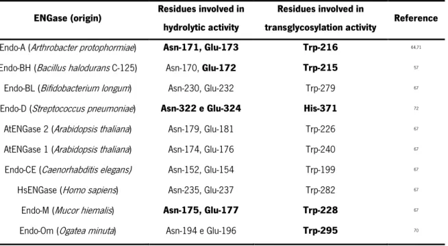
Several ENGases of the GH85 family have already been discovered and yet a few have been characterized. Table 1 shows the predicted catalytic residues involved in the hydrolytic and
14
transglycosylation activity of some representative ENGases. In the case of some ENGases, the residues have been experimentally confirmed, but in other cases, they were predicted based on multiple sequence alignments67. In addition to the residues described in Table 1, there are others at the catalytic centre
that are not directly involved in the hydrolytic or transglycosylation activity but are important for other molecular/biochemical functions related to catalysis. For example, the Trp-251 residue present in Endo-M is important for determining the specificity of the substrate. This residue is conserved in Endo-A (Trp-244), Endo-BH (Trp-243), Endo-CC1 (Trp-253) and Endo-Om (Trp-266)68. The Tyr-217 residue from
Endo-M is also highly conserved in Endo-A (Tyr-205) and Endo-Om (Tyr-231), and is thought to be involved in the formation of the product resulting from the hydrolysis of the intermediate oxazolinium ion62,69,70.
Table 1. Summary of the predicted catalytic residues involved in the hydrolytic and transglycosylation activity of some representative ENGases of the GH85 family. Residues that were experimentally determined to be
essential for hydrolytic or transglycosylation activity are represented in bold.
ENGase (origin) Residues involved in hydrolytic activity
Residues involved in
transglycosylation activity Reference Endo-A (Arthrobacter protophormiae) Asn-171, Glu-173 Trp-216 64,71
Endo-BH (Bacillus halodurans C-125) Asn-170, Glu-172 Trp-215 57
Endo-BL (Bifidobacterium longum) Asn-230, Glu-232 Trp-279 67
Endo-D (Streptococcus pneumoniae) Asn-322 e Glu-324 His-371 72
AtENGase 2 (Arabidopsis thaliana) Asn-179, Glu-181 Trp-226 67
AtENGase 1 (Arabidopsis thaliana) Asn-174, Glu-176 Trp-240 67
Endo-CE (Caenorhabditis elegans) Asn-152, Glu-154 Trp-199 67
HsENGase (Homo sapiens) Asn-235, Glu-237 Trp-282 67
Endo-M (Mucor hiemalis) Asn-175, Glu-177 Trp-228 67
Endo-Om (Ogatea minuta) Asn-194 e Glu-196 Trp-295 70
2.2.
ENGases applications
The use of enzymes for industrial applications increased over the years, since they have high catalytic efficiency, allow to operate in conditions that are not harmful to the environment and are easily produced by large-scale fermentation73.
Since glycoproteins are produced in the form of a heterogeneous mixture of glycoforms, currently all recombinantly produced therapeutic glycoproteins, including monoclonal antibodies, are produced and
15
administered as a mixture of glycoforms74.As such, there is an urgent need for access to unique and pure
glycoforms of glycoproteins and glycopeptides. ENGases have increasingly become a focus of research studies due to their application in the engineering of pharmaceutical glycoproteins, which are intended to present glycans with homogeneous structures, so that their pharmacokinetics and pharmacodynamics are not affected65. These potent biocatalysts have been used in the convergent synthesis of glycopeptides.
For example, Wang et al.75 performed the convergent synthesis of CD52 glycopeptides mediated by
Endo-A and Endo-M. In addition, ENGases have also been used in the remodelling of glycans linked to immunoglobulin G (IgG), to maximize the functions of these antibodies76. Great advances have been made
to make ENGases optimal and preferred biocatalysts for synthetic reactions of glycopeptides and glycoproteins77, among them: the development of synthetic oxazolines as activated donor substrates35 and
the production of glycosynthases that can use activated oxazolines as substrates for transglycosylation reactions and almost do not present hydrolytic activity in relation to the product generated72,78. However,
there are still few ENGases available commercially and they are quite costly, rendering their application at industrial level unbearable. Among these are Endo F1, Endo F2 and Endo F3 from Elizabethkingia myicola (GH18 family), Endo-H from Streptomyces plicatus (GH85 family) and Endo-M from M. hiemalis (GH85 family)73.
Given the great diversity of organisms that produce ENGases and the variety of N-glycans in which they can act, the discovery and identification of new enzymes is being expanded65. Recently, while studying
the N-glycan profile of A. gossypii, Aguiar et al.18 detected the presence of N-glycans with only one GlcNAc,
indicating that this fungus may produce an ENGase capable of cleaving β-1,4 glycosidic bonds between two residues of GlcNAc. Through a search in the CAZy database for genes that could code for glycosyl hydrolases and glycosyltransferases, they identified in this fungus a gene (AFR597W) that could code for an ENGase of the GH85 family, which has no homologue in the closely related yeast Saccharomyces cervisiae18.In addition, through transcriptomic studies, Aguiar et al.18 detected the presence of transcripts
of the AFR597W gene. Furthermore, Murakami et al.52 detected endogenous ENGase activity in crude
extracts of the yeast Zygosaccharomyces rouxii. As such, the characterization of a new ENGase from the GH85 family present in this yeast will also be important. UniProt Database presents a gene that could code for an ENGase in Z. rouxii, ZYRO0B07216g.
16
3.
A. gossypii
A. gossypii is a filamentous hemiascomycete belonging to the Saccharomycetaceae family. This fungus was first isolated and characterized by Nowell in 1916 as a phytopathogenic fungus of the cotton plant (Gossypium hirsutum)79. In the last 30 years, this fungus has been exploited industrially due to its
natural production capacity of riboflavin (vitamin B2)80–82. In addition, A. gossypii has been considered as
a cellular factory for the production of other value-added compounds of commercial interest83,84, such as
recombinant proteins85,86, SCO (Single Cell Oil)87,88, flavours (nucleoside monophosphate of inosine and
guanosine)93 and aromas (2-phenylethanol alcohol94 and γ-lactones91).
A. gossypii exhibits a very simple life cycle as depicted in Figure 8. The main developmental phases of this filamentous fungus, which presents multinucleated hyphae, are: i) the isotropic growth phase during germination, where a germinal spherical cell is formed, ii) the polar growth phase, which begins with the bipolar branch of the germ cell and formation of mycelium that in turn also undergoes polar and dichotomous branching (this only at the tips), and iii) the sporulation phase.The uninucleate spores of A. gossypii present a haploid genome92,93.
Figure 8. Life cycle of A. gossypii. The main phases of the filamentous fungus A. gossypii life cycle are illustrated: a) and b) represent
17
This fungus has a higher phylogenetic relationship with yeasts than with other filamentous fungi94,
which makes it a suitable model organism for cellular biology studies of multicellular filamentous fungi92,93.
The properties that make this fungus suitable as an experimental system include its small genome (which did not undergo whole genome duplication)95, ease of genetic manipulation and relatively fast growth in
defined liquid or solid culture media96.
The 9.12 Mb genome of A. gossypii consists of seven chromosomes, and 95 % of the protein coding sequences found in this fungus have homologues in the genome of S. cerevisiae, most of them (4324 open reading frames; ORF) in synthenic locations. This genetic similarity with the well-studied baker's yeast has facilitated the identification of genes encoding proteins with potential activity of biotechnological interest and allowed the reconstruction of the evolutionary past of both these organisms95.
Moreover, since 259 protein coding genes of A. gossypii (5.4 %) have no homologue in baker's yeast (NOHBY), the characterization of these ORFs is important to better understand the physiology of this fungus. Noteworthy, AFR597W was identified as NOHBY65597.
4.
Z. rouxii
The yeast Z. rouxii was first assigned as Saccharomyces rouxii98 and then classified as an
hemiascomycete. Based on rRNA sequence comparisons, Z. rouxii was found at the time to be one of the yeasts phylogenetically most closely related to S. cerevisiae99,100. Later, after genome sequencing, it
was demonstrated that this species did not undergo the whole genome duplication event that resulted in the genus Saccharomyces101. Z. rouxii and phylogenetically closely related species are now referred
as Z. rouxii complex, since many works suggested that they exhibit mosaic genome structure with respect to some nuclear and mitochondrial phylogenetic markers102,103.
At the biotechnological level, Z. rouxii is considered one of the main spoilage microorganisms in food industry, which contaminates many food products containing high concentration of sugar and/or salt or low pH, such as sugar syrups, honey, fruit juices, and salad dressings104.This is due to
its halotolerance and osmotolerance, which enables it to grow in environments with high concentration of salts and/or sugars that are usually hostile to most yeast growth105. Therefore, this yeast can be
employed in the elaboration of balsamic vinegar, soy sauce and miso paste106. Given its preference for
fructose over glucose, Z. rouxii is considered a fructophilic yeast. Moreover, since it can adapt and grow at low pH values, it is also considered an acid-tolerant yeast101.
18
Regarding the life cycle of Z. rouxii, it is known that this species exists as haploid cells in most of the vegetative phase. Wickerham and Burton107 suggested that this yeast is heterothallic and has the
ability to produce few spores from individual colonies. The haploid life cycle of Z. rouxii is schematized in Figure 9108.
Figure 9. Life cycle of Z. rouxii. In most of the life cycle, Z. rouxii cells remain in a haploid state and then reproduce by budding, like most conventional yeasts, leading to the formation of a transient heterokaryotic zygote. Afterwards, the diploid zygote undergoes meiosis
and sporulation and the haploid state is finally restored. Figure adapted from108.
The haploid vegetative cells reproduce by budding, like most conventional yeasts, leading to the formation of a transient heterokaryotic zygote. Afterwards, meiosis and sporulation occur and the haploid state is finally restored108. However, some haploid Z. rouxii yeasts can shift to diploidism and
ascosporulation can occur without the formation of the a transient heterokaryotic zygote109.
5. Recombinant protein production in
Escherichia coli
: advantages and
challenges
Currently, there are several hosts used for recombinant protein production, among them, bacteria, yeast, insect, plant and animal cells. E. coli is the most common host used for prokaryotic and eukaryotic recombinant protein production, due to its fast growth kinetics, low-cost and high yield of recombinant proteins, well-characterized genome and panoply of available molecular tools for its manipulation110,111. The ease to produce recombinant proteins in E. coli contributed to several
structure-activity studies70,112,113 and it was also remarkable for the development of the pharmaceutical industry114. In
19
H115, Endo-A116, Endo-BH57, Endo-CE54 and Endo-CC from Coprinopsis cinerea117. Note that among these,
Endo-M and Endo-CC belong to eukaryotic organisms.
Despite all the advantages of using E. coli as an expression system, there are still some drawbacks associated with the expression of recombinant proteins in this organism, among which, lack of enzymatic machinery to perform eukaryotic posttranslational modifications, inefficient formation of disulfide bonds, absence of chaperones to perform correct folding of the expressed proteins and lack of efficient secretion systems. Some of the above-mentioned limitations result, sometimes, in the production of proteins with low stability and solubility inside inclusion bodies110,118.
Over the years, many strategies have been developed to improve recombinant protein production in E. coli , among them: i) the use of genetically improved E. coli strains, such as, C41(DE3), Origami and SHuffle119,120; ii) optimization of the inducer concentration121–123; iii) optimization of the induction
temperature124; iv) co-expression of chaperones and foldases118; v) selection of the ideal growth phase to
induce the expression in each strain125; vi) the addition of glucose to the medium, resulting in catabolic
repression of the lac operon126; and vii) the addition of additives to the culture medium, such as Triton
X-100, urea, ammonium sulphate, arginine, glycerol, and ethylenediamine tetra acetic acid (EDTA)118.
Moreover, the expression of fusion proteins is often used to improve solubility and facilitate the purification of recombinant proteins. Common fusion tags used are maltose binding protein (MBP)127, glutathione
S-transferase (GST)128, small ubiquitin-like modifier (SUMO)129, thioredoxin A (TrxA)130, NusA (N-utilizing
20
OBJECTIVES
ENGases have been studied because of their usefulness in structural and functional glycobiology studies, as well as in the engineering of pharmaceutical glycoproteins, which need to present homogeneous glycans in their structure.However, few ENGases have been characterized thus far and even fewer are available commercially, which has limited their application at the industrial level.Therefore, it is necessary to continue the search for new ENGases with interesting properties, which can be produced at low-cost in enough quantity to be applied in the industry in an affordable way. Moreover, a better clarification of the physiological importance of ENGases is also necessary.
Recently, Aguiar et al.17identified a gene in the filamentous fungus A. gossypii that should code
for an ENGase of the GH85 family. Moreover, a gene homologue to that exists in the genome of the yeast Z. rouxii and endogenous ENGase activity was already detected in crude cell extracts of this yeast51.
Envisioning the characterization of the putative ENGases above-mentioned, the objectives of the present master thesis include: i) in silico analysis of the potential ENGase activity of these proteins, namely the construction of homology-based models and the identification of possible catalytic residues through multiple comparison alignments, ii) construction of expression plasmids for the recombinant production of these ENGases in E. coli , iii) optimization of their recombinant production and purification, iv) optimization of protocols for the analysis of ENGase activity, namely in A. gossypii strains, vi) disruption of the A. gossypii gene AFR597W, which should code a putative ENGase of the GH85 family, and assessment of physiological alterations in the generated mutant.
21