This work
relates the development and validation of a simple reversed-phase high-performance liquid chromatographic (HPLC) method for the analysis of xanthone (XAN) and 3-methoxyxanthone (3-MeOXAN) in poly(D,L-lactide-co-glycolide) (PLGA) nanocapsule formulations. This method does not require any complex sample extraction procedure. Chromatographic separation is made with a reversed-phase C18column, using methanol–water (90:10, v/v) as a mobile phase at a flow rate of 1 mL/min. Identification is made by UV detection at 237 nm. The isocratic system operates at ambient temperature and requires 7 min of chromatographic time. The developed method is statistically validated according to United States Pharmacopoeia 25 and International Conference on Harmonization guidelines for its specificity, linearity, accuracy, and precision. The assay method proposed in this study is specific for XAN and 3-MeOXAN in the presence of nanocapsule excipients. Diode-array analyses confirm the homogeneity of XAN and 3-MeOXAN peaks in stressed conditions. Standard curves are linear (r > 0.999) over the concentration range of 0.4–2.5 and 1.0–5.8 µg/mL for XAN and 3-MeOXAN, respectively. Recovery from nanocapsules ranges from 99.6% to 102.8% for XAN and 98.8% to 102.4% for 3-MeOXAN. Repeatability (intra-assay precision) is acceptable with relative standard deviation values of 1.2% for XAN and 0.3% for 3-MeOXAN.
Introduction
Xanthones represent a large group of heterocyclic compounds including natural, semisynthetic, and totally synthetic structures (1). Chemically, xanthonic nucleus corresponds to dibenzo- γ-pyrone. Xanthone (XAN) molecules, having a variety of sub-stituents on the different carbons of the nucleus, constitute a group of compounds with a broad spectrum of biological activi-ties. Among others, antitumoral (2,3), antibacterial (4),
anti-inflammatory (5), hepatoprotective (6), antimalarial (7), immunomodulatory (8,9), as well as inhibitory activities of angiotensin converting enzyme (10) and monoamine oxidase (MAO) (11,12) have been described. Xanthone itself was described as a good MAO-A inhibitor (13).
Poor aqueous solubility of XAN and many of its derivatives such as 3-methoxyxanthone (3-MeOXAN) (Figure 1) is a major obstacle for the assessment of the pharmacological activity of these compounds and their use in the therapy. In general, besides the difficulties of administration of drug substances, water in-solubility is often associated with poor bioavaliability (14).
One approach to overcome the difficulty of administration of poorly water-soluble compounds is by their incorporation in car-rier systems such as polymeric microparticles and nanoparticles. The efficiency of this approach was successfully proven for dif-ferent drugs (15–17). Nanoparticles are solid submicronic drug carriers of a polymeric nature in the nanometer size (18). According to the process used for preparation of nanoparticles, nanospheres or nanocapsules can be obtained. Nanocapsules are vesicular systems in which the drug is confined to a cavity sur-rounded by a polymeric membrane; nanospheres are matrix sys-tems in which the drug is dispersed throughout the particles (19). Besides the improvement of delivery of water-insoluble drugs, nanoparticles have afforded several advantages for different drugs such as reducing drug-associated adverse effects (19), protecting the compound from inactivation before reaching its site of action (20), and increasing the intracellular penetration (21).
By incorporating XAN or its derivatives (such as 3-MeOXAN) in nanoparticles, these poorly water-soluble compounds may be
Abstract
A Validated HPLC Method for the Assay of Xanthone
and 3-Methoxyxanthone in PLGA Nanocapsules
Maribel Teixeira1,2, Carlos M.M. Afonso1, Madalena M.M.M. Pinto1, and Carlos Maurício Barbosa3,4,*
1Centro de Estudos de Química Orgânica, Fitoquímica e Farmacologia da Universidade do Porto-Faculdade de Farmácia do Porto, R. Aníbal
Cunha, 164, 4050 Porto, Portugal;2Instituto Superior de Ciências da Saúde-Norte, R. Central de Gandra, 1317, 4580 Paredes, Portugal; 3CTMUP/ Faculdade de Farmácia do Porto, R. Aníbal Cunha, 164, 4050 Porto, Portugal;4CETMED-Centro Tecnológico do Medicamento,
R. do Passeio Alegre, 840, 4150- 574 Porto, Portugal
Author to whom correspondence should be addressed: email [email protected].
Figure 1. Chemical structure of xanthones: XAN (R = H) and 3-MeOXAN
administered as nanoparticle aqueous dispersions at concentra-tions higher than their maximum hydrosolubility. Moreover, the incorporation of these compounds in nanoparticles may also allow different ways of administration and, simultaneously, may afford their in vivo protection and targeting.
In our laboratory we routinely carry out in vitro studies with poly(D,L-lactide-co-glycolide) (PLGA) nanoparticles containing either xanthone or xanthone derivatives in order to test the use-fulness of these colloidal systems as potential carriers for this group of compounds. For this purpose, the estimation of nanopar-ticle content is an essential tool for guarantying the reliability of the results. The aim of this work was to develop and validate a spe-cific, sensitive, and simple high-performance liquid chromatog-raphy (HPLC) method for the quantitative analysis of XAN and 3-MeOXAN, which were entrapped in PLGA nanocapsules for the first time. PLGA has been selected because polyesters including poly(lactic acid), poly(glycolic acid), and their copolymers such as PLGA have emerged as the most widely studied class of biodegrad-able polymers for pharmaceutical use because of their biocompat-ibility and biodegradability (22). The procedures and parameters used for validation of the analytical method were those described in the International Conference on Harmonization (ICH) guide-lines (23,24), which are similar to the ones established by the United States Pharmacopoeia (USP) (25).
Experimental
Reagents and chemicals
XAN, PLGA (50:50, MW 50,000–75,000), Pluronic F-68, and
soybean lecithin (40% purity by thin-layer chromatography) were purchased from Sigma (Madrid, Spain). 3-MeOXAN was synthe-sized in our laboratory by alkaline cyclization of 2-hydroxy-2',4-dimethoxybenzophenone (26). Myritol 318 was kindly supplied by Henkel (Lisbon, Portugal). HPLC-grade methanol and acetoni-trile were obtained from Merck (Whitehouse Station, NJ). HPLC-grade water was obtained by a MilliQ system (Millipore, Billerica, MA). Other chemicals were of analytical grade.
Nanocapsule preparation and characterization
XAN and 3-MeOXAN nanocapsules of PLGA were prepared according to a modification of the interfacial polymer deposition method described by Fessi et al. (27). Approximately 50 mg of polymer and 100 mg of soybean lecithin were briefly dissolved in 10 mL of acetone. XAN (7.2 mg) or 3-MeOXAN (16.8 mg) was dis-solved in 0.6 mL of Myritol 318, and the obtained solution was added to the acetonic solution. The final solution was poured into 20 mL of an aqueous solution of pluronic F-68 (0.5%, w/v) under moderate stirring, leading to the formation of nanocapsules. Acetone was then removed under vacuum, and the colloidal dis-persion of nanocapsules was concentrated to 5 mL by evaporation under reduced pressure. Nonencapsulated xanthones (XAN or 3-MeOXAN) were separated by ultrafiltration/cetrifugation using centrifugal filter devices Centricon YM-50 (Millipore).
Empty nanocapsules were prepared according to the same pro-cedure, but omitting xanthones in the organic phase.
The mean size and polydispersity index of aqueous nanocapsule
dispersions were determined by laser light scattering (Zetasizer 5000, Malvern Instruments, Worcestershire, U.K.) generating a volume-average distribution for analyzed data.
Instrumental and chromatographic conditions
HPLC analysis was performed with a Jasco liquid chromato-graph (Easton, MD) equipped with a Jasco 880-PU pump and a Jasco 875-UV spectrophotometric detector. The separation was carried out on a 250-× 4.6-mm i.d. Nucleosil C18column (5 µm) (Macherey-Nagel, Düren, Germany). Liquid chromatography analysis was performed by isocratic elution. The mobile phase composition was methanol–water (90:10, v/v), and the flow rate was set at 1.0 mL/min. The injected volume was 20 µL, and the detection wavelength was set at 237 nm. CWS 1.7 software (DataApex, Prague, Czech Republic) managed chromatographic data.
Analysis of samples of XAN and 3-MeOXAN subjected to thermal, acid, and alkaline stress conditions was also performed by HPLC using a different system equipped with a diode-array detector. A Spectra System liquid chromatograph equipped with a Series II digital pump (Science Marketing International, Gloucester, U.K.) with diode-array UV6000LP detector (ThermoFinnigan, San Jose, CA) was used. Samples were chro-matographed using the same procedure described previously, including the used column, injected volume, and detection wave-length. Chromquest for Windows NT software (ThermoFinnigan) managed chromatographic data.
Preparation of sample solutions for determination of XAN and 3-MeOXAN in nanocapsules
Sample solutions were obtained by dissolving an aliquot of XAN or 3-MeOXAN nanocapsule dispersion in acetonitrile (corre-sponding to a dilution of 1/1000) and subjected to HPLC analysis. Considering 100% of entrapment of xanthones in nanocapsules, the obtained sample solutions had a maximum theoretical con-centration (MTC) of 1.44 and 3.36 µg/mL of XAN and 3-MeOXAN, respectively. All analyses were performed in triplicate, and the mean results are reported.
Preparation of XAN and 3-MeOXAN standard solutions Stock standard solutions of XAN and 3-MeOXAN (50 µg/mL) were prepared in acetonitrile. XAN and 3-MeOXAN standard solu-tions were obtained by dilution of the respective freshly prepared stock standard solution with acetonitrile to give six different con-centrations over the range of interest (0.4–2.5 µg/mL for XAN and 1.0–5.8 µg/mL for 3-MeOXAN).
Results and Discussion
Method development
The methanol–water ratio in mobile phase and flow rate were selected in order to find the best conditions for the determination and quantitation of XAN and 3-MeOXAN in nanocapsule formula-tions. A mobile phase of methanol–water (90:10, v/v) was selected to achieve good separation and sensitivity. A flow rate of 1.0 mL/min gave an optimal signal-to-noise ratio and a reasonable
separation time. Retention times were 5.3 min for XAN and 5.8 min for 3-MeOXAN. The total time required for analysis was 7 min. The maximum absorption of both compounds in the exper-imental conditions was found to be 237 nm. Therefore, this wave-length was chosen for the analysis.
For preparation of sample solution of nanocapsules containing XAN or 3-MeOXAN, different solvents (acetonitrile and dichloromethane) were evaluated in order to achieve a complete dissolution of nanocapsule aqueous dispersions. Acetonitrile was
found to afford a complete dissolution upon a 1000-fold dilution of aliquots of XAN or 3-MeOXAN nanocapsule dispersions. Validation study
Specificity
According to ICH guidelines, the specificity of an analytical method is the ability to assess unequivocally the analyte in the presence of components that may be expected to be present such as impurities, degradation products, excipients, etc. (23,24).
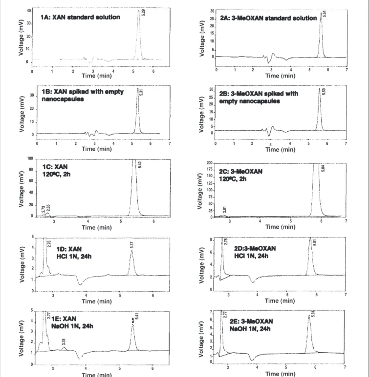
Figure 2. Representative chromatograms obtained following the injection of: (1A) XAN, 1.4 µg/mL; (2A) 3-MeOXAN, 1.0 µg/mL; (1B) XAN and (2B) 3-MeOXAN
stan-dard solutions spiked with empty nanocapsules; (1C) XAN and (2C) 3-MeOXAN subjected to thermal degradation, 120°C for 2 h; (1D) XAN and (2D) 3-MeOXAN sub-jected to acid degradation, HCl 1N for 24 h; and (1E) XAN and (2E) 3-MeOXAN subsub-jected to basic degradation, NaOH 1N for 24 h.
40 30 30 25 20 15 10 5 0 200 175 150 125 100 75 50 25 0 30 25 20 15 10 5 0 0 1 2 3 4 5 6 7 0 1 2 3 4 5 6 7 20 10 0 30 20 10 0 0 100 80 60 40 20 0 1 3 3 4 5 6 3 4 5 6 5 4 3 2 1 0 8 6 4 2 0 7 6 5 4 3 2 1 0 5 4 3 2 1 0 4 5 6 3 4 5 6 7 3 4 5 6 7 3 4 5 6 7 2 3 4 5 6 7 0 1 2 3 4 5 5.2 9 5.3 1 5.4 2 2.7 3 2.85 2.7 6 5.3 7 5.4 1 2.7 7 3.3 5 5.6 4 5.5 9 5.8 4 2.8 1 2.7 8 5.8 1 5.8 1 2.7 7 6
In the present study, the specificity of the analytical method was determined either in samples subjected to stress conditions or in samples containing nanocapsule excipients (i.e., spiked with empty nanocapsules). Samples of both xanthones were subjected to thermal, acidic, and alkaline stress conditions. For the evalua-tion of thermal degradaevalua-tion, porevalua-tions of XAN and 3-MeOXAN were placed in an oven at 120ºC for 2 h. Afterwards, samples were dissolved in methanol and subjected to HPLC analysis. For the evaluation of the degradation of xanthones in acidic and alkaline conditions, a known amount (1 mg) of XAN or 3-MeOXAN was mixed with 25 mL of HCl 1N or NaOH 1N, and the obtained prod-ucts were stored at room temperature for 24 h. After filtration, solutions were appropriately neutralized with HCl 1N or NaOH 1N and diluted with methanol for HPLC analysis. Controls and blank preparations were also prepared and assayed.
Figure 2 shows representative chromatograms of XAN (1A) and 3-MeOXAN (2A) standard solutions. Retention times were 5.3
min for XAN and 5.8 min for 3-MeOXAN. No peaks interfering with XAN or 3-MeOXAN could be detected. A peak with a reten-tion time between 2.8 and 3.0 min, because of the solvent front, was observed. Chromatograms corresponding to xanthone sam-ples subjected to thermal, acidic, and alkaline stress conditions are also shown in Figure 2 (1C–E and 2C–E). No interfering peaks with retention times similar to those of XAN or 3-MeOXAN were observed from any of the stressed samples. Comparison of diode-array spectra, which were obtained at the leading edge, apex, and trailing edge of the XAN and 3-MeOXAN peaks, were identical. This peak purity check, made possible by diode-array technology, is a good indication for the absence of interfering peaks.
In order to evaluate the specificity of the analytical method con-cerning the presence of nanocapsule excipients (i.e., the potential interference of the excipients), a comparison of the test results from the analysis of XAN and 3-MeOXAN standard solutions spiked with empty nanocapsules with those obtained from the analysis of XAN and 3-MeOXAN standard solutions alone was carried out. Obtained chromatograms (Figure 2, 1B and 2B) show the absence of any peak in the region where xanthones elute, which indicates that the method is specific concerning to nanocapsule excipients.
Linearity and range
According to ICH guidelines, the linearity of an analytical method is its ability (within a given range) to obtain test results that are directly pro-portional to the concentration of analyte in the sample (23,24).
Linear regression analysis was carried out by plotting average peak area (y) versus analyte
con-centration (x) in the concentration range of
0.4–2.5 µg/mL for XAN and 1.0–5.8 µg/mL for 3-MeOXAN. Calibration curves were constructed at six concentration levels using the linear-squares regression procedure. The overall procedure was repeated three times on different days. The peak
Table II. Results of Accuracy Determinations
XAN 3-MeOXAN
Theoretical Experimental Theoretical Experimental
concentration concentration concentration concentration
(µg/mL) %MTC (µg/mL) Recovery (%) (µg/mL) %MTC (µg/mL) Recovery (%) 0.4 28 0.410 102.58 1.0 30 1.024 102.38 0.4 28 0.411 102.75 1.0 30 1.017 101.66 0.4 28 0.401 100.25 1.0 30 1.011 101.14 1.0 69 1.004 100.40 2.4 71 2.371 98.77 1.0 69 1.001 100.09 2.4 71 2.416 100.69 1.0 69 1.000 100.01 2.4 71 2.428 101.16 1.4 97 1.395 99.64 3.4 101 3.398 99.95 1.4 97 1.412 100.86 3.4 101 3.423 100.67 1.4 97 1.394 99.61 3.4 101 3.454 101.58
Mean recovery (%) 100.69 Mean recovery (%) 100.89
RSD (%) 1.18 RSD (%) 1.04
Table I. Summary of Standard Curve Results
XAN 3-MeOXAN
Average peak Average peak
Concentration area response Concentration area response
(µg/mL) (mV) RSD (%) (µg/mL) (mV) RSD (%) 0.4 126.50 1.74 1.0 260.55 2.67 0.6 187.75 1.34 1.7 442.67 1.30 0.7 217.55 2.00 2.4 628.46 2.56 1.0 312.27 3.74 3.4 905.49 1.77 1.4 426.89 3.74 4.0 1054.74 2.39 2.5 777.79 1.37 5.8 1554.73 2.69 Y-intercept 0.9386 ± 3.925 –14.66 ± 10.30* Slope 309.6 ± 3.01 269.6 ± 3.00* Correlation coefficient (r) 0.999246 0.999014 Coefficient of 0.998492 0.998029 determination (R2) * Confidence limits (P = 0.05).
area values obtained for three replicate analyses were averaged at each concentration.
Obtained calibration curves showed to be linear over the con-centration ranges examined for XAN and 3-MeOXAN, giving cor-relation coefficients (r) greater than 0.999 and coefficients of
determination (R2) greater than 0.9980 (i.e., over a 99.8% rela-tionship betweenx and y) (Table I).
Accuracy
According to ICH guidelines, the accuracy of an analytical method expresses the closeness of agreement between the value that is accepted either as a conventional true value or an accepted reference value and the value found (23,24). Accuracy is often cal-culated as percent recovery by the assay of known, added amounts of analyte to the sample. Accuracy should be assessed using a minimum of nine determinations over a minimum of three con-centration levels.
Accuracy was determined by spiking known amounts of XAN or 3-MeOXAN to aqueous dispersions of empty nanocapsules in order to obtain XAN concentrations of 0.4, 1.0, and 1.4 µg/mL (corresponding approximately to 28%, 69%, and 97% of MTC)
and 3-MeOXAN concentrations of 1.0, 2.4, and 3.4 µg/mL (corre-sponding approximately to 30%, 71%, and 101% of MTC). Table II summarizes the accuracy results, expressed as percent recovery and relative standard deviation (RSD). Recovery data were within the range of 99.61–102.75% (RSD = 1.18%) and 98.77–102.38% (RSD = 1.04%) for XAN and 3-MeOXAN, respectively. Overall mean recovery values were 100.69% (n= 9) for XAN and 100.89%
(n = 9) for 3-MeOXAN. Because the mean recovery results were
within an acceptable ± 3% range, according to Segall et al., the method was deemed to be accurate (28).
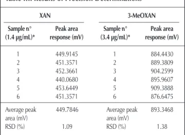
Precision
According to ICH guidelines, the precision of an analytical method expresses the closeness of agreement between a series of measurements obtained from multiple sampling of the same homogeneous sample under the prescribed conditions (23,24). Precision may be measured as repeatability (also termed “intra-assay precision”). Repeatability expresses the precision under the same operating conditions over a short interval of time. ICH guidelines recommend that repeatability should be assessed using a minimum of nine determinations covering the specified range (i.e., three concentrations and three replicates for each concentration) or a minimum of six determinations of 100% of the test concentration.
In the present study, precision of the analytical method was determined by the analysis, on the same day, of six independently prepared standard solutions (1.4 µg/mL for XAN and 3.4 µg/mL for 3-MeOXAN), corresponding to approximately 100% of MTC. Obtained RSD values (1.09% for XAN and 1.38% for 3-MeOXAN) indicate that the proposed HPLC method shows acceptable repeatability (Table III). These results are in agreement to the cri-teria proposed by Green for the precision of an analytical method, for which the RSD should be lower than 2% (29).
Application of the developed method to the quantitative determination of XAN and of 3-MeOXAN in nanocapsules
The present validated method was used to determine either XAN or 3-MeOXAN encapsulated in different nanocapsule batches. Table IV shows encapsulation parameters and particle size of prepared PLGA nanocapsule formulations. High encapsu-lation efficiency values of xanthones in PLGA nanocapsules (84.1% for XAN and 81.2% for 3-MeOXAN) were obtained. The mean particle size of XAN and 3-MeOXAN nanocapsules was 231 nm and 245 nm, respectively.
Conclusion
A simple isocratic reversed-phase HPLC method was developed for the determination of XAN and 3-MeOXAN in PLGA nanocap-sule formulations. The method was validated according to ICH guidelines and USP 25 for its specificity, linearity, accuracy, and precision. Results of validation showed that the proposed method is specific, linear, accurate, and precise either for XAN or 3-MeOXAN, within the established ranges.
XAN and 3-MeOXAN nanocapsule formulations were prepared for the first time. No degradation of the compounds was found Table IV. Encapsulation Parameters and Mean Particle
Size of PLGA Nanocapsule Formulations Containing XAN or 3-MeOXAN*
Theoretical
concentration Encapsulation Polidispersity (mg/mL)† efficiency (%)‡ Diameter (nm) index§
XAN nanocapsules
1.4 84.1 ± 4.9 230.6 ± 11.2 0.483
3-MeOXAN nanocapsules
3.4 81.2 ± 3.0 244.5 ± 29.8 0.431
* Values express the mean results ± standard deviation values of different batches (n = 4 for XAN nanocapsules and n = 3 for 3-MeOXAN nanocapsules.
†Amount of compound (XAN or 3-MeOXAN) used to prepare nanocapsule/volume of nanocapsule dispersion.
‡(Actual concentration/theorectical concentration)× 100.
§Varies from 0.0 corresponding to a perfect homogeneous dispersion to 1.0 corre-sponding to a complete heterogeneous dispersion.
Table III. Results of Precision Determinations
XAN 3-MeOXAN
Sample n° Peak area Sample n° Peak area
(1.4 µg/mL)* response (mV) (3.4 µg/mL)* response (mV) 1 449.9145 1 884.4430 2 451.3571 2 889.3809 3 452.3661 3 904.2599 4 440.0680 4 895.9607 5 453.6449 5 909.3888 6 451.3571 6 876.6475
Average peak 449.7846 Average peak 893.3468
area (mV) area (mV)
RSD (%) 1.09 RSD (%) 1.38
upon nanocapsule preparation by the adopted interfacial polymer deposition method, and high encapsulation efficiencies were obtained. Results clearly demonstrate the suitability of this method for incorporating both poorly water-soluble compounds in PLGA nanocapsules. In vitro studies are being undertaken in different cell lines in order to test the potential of these colloidal delivery systems for xanthone and its derivatives.
The presently developed and validated HPLC method was suc-cessfully used to determine either XAN or 3-MeOXAN content in nanocapsule formulations, affording a very important tool for the evaluation of the finished products. The method could also be useful for the quantitation of different xanthone derivatives encapsulated in nanocapsules. However, further validation studies should be performed for each compound.
Acknowledgments
To Fundação para a Ciência e a Tecnologia (FCT) (Unidade de I&D nº226/94), POCTI (QCA III), FEDER, and Praxis XXI (grant to Maribel Teixeira) for financial support.
References
1. V. Peres, T.J. Nagem, and F.F. de Oliveira. Tetraoxygenated naturally
occurring xanthones. Phytochem.55: 683–710 (2000).
2. C.N. Lin, S.J. Lion, T.H. Lee, and S.J. Won. Xanthone derivatives as
potential anti-cancer drugs. J. Pharm. Pharmacol.48: 539–44 (1996).
3. H. Kamei, T. Koide, T. Kojima, J. Hashimoto, and Y. Hasegawa. Inhibition of cell growth in culture by quinones. Cancer Biotherapy
& Radiopharmaceuticals13: 185–88 (1998).
4. M. Hnuma, H. Tosa, F. Asai, Y. Kobayashi, R. Shimano, and K.I. Miyandis. Antibacterial activity of xanthones from Guttiferaeous plants against methicillin-resistant Staphylococcus aureus. J. Pharm.
Pharmacol.48: 861–65 (1996).
5. C.N. Lin, M.I. Chung, S.J. Lion, T.H. Lee, and J.P. Wang. Synthesis and anti-inflammatory effects of xanthone derivatives. J. Pharm.
Pharmacol.48: 532–38 (1996).
6. E.R. Fernandes, F.D. Carvalho, F.G. Remião, M.L. Bastos, M. Pinto, and O.R. Gottlieb. Hepatoprotective activity of xanthones and xan-thonolignoids against ter-butylhydroperoxide-induced toxicity in
iso-lated rat hepatocytes—comparision with silybin. Pharm. Res.12:
1756–60 (1995).
7. M.V. Ignatuschenko, R.W. Winter, H.P. Bachinger, D.J. Hinrich, and M.K. Riscoe. Xanthones as antimalarial agents; studies of a possible
mode of action. FEBS Lett.409: 67–73 (1997).
8. M.J. Gonzales, M.S.J. Nascimento, H. Cidade, M.M. Pinto, A. Kijjoa, C. Anantachoke, A.M.S. Silva, and W. Herz. Immunomodulatory activity of xanthones from Calophyllum teysmannii var.
inuphyl-loide. Planta Médica65: 368–71 (1999).
9. M. Pinto and M.S.J. Nascimento. Anticomplementary activity of
hydroxy- and methoxyxanthones. Pharm. Pharmacol. Lett.2/3:
125–27 (1997).
10. C.H. Chen and J.Y. Lin. Inhibition of angiotensine-I-converting enzyme by tetrahydroxyxanthones isolated from Tripterospermum
lanceolatum. J. Nat. Product.55: 691–95 (1992).
11. U. Thull, S. Kneuber, B. Testa, M.F.M. Borges, and M.M. Pinto. Substituted xanthones as selective and reversible monoamine
oxi-dase A (MAO-A) inhibitors. Pharm. Res.10: 1187–90 (1993).
12. C. Gnerre, U. Thull, P. Gaillard, P.-A. Carrupt, B. Testa, E. Fernandes, F. Silva, M. Pinto, M.M.M. Pinto, J.-L-. Wolfender, K. Hostettmann, and G. Cruciani. Natural and synthetic xanthones as monoaminoxi-dase inhibitors: biological assays and 3D-QSAR. Helv. Chim. Acta
84: 552–70 (2001).
13. U. Thull and B. Testa. Screening of unsubstituted cyclic compounds
as inhibitors of monoamino oxidases. Biochem. Pharmacol.47(12):
2307–10 (1994).
14. P. Speiser. “Poorly soluble drugs, a challenge in drug delivery”. In
Emulsions and Nanosuspensions for the Formulation of Poorly Soluble Drugs. R.H. Müller, S. Benita, and B. Böhm, Eds. Scientific
Pub., Stuttgard, Germany, 1998, pp. 15–19.
15. A. Sanches, J.L. Villa-Jato, and M.J. Alonso. Development of biodegradable microspheres and nanospheres for the controlled
release of cyclosporine A. Int. J. Pharm.99: 263–73 (1993).
16. Y.I. Kim, L. Fluckiger, M. Hoffman, I. Lartaud-Idjouadiene, J. Atkinson, and P. Maincent. The antihypertensive effect of orally administered nifedipine-loaded nanoparticles in spontaneously
hypertensive rats. Brit. J. Pharmacol.120: 399–404 (1997).
17. C. Mallard, J. Coudane, I. Rault, and M. Vert. In vitro delivery of a sparingly water soluble compound from PLA50 microparticles.
J. Microencapsul.17(13): 13–28 (2000).
18. B. Magenheim and S. Benita. Nanoparticle characterization: a
com-prehensive physicochemical approach. S.T.P. Pharma. Sci. 1:
221–41 (1991).
19. P. Couvreur, C. Dubernet, and F. Puisieux. Controlled drug delivery with nanoparticles: current possibilities and future trends. Eur.
J. Biopharm.41: 2–13 (1995).
20. P.J. Lowe and C.S. Temple. Calcitonin and insulin in isobutyl-cyanoacrylate nanocapsules: protection against proteases and effect
on intestinal absorption in rats. J. Pharm. Pharmacol.46: 547–52
(1994).
21. P. Couvreur, E. Fattal, H. Alphandary, F. Puisieux, and A. Andremont. Intracellular targeting of antibiotics by means of biodegradable
nanoparticles. J. Control. Rel.19: 259–68 (1992).
22. R. Jain, N.H. Shah, A.W. Malick, and C. Rhodes. Controlled drug delivery by biodegradable poly(ester) devices: different preparative
approaches. Drug Dev. Ind. Pharm.24: 703–27 (1998).
23. European Commission, Directorate General III–Industry
Pharmaceuticals and Cosmetics. “Validation of analytical proce-dures: methodology”. In The Rules Governing Medicinal Products in
European Union.3A: 107–17 (1998).
24. European Commission, Directorate General III–Industry
Pharmaceuticals and Cosmetics. “Validation of analytical proce-dures: definition and terminology”. In The Rules Governing
Medicinal Products in European Union.3A: 119–25 (1998).
25. United States Pharmacopeia 25/NF 20. United States Pharmacopeial Convention, Rockville, MD, 2002, pp. 2256–59.
26. E.G.R. Fernandes, A.M.S. Silva, J.A.S. Cavaleiro, F.M. Silva, M.F. Borges, and M.M.M. Pinto. Hepatoprotective activity of xanthones and xanthonolignoids against tert-butylhydroperoxide-induced toxi-city in isolated rat hepatocytescomparision with silybin Magn.
Reson. Chem.36: 305–09 (1998).
27. H. Fessi, F. Puisieux, J. Ph. Devissaguet, N. Ammoury, and S. Benita. Nanocapsule formation by interfacial polymer deposition following
solvent displacement. Int. J. Pharm.55: R1–R4 (1989).
28. A. Segall, F. Hormaechea, M. Vitale, V. Perez, and M.T. Pizzorno. Development and validation of a reverse-phase liquid chromato-graphic method for analysis of estradiol valerate and
medroxypro-gesterone acetate in a tablet formulation. J. Pharm. Biomed. Anal.19:
803–808 (1999).
29. J.M. Green. A practical guide to analytical method validation. Anal.
Chem.68: 305A–309A (1996).