RESUMO.- [Valores de normalidade das respostas da onda-b do eletrorretinograma de campo total (ERG) em cães da raça Lhasa Apso com catarata de acordo com a faixa etária.]Cães da raça Lhasa Apso com catarata imatu-ra, madura e hipermadura foram subdivididos em 4 grupos (G1: 1 a 3 anos, G2: 4 a 7 anos, G3: 8 a 11 anos, G4: acima de 12 anos), submetidos ao mesmo protocolo de sedação para a realização do exame de eletrorretinograma (ERG),com o objetivo de determinar o valor de normalidade da resposta da onda-b do ERG de campo total. Três respostas foram ob-tidas: resposta de bastonetes, máxima resposta e resposta de cones. Os valores da amplitude pico a pico e do tempo de culminação da onda-b dos grupos foram comparados e
ana-lisados pelo teste de Kruskal-Wallis (análise de variância por medidas não repetidas), seguido pelo teste de Dunn (quan-do p<0,05). Quan(quan-do compara(quan-do G4 ao G1 e G2, observou-se diminuição significantiva na amplitude da máxima respos -ta, sugerindo influência da idade nos parâmetros eletrore -tinográficos. Não foi observada diferença significativa nos valores obtidos do tempo de culminação em nenhuma das respostas dos quatro grupos analisados. Este estudo deter-minou os parâmetros normais da onda b no ERG dos cães da raça Lhasa Apso com catarata de acordo com a faixa etária. TERMOS DE INDEXAÇÃO: Eletrorretinograma, catarata, caninos, LhasaApso.
INTRODUCTION
The full-field electroretinogram (ERG) is a complex recor -ding of electrical potentials, originated from the retinal res-ponse to light stimulation (Pereira et al. 2003). It is a very precise electrophysiological test of retinal function evalua-tion (Komáromy et al. 1998) and has the advantage to be a noninvasive technique (Safatle et al. 2005).
Normal values of b-wave responses of full-field electroretinogram
in Lhasa Apso dogs with cataracts according to age
1Ana C.A. Góes2, Tatiane Villar2, Denise A. Otsuki3, Ricardo Lisak2, Ricardo A. Pecora2 and Angélica M.V. Safatle2*
ABSTRACT.- Góes A.C.A., Villar T., Otsuki D.A., Lisak R., Pecora R.A. & Safatle A.M.V. 2015.
Normal values of b-wave responses of full-field electroretinogram in Lhasa Apso
dogs with cataracts according to age. Pesquisa Veterinária Brasileira 35(3):274-280. Clí-nica Veterinária Vetmasters, Avenida Pacaembu 1166, São Paulo, SP 01234-000, Brazil. E-mail: [email protected]
Lhasa Apso dogs with immature, mature or hypermature cataracts were divided into four groups according to their age (G1: 1 to 3 years old, G2: 4 to 7 years old, G3: 8 to 11 years old, G4: more than 12 years old). All animals were evaluated under the same seda-tion protocol to allow the performance of the electroretinogram (ERG) exam to determine normal value of b-wave response of the full-field ERG according to age. Three ERG respon -ses were recorded: rod, maximal and cone respon-ses. The amplitude values and b-wave implicit time of the responses of all groups were compared and analyzed by Kruskal-Wallis test (variance analysis for non-repeated measures), followed by the Dunn post-test (when p<0,05). A significant decrease was observed in maximal responses’ amplitude, when com -paring the G4 group with G1 and G2. No statistically relevant differences were observed in the b-wave implicit time values between groups. The ERG values are directly influenced by the animal’s age. Older patients presented a decrease in the amplitude of the maximal res -ponse. The study determined the normal parameters of ERG b-waves for Lhasa Apso dogs with cataract according to their age group.
INDEX TERMS: Electroretinogram, cataract, dogs, Lhasa Apso.
1 Received on October 9, 2014.
Accepted for publication on February 25, 2015.
2 Clínica Veterinária Vetmasters, Avenida Pacaembu 1166, São Paulo, SP 01234-000, Brazil. *Corresponding author: [email protected]
The main indication for the assessment of ERG in ve-terinary ophthalmology is as a preoperative examination in dogs with cataracts (Safatle et al. 2010a), which is the most common intraocular disease in dogs (Davidson & Nel-ms 1999) and one of the main causes of blindness in this species (Glover & Constantinescu 1997, Adkins & Hendrix 2003, Laus 2009).
The cataract is a relatively common affection in Lhasa Apso (LA) dogs. Therefore, the ERG has been frequently performed in these animals. However, normal ERG values are still not established for the breed.
Determination of normal ERG values is of great im-portance in order to make this exam of real usefulness in clinical practice (Paranhos et al. 2002). Considering that there are variable intrinsic and extrinsic factors that con-tribute to interfere in the values obtained from ERG re-cording, it is mostly difficult to establish a normal range. The main intrinsic factors that may interfere in the results are the retinal function, dark/light adaptation, species, age, opaque media and pupillary dilation. The extrinsic factors consist mainly of the duration and intensity of light stimuli, dark/light adaptation period, anesthetic protocol, positioning and electrode type, recording equi-pment and environmental condition. All these factors are variables among different veterinary clinics, laboratories or hospitals (Ekesten 2007, Lee et al. 2009). Therefore, it is highly recommended that each laboratory establishes its own normal parameters to avoid misinterpretation of results (Mentzer et al., 2006, Noman et al. 2008).
Thereby, as the use of this exam becomes more frequent in the clinical practice of veterinary medicine, notably in dogs of the LA breed and by the absence of related data in the literature, it was conceived to determinate the values of b-wave responses of full-field ERG in Lhasa Apso dogs of di -fferent ages and with cataract of stages where lens opacity disturbs the performance of a good fundoscopy (immature, mature and hypermature stages), using the portable device BPM 200 (RetinoGraphics, Inc., Norwalk, CT, USA).
MATERIALS AND METHODS
A total of 86 electroretinograms were performed in LA dogs with cataracts using the BPM 200 Electroretinographic System (Reti-noGraphicsInc, Norwalk, CT, USA), at the Clínica Veterinária Vet-masters, located in São Paulo, Brazil, during the period from Fe-bruary 2000 to December 2012. Among these 86 dogs included in the study, 36 were females and 50 were males, and their ages varied between one and thirteen years old. Inclusion criteria for this study were Lhasa Apso dogs with cataracts varying between immature, mature and hypermature stages and without any other ocular disorders in at least one eye.
Only one eye of each animal was considered in the statistical analysis. The eye of each animal was chosen based to the absence of other abnormalities, ocular ultrasound results with no altera-tion, good waveforms and without excessive noise in the results than the other eye.
An actual total of 106 Lhasa Apso dogs were examined during that period, but twenty of these animals were excluded from this study, due to the presence of other ocular associated disorders besides the cataract, as for example glaucoma and uveitis, and as well as those patients whose indication for the ERG exam was
other than a preoperative examination for the cataract surgery, as for example the SARDS (Sudden Acquired Retinal Degeneration Syndrome) or PRA (Progressive Retinal Atrophie).
The animals were divided into four groups, according to their age: G1 from one year to three years and eleven months old (14 eyes), G2 from four years to seven years and eleven months old (40 eyes), G3 from eight years to eleven years and eleven months old (26 eyes), and G4 from over twelve years old (6 eyes).
The exams were performed in a surgery room, where no ex-ternal light could enter the room, with the animal placed on a surgery table covered by a rubber mat to reduce electrical inter-ference. When necessary, a dim red light was used to place, check or move the electrodes.
The anesthetic protocol was the same to all animals: 0.3mg/ kg xylasine hydrochloride 2% (Xilazin 2%®, Syntec, Hortolândia,
SP, Brasil) and 0.2mg/kg butorphanol (Turbogesic 10mg®, Fort
Dodge Saúde Animal Ltda, Campinas, SP, Brazil), both via intra-muscular. Pupillary dilation was obtained with one drop of topi-cal tropicamide 1% eye drop (Mydriacyl 1%®, Alcon Laboratórios
Ltda, São Paulo, SP, Brazil) applied 30 minutes prior to the exam. Also, the inner face of the earlobes were clipped and cleaned prior to the exam.
Topical anesthesia was performed with proxymetacainechlo-ridate 5% eye drop (Anestalcon®, Alcon Laboratórios Ltda, São
Paulo, SP, Brazl) before placement of the ERG-jet contact lens electrodes.
After preparation and sedation of the patients, the animals were taken into a dark room, where dark adaptation was perfor-med for 30 minutes with the aid of an occlusive bandage around
the eyes to ensure full darkness. The bandage was then removed and topical anesthesia applied. The patients were then positioned
in lateral recumbency, first the right side and then the left side, to
permit the execution of the scotopic phase of the exam without jeopardizing the dark adaptation of the other eye.
The ERG-jet contact lens electrodes were filled with 2%
methylcellulose (Metilcelulose 2%®, Ophthalmos Ind.
Farma-cêutica, São Paulo, SP, Brazil) prior to its placement, in order to improve electrical contact and protect ocular surface. The
refe-rence electrode was filled with electrolytic gel (Ten20®
Conduc-tive Paste, Weaver and Company, Aurora, CO, USA) and placed at the temporal canthus of the eye. The ground electrode was also
filled with electrolytic gel and fixed at the internal face of the
earlobe that were previously clipped and cleaned (Fig.1). With
the animal in lateral recumbency, the right eye was the first to
be examined.
Dark-adapted rod response was the first to be recorded, using
maximal intensity white light (0.01 cd.s.(m-2)-1) and a
inter-stimu-li interval o 2s. Then, mixed dark-adapted rod and cone respon-se, also denominated maximal scotopic responrespon-se, was recorded using high intensity stimuli (2.5 cd.s-1(m-2)-1) and a inter-stimuli
interval of 10 s.
After the scotopic phase, light adaptation was performed for 10 minutes with external light of 30 cd.s.(m-2)-1, as a preparation
for the photopic phase of the exam. Finally, cone response was ob-tained using maximum intensity light stimulation (2.5 cd.s.(m-2)-1) with an inter-stimuli of 1s. In this phase, the left eye was first eva -luated then the right, to facilitate recumbence side alternations.
For the three responses, an average of 16 stimulations was performed. At the end of the exam, the b-wave amplitude (µV) was measured from the trough of the a-wave to the peak of the b-wave and the b-wave implicit time (ms) from the onset of the stimulus to the peak of the b-wave. Both measurements were obtained in all three responses of each animal. Attenuated or extinct respon-ses were excluded from this study.
It was verified that the variables did not present a normal dis -tribution, when analyzed by the Kolmogorov-Smirnov test. The responses of all different groups were analyzed by the Kruskal--Wallis test (variance analysis for non-repeated measurements), followed by the Dunn post-test (when p<0.05) to allow further
comparison between groups. Statistical significance was set at
5% (p<0.05). The statistical analysis were performed in computer programs (SigmaStat 11.0 and GraphPad Prism 5).
RESULTS
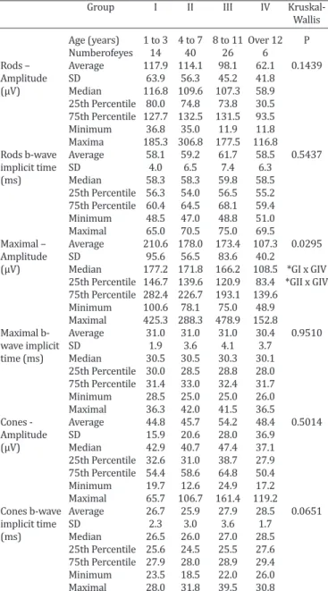
The measurements of the wave amplitude and of the b--wave implicit time of rod response, maximal response and cone response are presented in Table 1, along with average, standard deviation, median, 25th percentile, 75th percenti-le and the minimum and maximal values of each response. The data are subdivided into groups according to the age group of the animals.
A lower amplitude in maximal-responses was observed, when comparing G4 with G1 and G2 (Fig.2). Associating age with rod-responses, we detected a correlation, althou-gh not significant, of a progressive lower response with the progression of age. B-wave cone implicit time was delayed with the progression of age and maximal B-wave implicit time remained unaltered regardless of age. No other res-ponses had evidence of any statistical significance.
DISCUSSION
The full-field ERG is the most used electrophysiological test in veterinary ophthalmology, since it is objective (Tzekov & Arden 1999) and more sensitive than other examination techniques, such as the fundoscopy. Therefore, it allows the diagnosis of retinal alterations on early stages of the disea-se or, even, when fundoscopy is impaired due to media opa-cities, as, for example, the cataract (Komáromy et al. 1998). Retinal degeneration is a much less commonly observed disease in Lhasa Apso dogs in our clinical practice, contra-ry to what is observed in Poodle or Cocker Spaniel dogs, who are genetically predisposed to cataracts and retinal progressive atrophy (Adkins & Hendrix 2005). In the me-antime, as cataract in this breed is becoming more
frequen-Table 1. Amplitude (μV) and implicit time (ms) of rod, maximal and cone responses, of the four age groups of Lhasa
Apso dogs with cataracts
Group I II III IV
Wallis
Age (years) 1 to 3 4 to 7 8 to 11 Over 12 P Numberofeyes 14 40 26 6
Rods – Average 117.9 114.1 98.1 62.1 0.1439 Amplitude SD 63.9 56.3 45.2 41.8
(μV) Median 116.8 109.6 107.3 58.9 25th Percentile 80.0 74.8 73.8 30.5 75th Percentile 127.7 132.5 131.5 93.5 Minimum 36.8 35.0 11.9 11.8 Maxima 185.3 306.8 177.5 116.8
Rods b-wave Average 58.1 59.2 61.7 58.5 0.5437 implicit time SD 4.0 6.5 7.4 6.3
(ms) Median 58.3 58.3 59.8 58.5 25th Percentile 56.3 54.0 56.5 55.2 75th Percentile 60.4 64.5 68.1 59.4 Minimum 48.5 47.0 48.8 51.0 Maximal 65.0 70.5 75.0 69.5
Maximal – Average 210.6 178.0 173.4 107.3 0.0295 Amplitude SD 95.6 56.5 83.6 40.2
(μV) Median 177.2 171.8 166.2 108.5 *GI x GIV 25th Percentile 146.7 139.6 120.9 83.4 *GII x GIV 75th Percentile 282.4 226.7 193.1 139.6
Minimum 100.6 78.1 75.0 48.9 Maximal 425.3 288.3 478.9 152.8
Maximal b- Average 31.0 31.0 31.0 30.4 0.9510 wave implicit SD 1.9 3.6 4.1 3.7
time (ms) Median 30.5 30.5 30.3 30.1 25th Percentile 30.0 28.5 28.8 28.0 75th Percentile 31.4 33.0 32.4 31.7 Minimum 28.5 25.0 25.0 26.0 Maximal 36.3 42.0 41.5 36.5
Cones - Average 44.8 45.7 54.2 48.4 0.5014 Amplitude SD 15.9 20.6 28.0 36.9
(μV) Median 42.9 40.7 47.4 37.1 25th Percentile 32.6 31.0 38.7 27.9 75th Percentile 54.4 58.6 64.8 50.4 Minimum 19.7 12.6 24.9 17.2 Maximal 65.7 106.7 161.4 119.2
Cones b-wave Average 26.7 25.9 27.9 28.5 0.0651 implicit time SD 2.3 3.0 3.6 1.7
(ms) Median 26.5 26.0 27.0 28.5 25th Percentile 25.6 24.5 25.5 27.6 75th Percentile 27.9 28.0 28.9 29.4 Minimum 23.5 18.5 22.0 26.0 Maximal 28.0 31.8 39.5 30.8
tly observed in our daily routine, the ERG is important for testing normal retinal activity, once the cataract consists in a physical impediment to the ophthalmoscopy.
Thereby, ERG is a mandatory exam in those patients, who are candidates for cataract surgery, constituting an important pre-operatory exam in these animals. In a recent study, Chiu et al (2009) evidenced that 27% of dogs with cataracts, that underwent ERG examination, had a certain degree of retinal dysfunction, and correlated this fact to inherited PRA as the most likely cause, due to a large popu-lation percentage of Schnauzers, Poodles, Pomeranians and Cocker Spaniels in the study. Therefore, the importance of a full-field ERG long protocol, which offers a more thorough assessment of retinal responses and function, is to provide the clinician and surgeon with more precise information to decide whether elective surgery should be performed. In addition, the concept of yes-or-no ERG protocol, used as a quick determination for whether retinal function is pre-sent or abpre-sent before cataract surgery, has been questioned (Ekesten et al. 2012).
Some precautions must be taken in order to perform the ERG, such as the maintenance of a fasting period of food and water prior to the exam, due to the sedation required. Furthermore, the patient’s cooperation is also required, since the patients must remain motionless (Narfström 2006), a condition which was successfully achieved by the sedation protocol used in this study, without disturbing or compromising the results.
Recently, a study experimented performing the yes-or--no ERG protocols in dogs under three different consciou-sness state (awake, sedated and anesthetized) and results demonstrated that the awake animals had higher a- and b-wave amplitudes and shorter a-wave implicit time than those from sedated or anesthetized dogs. Also, the b-wave implicit time was shorter in awake animals than in sedated, but not significantly different from the anesthetized. No
significant difference was observed in results between se -dated or anesthetized dogs or among consciousness state for high or low frequency noise (Freeman et al. 2013).
ERG exams have been performed in the latest years with the use of xylazine, ketamine and medetomidine as anesthetic agents (Chaudieu 2004, Rosolen et al. 2004). The combination of xylazine and butorphanol in this study, provided a good sedation and allowed adequate recording and low noise interference. No alterations were observed on ERG results due to the sedation protocol, as it may be observed when conducted with barbiturate or inhalation anesthetic agents (Norman 2008, Ekesten et al. 2012).
Satisfactorily pupil dilatation, to allow adequate reti-nal stimulation, proved to be important for the success of the exam in our study, in agreement to the studies of Birch (1989).
According to Mentzer et al. (2006), ERG results inter-pretation must consider the species, breed and age of the animal, thus the presence of mean opacities, electrode kind, electroretinogram system used and the sedation protocol. In this study, the animals were divided into groups accor-ding to their age, the electrodes and electroretinogram system used were of the same type and sedation protocol performed was identical to all animals.
The assessment of a unilateral or bilateral ERG depends on the species, the equipment used and the purpose of the exam (Narfström 2006). In this study, each eye was indivi-dually examined with the animal on lateral recumbence to avoid the light stimulation to reach the other eye.
In comparison with another similar study from the same authors on Poodle dogs, the Lhasa Apso dogs presented ex-pressively higher mean values of maximal amplitude than the other measured responses. As an example, the mean values of amplitude in Poodle dogs with cataract divided into groups according to their age exactly as in this present study (from one to 3 years, 4 to 7 years, 8 to 11 years, and Fig.2. Graphic representation of the b-wave amplitude (µV) and implicit time (ms) values of rods,
over 12 years old), were, respectively, of 152.3µV, 145.42µV, 128.16µV and 105.7µV (Safatle et al. 2010b). Meanwhile, the mean values of maximal amplitude in the Lhasa Apso dogs, were of 210.6µV, 178.0µV, 173.4µV and 107.3µV, res-pectively, using the same electroretinogram system, at the same location, with the same veterinarians andsedation protocol. Therefore, it may be inferred that the response’s values of the Lhasa Apso dogs are substantially higher, in particular of the youngest animals. The implicit time of the responses, however, remain similar between breeds.
In the same study of Safatle et al. (2010b), a decrease in the amplitude values was observed in all responses, when comparing dogs from G1 (with ages up to 3 years old) with the other groups in the study, meaning that the amplitude of the responses decreases with the animal’s aging (Fig.3). Moreover, the implicit time of rod response observed in this group was delayed when compared to the rest. Such findings contrast with those of this present study, in whi -ch a statistically significant difference was verified only in the maximal response when comparing groups G1 with G4, and G2 with G4. Furthermore, no statistically relevant al-terations were observed in the b-wave implicit time of this study.
Nevertheless, if comparing the dogs in this study with Cocker Spaniel dogs in another study from Safatle et al. (2010a), the same situation is verified: higher mean values of amplitude in Lhasa Apso dogs.
In a study to standardize the ERG values in Shih Tzu dogs, Lee et al. (2009) also demonstrated lower values of amplitude, when confronted with the same data of the Lha-sa Apso dogs. However, the legitimacy of such comparison is questionable, not only due to differences in the sedation protocol, in the adaptation time and in the response pat-tern recorded, but also due to the electroretinograph (Han-dheld Multispecies ERG) used. Moreover, the sample was composed by young dogs without any ophthalmic disorder, what also differs from our study, which included only dogs with cataracts. According to Maehara et al. (2007), the am-plitude of all responses increases substantially after the ca-taract removal, in comparison with the amplitude prior to the surgery.
In this study, a distinguished heterogeneity of the sam-ple was observed, mainly regarding to the group division by age. It is, consequently, assumed that cataract in this breed is a disorder that affects mainly adults, what would explain the larger number of animals grouped in G2 and G3. Other hypothesis could explain the lowest amount of ani-mals grouped in G4 besides the lower frequency of cataract in older animals of this breed, as, for example, the owner’s reluctance in submitting his animal to an exam that requi-res sedation.
CONCLUSIONS
The electroretinogram response is directly influenced by the animal’s age;
Older patients present a decrease in the amplitude of maximal response;
No detectable alterations were observed in the implicit time of all different age groups;
This study also determined the normal parameters of Lhasa Apso dogs with cataracts, demonstrating the lowest and maximal values for each response in all different age groups, and also allows to infer that Lhasa Apso dogs pre-sent considerably higher response values than other bree-ds whose data have already been determined;
More studies are necessary in order to standardize the full-field ERG results in the Lhasa Apso breed, considering the limitations of this study, such as the low amount of pa-tients over 12 years old and of the sample as a whole;
Furthermore, it is suggested that other subsequent stu-dies may clarify the possible existence of a relation betwe-en the animal’s pelage and the electroretinogram respon -ses, as an example of what occurs in albino animals;
Regardless of the referred limitations, this study fills part of a lacuna in the knowledge of electrophysiology in Lhasa Apso dogs, whose specific data has not been establi -shed until the present time.
REFERENCES
Adkins E.A. & Hendrix D.V.H. 2003. Cataract evaluation and treatment in dogs. Compendium on Cont. Educ. Pract. Vet. 25(11):812-824.
Adkins E.A. & Hendrix D.V.H. 2005. Outcomes of dogs presented for cata-ract evaluation: a retrospective study. J. Am. Anim. Hosp. Assoc. 41:235-240.
Birch D.G. 1989. Clinical electroretinography. Ophthalmol. Clin. North Am. 2(3):469-497.
Chaudieu G. & Molon-Noblot S. 2004. Early retinopathy in the Bernese Mountain Dog in France: preliminary observations. Vet. Ophthalmol. 7(3):175-184.
Chiu En-I., Fei A.C.Y. & Lin C.T. 2009. Characteristics of electroretinograms in canine eyes with cataracts. Taiwan Vet. J. 35(4):225-232.
Davidson M.G. & Nelms S.R. 1999. Disease of the lens and cataract forma-tion, p.797-825. In: Gelatt K.N. (Ed.), Veterinary Ophthalmology. 3rd ed. Williams and Wilkins Lippincot, Philadelphia.
Ekesten B. 2007. Electrodiagnostic evaluation of vision, p.520-535. In: Ge-latt K.N. (Ed.), Veterinary Ophthalmology. 4th ed. Blackwell Publishing Ltd, Oxford.
Ekesten B., Komáromy A.M., Ofri R., Petersen-Jones S.M. & Narfström K. 2012. Guidelines for clinical electroretinography in the dog: 2012 upda-te. Doc. Ophthalmol. 127:79-87.
Freeman K.S., Good K.L., Kass P.H., Park S.A., Nestorowicz N. & Ofri R. 2013. Effects of chemical restraint on electroretinograms recorded sequen-tially in awake, sedated, and anesthetized dogs. Am. J. Vet. Res. 74(7): 1036-1042.
Glover T.D. & Constantinescu G.M. 1997. Surgery for cataracts. Vet. Clin. North Am., Small Anim. Pract. 27(5):1143-1173.
Komáromy A.M., Smith P.J. & Brooks D.E. 1998. Electroretinography in dogs and cats. Part I. Retinal morphology and physiology. Compend. Contin. Educ. Pract. Vet. 20:343-354.
Laus J.L. 2009. Oftalmologia clínica e cirúrgica em cães e gatos. Roca, São Paulo. 230p.
Lee J.S., Kim K.H., Jang H., Lee B., Kim J.Y. & Jeong S.W. 2009. The normal electroretinogram in adult healthy Shih Tzu dogs using the HMsERG. J. Vet. Sci. 10(3):233-238.
Maehara S., Itoh N., Wakaiki S., Yamasaki A., Tsuzuki K. & Izumisawa Y. 2007. The effects of cataratact stage, lens-induced uveítes and catara-tact removal on ERG in dogs with cataract. Vet. Ophthalmol. 10(5):308-312.
Ophthalmol. 111(2):95-106.
Narfström K. 2006. Electroretinographic testing in large animals, p.923-933. In: Heckenlively J.R. & Arden G.B. (Eds), Principles and Practice of Clinical Electrophysiology of Vision. Mit Press, Cambridge.
Norman J., Narfström K. & Barrett P.M. 2008. The effects of medetomidine hydrochloride on the electroretinogram of normal dogs. Vet. Ophthalmol. 11(5):299-305.
Paranhos F.R.L., Paranhos J.R.A. & Nehemy M.B. 2002. Eletrorretinograma: considerações a respeito dos limites de normalidade e comparação en-tre valores normais de dois diferentes laboratórios. Arq. Bras. Oftalmol. 65:213-216.
Pereira J.M., Mendieta L., Sacai P.Y., Salomão S.R. & Berezovsky A. 2003. Estudo normativo do eletrorretinograma de campo total em adultos jo-vens. Arq. Bras. Oftalmol. 66:137-144.
Rosolen S.G., Rigaudiere F., LeGargasson J.F., Chalier C., Rufiange M., Racine
J., Joly S. & Lachapelle P. 2004. Comparing the photopic ERG i-wavein diferente species. Vet. Ophthalmol. 3:189-192.
Safatle A.M.V., Hvenegaard A.P., Gomes D., Leandro D.C., Otsuki D.A. & Lisak R. 2010a. Importância do eletrorretinograma de campo total (fullfield ERG) em cães da raça Cocker Spaniel Inglês portadores de catarata. Pesq. Vet. Bras. 11(2):149-154.
Safatle A.M.V., Lisak R., Otsuki D.A. & Gomes D. 2010b. Determinação dos valores normais do eletrorretinograma de campo total em cães da raça Poodle portadores de catarata de acordo com a faixa etária. Ciência Ru-ral 40(3):587-593.
Safatle A.M.V., Salomão S.R., Berezovsky A., Sakai P.Y., Fantoni D.T., Yasbek K.V. & Barros P.S.M. 2005. Retinal degeneration in a Pit Bull dog: electro-retinographic findings. Arch. Vet. Sci. 10(2):119-124.