CLINICAL SCIENCE
DiGeorge Syndrome: a not so rare disease
Angela BF Fomin,IAntonio Carlos Pastorino,IChong Ae Kim,IIAlexandre C Pereira,IIIMagda Carneiro-Sampaio,ICristina Miuki Abe JacobI
IAllergy and Immunology Unit, Instituto da Crianc¸a, Hospital das Clinicas, Faculdade de Medicina, Universidade de Sa˜o Paulo, Sa˜o Paulo, SP, Brazil.
IIGenetics Unit, Instituto da Crianc¸a, Hospital das Clinicas, Faculdade de Medicina, Universidade de Sa˜o Paulo, Sa˜o Paulo, SP, Brazil.IIIGenetics and
Molecular Cardiology Laboratory, Instituto do Corac¸a˜o, Hospital das Clinicas, Faculdade de Medicina, Universidade de Sa˜o Paulo, Sa˜o Paulo, SP, Brazil.
INTRODUCTION:The DiGeorge Syndrome was first described in 1968 as a primary immunodeficiency resulting from the abnormal development of the third and fourth pharyngeal pouches during embryonic life. It is characterized by hypocalcemia due to hypoparathyroidism, heart defects, and thymic hypoplasia or aplasia. Its incidence is 1:3000 live births and, despite its high frequency, little is known about its natural history and progression.rThis is probably
due to diagnostic difficulties and the great variety of names used to describe it, such as velocardiofacial, Shprintzen, DiGeorge, and CATCH 22 Syndromes, as well as conotruncal facial anomaly. All represent the same genetic condition, chromosome 22q11.2 deletion, which might have several clinical expressions.
OBJECTIVES: To describe clinical and laboratorial data and phenotypic characteristics of patients with DiGeorge Syndrome.
METHODS:Patients underwent standard clinical and epidemiological protocol and tests to detect heart diseases, facial abnormalities, dimorphisms, neurological or behavioral disorders, recurrent infections and other comorbid-ities.
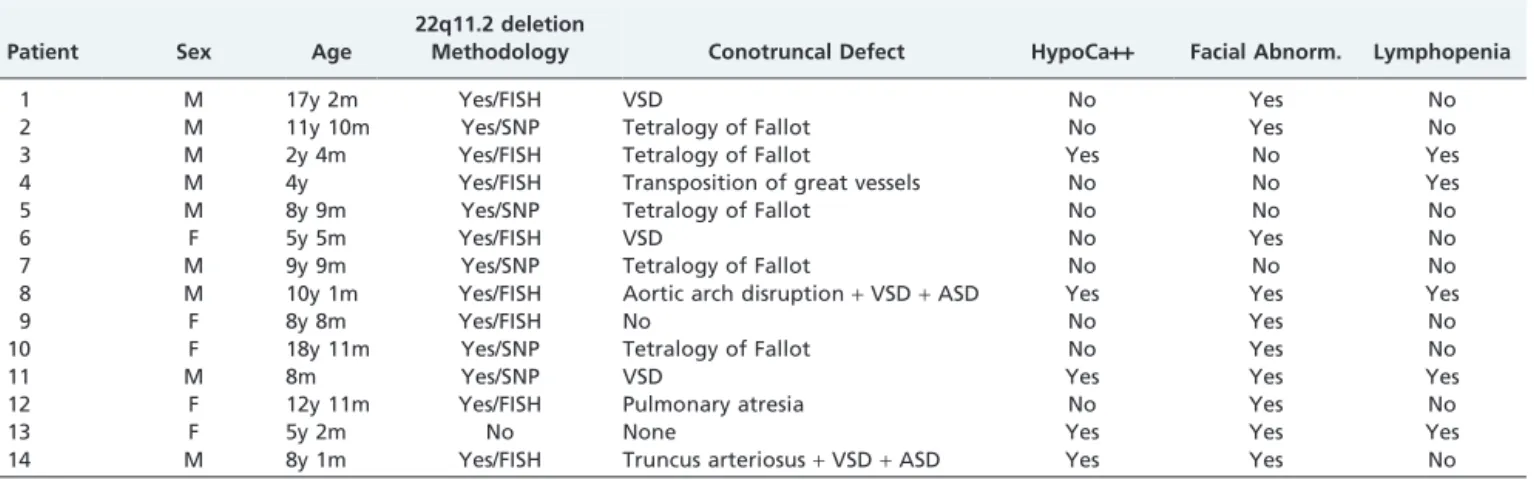
RESULTS:Of 14 patients (8m – 18y11m), only one did not have 22q11.2 deletion detected. The main findings were: conotruncal malformation (n = 12), facial abnormalities (n = 11), hypocalcemia (n = 5) and low lymphocyte count (n = 2).
CONCLUSION:The authors pointed out the necessity of DGS suspicion in all patient presenting with heart defects, facial abnormalities (associated or not with hypocalcemia), and immunological disorders because although frequency of DGS is high, few patients with a confirmed diagnosis are followed up.
KEYWORDS: DiGeorge syndrome; Immunologic deficiency syndromes; Thymus; 22q11.2 deletion.
Fomin ABF, Pastorino AC, Pereira AC, Kim CA, Carneiro-Sampaio M, Abe Jacob CM. DiGeorge Syndrome: a not so rare disease. Clinics. 2010;65(9):865-869.
Received for publication onMarch 18, 2010;First review completed onApril 5, 2010;Accepted for publication onJune 22, 2010
E-mail: [email protected]
Tel.: 55 11 3096-8585
INTRODUCTION
In spite of the high frequency of DiGeorge Syndrome (DGS), the commonest chromosome deletion syndrome, its diagnosis is often not suspected. Variable clinical pheno-types and different abnormalities may be caused by 22q11.2 deletion: thymus dysfunction, cardiac diseases, immunode-ficiency, and other clinical problems.1 In our country, few cases have been reported, but the clinical phenotypes associated with DiGeorge Syndrome should be well known so that patients receive an early diagnosis and correct treatment.2,3
Most patients with DGS have a partial form of the disease and thymic hypoplasia. This defect results in cellular immunodeficiency, although humoral defects have also
been described. Autoimmune diseases have been associated with DGS, probably in consequence of T cell regulatory defects and impaired central tolerance.4
Cardiac abnormalities, mainly conotruncal defects, have been found in DGS, and several authors have pointed out that isolated congenital heart disease may be associated with it.5
Parathyroid dysfunction may cause hypocalcemia and seizures in the neonatal period. This is considered a warning sign suggestive of DGS. Several authors recom-mend that newborns undergo screening for lymphopenia, which may assist in the early diagnosis of severe combined immunodeficiency and DGS.6
Most DGS patients are hemizygous for a 3Mb region on human chromosome 22 while others have a smaller 1.5Mb nested deletion. These observations suggest that haploin-sufficiency of one or more genes on human chromosome 22 are responsible for its etiology. This study describes the most important clinical phenotypes resulting from DGS and draws attention to the frequency of this disease and its diagnosis.
METHODS AND MATERIALS
Fourteen (14) patients with DGS were evaluated (8 males). The mean age was 8y10m (8m – 18y11m). All patients met the following inclusion criteria: small or normal number of circulating T cells, normal number of circulating B cells, normal or low serum immunoglobulin level, hypoparathyr-oidism, conotruncal defect, facial abnormalities, and 22q11.2 or 10p deletions.7 The patients were referred from the Genetic Unit and Instituto do Corac¸a˜o of Hospital das Clı´nicas of Universidade de Sa˜o Paulo (HC-FMUSP), Sa˜o Paulo, Brazil. This study was approved by the Ethics Committee of the institution where it was conducted, and all patients and controls signed an informed consent to participate in this study.
Clinical evaluation
All patients underwent a clinical and epidemiological protocol to collect information about personal characteristics and history, family history, heart disease, facial abnormal-ities, dimorphisms, neurological or behavioral disorders, recurrent infections, and other comorbidities. Patients were examined by the same investigators (an immunologist and a geneticist) and their anthropometric measurements were recorded.
Laboratory tests
The levels of serum triiodothyronine (T3), thyroxin (T4), free thyroxin (FT4), TSH, and thyroid antibodies (thyroglo-bulin and peroxidase) were measured. Parathyroid hor-mone, ionic or total calcium, and phosphorus levels were also measured. All patients had a complete blood count, and the Comans-Bitter values were used as reference to lymphocytes.8
Detection of 22q11.2 deletion
Eight (8) patients (described in Table 1) had microdele-tions in 22q11.2, detectable only by FISH (Fluorescence In Situ Hybridization), a technique that integrates the use of classical cytogenetics with molecular genetics, using DNA probes labeled with fluorescent material that identify specific regions of the genome.9,10 Two (2) were used as probes for this analysis: the N25 (which hybridizes at
D22S75, located within the region commonly deleted in SD22q11) and TUPLES 1 (which binds to any gene TUPLES and a part of the DNA located close to their ends).
The lost of heterozygosity detected by these markers could be used as a real tool of diagnosis in patients suspected of SD22q11. Gioli-Pereira et al.,11 researching
SNPs (single nucleotide polymorphisms) located in 22q11.2, in a Brazilian population, found these markers are efficient to determine the loss of heterozygosity in up to 92.9% of cases. Five (5) patients (IGBS, MASS, ASI, CSS and TPS) were referred from Instituto do Corac¸a˜o, their mutations having been detected by the SNPs technique following the procedures described by Gioli-Pereira et al.11
RESULTS
Table 1 shows clinical and laboratory data regarding diagnostic criteria. One patient (ABARS) did not have 22q11.2 deletion. She was included in the study because she met other criteria at the time of diagnosis: hypocalcemia, facial dimorphism, and lymphopenia, according to Notarangelo et al.7
Isolated or combined cardiac malformations were the most frequent findings, and 12 patients (86%) were affected with the followed distributions: 5 had tetralogy of Fallot; 5 ventricular septal defect (3 isolated and 2 associated with atrial septal defects); 1 isolated transposition of great vessels; 1 aortic arch disruption associated with septal defects; 1 truncus arteriosus associated with septal defects; and 1 pulmonary atresia. Only 4 patients did not need to undergo surgical correction of these malformations.
Eleven (11) patients (85.7%) had facial dimorphisms, and the most prevalent were oral defects, alone or in combina-tions. Microstomia was found in 64.3% (9/14) of the patients, and 57.2% (8/14) had palate defects, micrognathia, and short stature. Elongated fingers were observed in 50% of the patients; straight facial profile, dental abnormalities and over-folded ear helix in 42.8% (6/14); hypertelorism in 35.8% (5/14); and strabismus in 21.5% (3/14). One patient had a congenital clubfoot, which was corrected surgically before this study.
Seven (7) patients had recurrent infections, mainly pneumonia, and only one of them was free of any cardiac defect that might explain lung infections. Most infections
Table 1- Diagnostic criteria in 14 patients with DGS.
Patient Sex Age
22q11.2 deletion
Methodology Conotruncal Defect HypoCa++ Facial Abnorm. Lymphopenia
1 M 17y 2m Yes/FISH VSD No Yes No
2 M 11y 10m Yes/SNP Tetralogy of Fallot No Yes No
3 M 2y 4m Yes/FISH Tetralogy of Fallot Yes No Yes
4 M 4y Yes/FISH Transposition of great vessels No No Yes
5 M 8y 9m Yes/SNP Tetralogy of Fallot No No No
6 F 5y 5m Yes/FISH VSD No Yes No
7 M 9y 9m Yes/SNP Tetralogy of Fallot No No No
8 M 10y 1m Yes/FISH Aortic arch disruption+VSD+ASD Yes Yes Yes
9 F 8y 8m Yes/FISH No No Yes No
10 F 18y 11m Yes/SNP Tetralogy of Fallot No Yes No
11 M 8m Yes/SNP VSD Yes Yes Yes
12 F 12y 11m Yes/FISH Pulmonary atresia No Yes No
13 F 5y 2m No None Yes Yes Yes
14 M 8y 1m Yes/FISH Truncus arteriosus+VSD+ASD Yes Yes No
occurred in the first year of life, before cardiac surgery. Currently, none of them has any infection.
Laboratory findings
Five (35.8%) patients had hypocalcemia and four of them were under treatment for hypoparathyroidism. One had neonatal hypothyroidism since birth and is currently receiving hormonal treatment. No thyroid autoantibodies were found in any patients. All patients currently have normal leukocyte counts, but two have low lymphocyte counts, below the normal range for their age. These data are shown in Table 2.
DISCUSSION
Patients with 22q11.2 deletion syndromes, such as the velocardiofacial, Shprintzen and DiGeorge Syndromes may have several clinical abnormalities and different degrees of organ commitment.12The syndromes received the name of
the author that first described each one separately. Later, with the advance of the diagnostic methods in the field of genetics, it was found that the 22q11.2 chromosome was deleted in all of them. They are currently grouped as ‘‘the 22q11.2 deletion syndromes’’ because of the difficulties in choosing a single term.13
The 22q11.2 deletion is the most common human chromosome deletion, and its incidence is about 1:3000 live births.13 In this study, held in a teaching hospital and reference center for primary immunodeficiency, the number of patients available for inclusion in the study was small, which confirmed the difficulty that physicians have in diagnosing it and determining phenotypes.
The distribution of patient age in this series corroborates the difficulty of diagnosing DGS: only 28.5% of the patients were younger than 4 years, and most were older.
Currently, 180 clinical signs and symptoms, such as physical and behavioral abnormalities, have been described, but typical findings are not found in all cases.14 The facial phenotype, although easily recognized, may be subtle in some patients, and growth of the face and consequent accentuation of abnormalities become more noticeable as they grow older. DGS diagnoses are based on a set of clinical findings or 22q11.2 deletion, but 22q11.2 deletion is only investigated when there are signs suggesting that the patient may have the syndrome.
During the selection of patients in this study, the 22q11.2 deletion was detected in all but 1 subject. This patient, without heart disease, had a severe cleft palate that required several corrective surgeries, a small number of lymphocytes, and a history of neonatal hypocalcemia.
Diagnosis becomes more difficult when a patient with classic features of velocardiofacial syndrome or DGS has no evidence of deletion by FISH. A point mutation, which has been described in a few patients, might be present in T-box 1 (TBX1).15 This mutation, a deletion that is too small to be detected by standard FISH, or a non-chromosome 22 cause can also be associated with the same clinical manifestations as in chromosome 22q11.2 deletion syndrome.
Recently, MLPA–HD (multiplex ligation dependent probe amplification with high-density) was reported16 as a technique that detects copy number changes at 37 loci on velocardiofacial syndrome, the cat eye syndrome, and more distal regions in 22q11 that have been deleted. Novel and variant chromosome 22 aberrations have been detected by this methodology along with the common recurrent dele-tions associated with DiGeorge syndrome and velocardio-facial syndrome. Some authors described patients with relevant phenotypic characteristics where karyotype and FISH were normal. Only MLPA with high density analysis can detect uncommon deletions and establish the clinical diagnosis of 22q11 deletion.16,17
The International Union of Immunological Societies Primary Immunodeficiency Diseases Classification Commit-tee7proposed criteria to help to establish the diagnosis of DGS. These criteria refer primarily to immune disorders and include the number of circulating T cells and the most frequent clinical characteristics, such as hypoparathyroi-dism, conotruncal malformation, facial abnormalities, and chromosomes 22q11.2 or 10p deletion.
Oskarsdottir et al.18conducted a retrospective study with 100 patients that had DGS and reported the clinical findings of patients diagnosed before and after 2 years of age. The frequency of delayed diagnoses, also reported in their study, according to age groups was: 26% in the neonatal period; 17% between 2-5 years; 41% between 6-12 years; and 16% in adolescence, i.e., between 13-16 years. The mean age at diagnosis was 6.7 years. Heart disease was the finding that most frequently led to the diagnosis of DGS at any age, which was similar to our findings. For children diagnosed before the age of 2, recurrent infections and thymic
Table 2- Laboratory data of 14 patients with DGS.
Patient Free-T4 ng/dL TSHmU/mL
Parathyroid hormone pg /Ml
Ionic calcium mol/L
Total calcium mg/dL
Phosphorus mg/dL
Leucocytes Cels/mL
1 0.98 1.49 36 1.18 NP 6.1 9600
2 1.03 1.55 34 NP 8.9 4.9 9500
3 1.16 2.98 9* 1.29 8.9 6.6 2900
4 1.37 2.04 59 NP NP NP 5900
5 1.41 5.29 48 NP 8.9 5.3 6400
6 1.30 3.01 53 1.29 NP 4.4 13400
7 1.23 1.15 62 NP 8.5 5.2 4900
8 1.08 4.53 8* 1.22 NP 5.5 7500
9 1.22 5.30 48 1.25 NP 5.6 8000
10 1.07 1.75 NP NP 8.4 4.3 9400
11 1.23 0.90 15* 1.09 NP 7.01 12400
12 1.48 3.81 58 1.16 NP 4.5 5780
13 1.21 4.83 13* 1.18 8.6 6.5 6050
14 1.16 0.76 39 1.31 NP 4.7 5100
hypoplasia were the most frequent findings, followed by hypocalcemia. In the group that was diagnosed later, recurrent infections, soft palate defects, and delayed language acquisition were the most important signs and symptoms. Facial abnormalities were the least frequent in the two groups, which confirms the observation that these changes are established over a span of years.18
The comparison of our results with findings reported by Oskarsdottir18 revealed that only 28% of the patients were
diagnosed before the age of four years, and that heart malformations were the most prevalent findings, although cardiac defects were not the reason for referral to our institution. Jiang et al. suggested that even isolated congenital heart disease has a strong association with DGS, and that 22q11.2 deletion should be investigated in such cases.5
The frequency of cardiac malformations in patients with DGS ranges from 49% to 83%. Conotruncal defects are the most commonly-seen heart diseases, which suggests that patients may have the tetralogy of Fallot. In the Brazilian patients described here, the tetralogy of Fallot was found in 50% of the patients, in agreement with data in the literature.19,20The 22q11.2 deletion should be investigated in all patients with heart malformations, such as tetralogy of Fallot, disrupted aortic arch, septal defects, and truncus arteriosus. This is one of the reasons why pediatric cardiology centers, where the earliest diagnoses are made, often report case series.21,22This practice does not seem to
be usual in our country yet; the number of patients that undergo cardiac surgery to correct conotruncal defects is significant, the number of referrals to our service is small, and reports of patients with DGS are still scarce.
Our patients were older, and mouth, eye, and tooth abnormalities, as well as the perception of impaired speech development and behavior, are more easily noticed, as related by Butts.23 In schoolchildren, short stature, long fingers, long face, and a cylindrically-shaped nose are enough to raise the hypothesis of DGS.
Behavioral, neurological and psychiatric disorders are frequent in this syndrome. Two patients described here had symptoms of depression and required medication. Both patients underwent psychotherapy; despite treatment, they still do not have good school performance, and their family relations are inadequate. According to some authors, these patients should be followed for a long time, because depression and anxiety may precede the establishment of psychoses.24
When first described, DGS was characterized by severe immunodeficiency at birth. Other clinical features were not given much attention. The early onset of recurrent infections due to severe immunodeficiency is no longer the main clinical feature of DGS, because only patients with complete DGS have an increased susceptibility to infection. The number of lymphocytes may be normal, and immune disorders may appear only in adulthood. Currently, other clinical signs and symptoms are also important, such as neonatal hypocalcemia, hypothyroidism, and conotruncal defects, even in the absence of facial abnormalities or recurrent infections.
Infants with DGS may have breastfeeding difficulties due to several factors, such as hypotonia, cardiac problems, and airway obstruction secondary to jaw malformations. Many cases are not detected at birth or in childhood because DGS is a progressive disease and clinical signs that may appear
later in life. At least 30% of the patients do not have heart defects, and many cardiac malformations, such as right-sided aorta, are ‘‘silent’’ anomalies and have no obvious clinical signs or symptoms. Therefore, a diagnosis is only made if a specific genetic study is performed.14
CONCLUSION
Pediatricians, neonatologists, ENTs, and cardiac surgeons should be aware of the possible presence of congenital heart defects, especially conotruncal defects, neonatal hypocalce-mia, and defects in the palatal region at birth, which may be detected soon after birth. Other signs and symptoms, such as facial abnormalities and impaired development, may appear only years later and delay the diagnosis. This is the first report of Brazilian patients with DGS and, although the frequency of DGS is high, few patients with a confirmed diagnosis are followed up.
ACKNOWLEDGMENT
This work was supported by grants from Fundac¸a˜o Amparo a Pesquisa do Estado de Sa˜o Paulo (FAPESP 2008/58238-4).
REFERENCES
1. Kobrynski LJ, Sullivan KE. Velocardiofacial syndrome, DiGeorge syndrome: the chromosome 22q11.2.2 deletion syndrome. Lancet. 2007;370:1443-52, doi: 10.1016/S0140-6736(07)61601-8.
2. Araujo AC, Fortuna ES, Carneiro-Sampaio MMS, Grumach AS. Primary Immunodeficiency diseases: a presentation of 6 cases. Braz J med Biol Res. 1988;21:915-7.
3. Kokitsu-Nakata MN, Guion-Almeida ML, Richieri-Costa A. 22q11 deletion Syndrome and limb anomalies: report on two Brazilian patients. Cleft palate- Craniofacial J. 2008;45:561-6, doi: 10.1597/06-170.1. 4. Piliero LM, Sanford AN, McDonald-McGinn DM, Zackai EH, Sullivan
KE. T-cell homeostasis in humans with thymic hypoplasia due to chromosome 22q11.2 deletion syndrome. Blood. 2004;103:1020-25, doi: 10.1182/blood-2003-08-2824.
5. Jiang L, Hou Z, Duan C, Chen B, Chen Z, Li Y, Huan Y, et al. Isolated congenital heart disease is associated with the 22q11.2 deletion even though it is rare. Int J Cardiol. 2009 Nov 16. [Epub ahead of print] PubMed PMID: 19923022.
6. Routes JM, Grossman WJ, Versky J, Laessing RH, Hoffman GL, Brokopp CD. Statewide Newborn Screening for Severe T-Cell Lymphopenia. JAMA 2009;302:2465-70, doi: 10.1001/jama.2009.1806.
7. Notarangelo L, Casanova JL, Conley ME, Chapel H, Fischer A, Puck J, et al. Primary immunodeficiency diseases: An update from the I nte rn atio nal U ni on of I mm u nol o gi c a l So ci et ie s P ri mar y Immunodeficiency Diseases Classification Committee Meeting in Budapest, 2005. J Allergy Clin Immunol. 2006;117:883-96, doi: 10.1016/ j.jaci.2005.12.1347.
8. Comans-Bitter WM, De Groot R, Van den Beemd R, Neijens HJ, Hop WCJ, Groenveld K, et al. Immunophenotyping of blood lymphocytes in childhood. References values for lymphocytes subpopulations. J Pediatr. 1997;130:388-93.
9. Thomas JA, Graham JM Jr. Chromosome 22q11 deletion syndrome: an update and review for the primary pediatrician. Clin Pediatr (Phila). 1997;36:253-66, doi: 10.1177/000992289703600502.
10. Matsuoka R, Kimura M, Scambler PJ, Morrow BE, Imamura S, Minoshima S, et al. Molecular and clinical study of 183 patients with conotruncal anomaly face syndrome. Hum Genet.1998;103:70-80, doi: 10. 1007/s004390050786.
11. Gioli-Pereira L, Pereira AC, Mesquita SM, Lopes AA, Krieger JE. PCR screening for 22q11.2.2 microdeletion: development of a new cost-effective diagnostic tool. Clin Chim Acta. 2006;369:78-81, doi: 10.1016/j. cca.2006.01.005.
12. Shprintzen RJ, Higgins AM, Anstshel K, Fremont W, Roizen N, Kates W. Velo-cardio-facial syndrome. Cur Opin Pediatr. 2005;17:725-30, doi: 10. 1097/01.mop.0000184465.73833.0b.
13. Goodship J, Cross L, LiIing J, Wren C. A population study of chromosome 21q11 deletions in infancy. Arch Dis Child. 1998;79:348-51, doi: 10.1136/adc.79.4.348.
15. Yagi H, Furutani Y, Hamada H, Sasaki T, Asakawa S, Minoshima S, et al. Role of TBX1 in human del 22q11.2 syndrome. Lancet. 2003;362:1366–73, doi: 10.1016/S0140-6736(03)14632-6.
16. Jalali GR, Vorstman JAS, Errami AB, Vijzelaar R, Biegel J, Shaikh T, et al. Detailed analysis of 22q11.2 with a high density MLPA probe set. Human Mutation. 2008; 29:433-40, doi: 10.1002/humu.20640.
17. Fernandez L, Nevado J, Santos F, Heine-Sun˜er D, Martinez-Glez V, Garcia-Min˜aur S, et al. A deletion and a duplication in distal 22q11.2 deletion syndrome region. Clinical implications and review. BMC Medical Genetics. 2009;10:48, doi: 10.1186/1471-2350-10-48.
18. Oskarsdottir S, Vujic M, Fasth A. Incidence and prevalence for the 22q11.2 deletion syndrome: a population-based study in Western Sweden. Arch Dis Child. 2004;89:148-51, doi: 10.1136/adc.2003.026880. 19. Goldmuntz E, Clark BJ, Mitchell LE, Jawad AF, Cuneo BF, Reed L, et al.
Frequency of 22q11.2 deletions in patients with conotruncal defects. J Am Coll Cardiol. 1998;32:492-8, doi: 10.1016/S0735-1097(98)00259-9. 20. Sullivan KE. The clinical, immunological and molecular spectrum of
chromosome 22q11.2.2 deletion syndrome and DiGeorge syndrome.
Curr Opin Allergy Clin Immunol. 2004; 4:505-12, doi: 10.1097/00130832-200412000-00006.
21. Sullivan KE, McDonald-McGinn DM, Driscoll D, Emanuel BS, Zackai EH, Jawad AF. Longitudinal analysis of Lymphocyte function and numbers in the first year of life and chromosome 22q11.2.2 deletion syndrome (DiGeorge syndrome/velocardiofacial syndrome). Clin Labor Diag Immunol. 1999;6:906-11.
22. Chinen J, Rosenblatt HM, Smith EO, Shearer WT, Noroski LM. Long-term assessment of T-cell population in DiGeorge syndrome. J Allergy Clin Immunol. 2003;111:573-9, doi: 10.1067/mai.2003.165.
23. Butts SC. The facial phenotype of the velo-cardio-facial syndrome. Int J Pediatr Otorhinolaryngol. 2008; 73:343-50, doi: 10.1016/j.ijporl.2008. 10.011.