CdMoO
4Micro-ellipsoids: Controllable Synthesis, Growth Mechanism, and Photocatalytic
Activity
Ke Daia, Hui Liub, Tianyu Gaoa, Qi Wangc, Hao Chenb*
Received: November 30, 2015; Revised: February 20, 2016; Accepted: November 3, 2016
CdMoO4 micro-ellipsoids were synthesized by a simple hydrothermal route with the assistance of
nonionic surfactant Triton X-100 and characterized by X-ray difraction, scanning electron microscopy and UV-Vis difuse relectance spectroscopy. The efects of hydrothermal pH, temperature, and time on the morphology and photocatalytic activity of CdMoO4 were investigated. With an initial hydrothermal
pH of 5.00, CdMoO4 micro-ellipsoids were obtained at 180 °C for 24 h and found to possess the
highest photocatalytic activity—89% Rhodamine B can be degraded for 30 minutes presented in the 0.4 g/L CdMoO4 suspension. The formation mechanism of the CdMoO4 micro-ellipsoids was initiated by the formation of small nanoparticles and bulk structures afterwards, which was followed by the
growth of micro-ellipsoids. Experiment results showed that the evolution of the micro-ellipsoids was an Ostwald ripening process.
Keywords: photocatalysis, CdMoO4, controllable synthesis, Ostwald ripening
* e-mail: [email protected]
1. Introduction
The controllable synthesis of microscale and nanoscale
materials with special morphology, architecture and properties have rapidly developed into a promising ield in material
chemistry1-7.Controlling synthesis conditions can generate
diferent material structures, and changes in these structure directly afect their properties. Therefore, preparing materials with special morphologies and illustrating the growth mechanisms are particularly interesting, and vice versa, we
can control synthesis of the required materials once growth mechanisms are clear.
Metal molybdates (MMoO4, M=Zn2+, Ni2+, Pb2+, Cd2+, Sr2+,
et al.), as a kind of novel materials, are attracting increasing attention because of their excellent optical and electrical properties, high surface energy, numerous active sites, and high selectivity8-11, As an important metal molybdate, CdMoO
4
is receiving considerable attention because of its electronic excitation with vacuum ultraviolet synchrotron radiation12, pressure-induced phase transformations13, and photocatalytic activities14,15. Therefore, synthetic methods and conditions
of cadmium molybdate with high photocatalytic activity are signiicant to explore. Over the last few years, a few eforts have been exerted to the exploration some approaches
to the fabrication of CdMoO4 micro/nanostructures with special characters16,17. For example, Khademolhoseini et al.18 synthesized CdMoO4 with an ultrasonic method and found
that the size and morphology of the products were greatly inluenced by the dosage of ultrasonic power. Wang et al.19
prepared 3D CdMoO4 hierarchical structures from nanoplates
with enhanced photocatalytic eiciency in the degradation of Rhodamine B under ultraviolet (UV) light irradiation. Li et al.20 introduced Cl- to the hydrothermal process and successfully obtained self-assembled CdMoO4 microspheres that were used
to degrade methylene blue. Li and Gong21 prepared octahedral CdMoO4 through diferent microemulsion systems. Wang et
al.22 designed a kind of CdMoO
4 microspheres with a
core-shell structure and investigated the photocatalytic activity and luorescence properties of the microspheres. Liu et al.23 synthesized CdMoO4 nanorods with the diameter of 30-50
nm at a relatively low temperature.
Based on the above mentioned studies, considerable
attention has been paid to the controllable synthesis of
molybdate cadmium with special morphology. However, studies on the relationship among the synthesis conditions (pH, temperature, and reaction time), morphologies, and photocatalytic activity of molybdate cadmium are few. In this work, we demonstrated a simple and eicient hydrothermal procedure for fabricating micro-ellipsoids CdMoO4 by
simply reacting ammonium molybdate with cadmium nitrate aqueous solution in the presence of Triton X-100.
Micro-ellipsoids CdMoO4 were selectively prepared by
adjusting the hydrothermal temperature, reaction time, and pH. The photocatalytic activities of CdMoO4 with various
morphologies synthesized under diferent conditions were comparatively investigated by Rhodamine B (RhB) degradation. The growth process of micro-ellipsoids CdMoO4 was also
investigated in detail.
a College of Resources and Environment, Huazhong Agricultural University, Hubei, Wuhan, China
b College of Science, Huazhong Agricultural University, Hubei, Wuhan, China
c School of Environment Sciences and Engineering, Zhejiang Gongshang University, Zhejiang,
37 CdMoO4 Micro-ellipsoids: Controllable Synthesis, Growth Mechanism, and Photocatalytic Activity
2. Experimental
2.1. Materials
Cadmium acetate (Cd(NO3)2•4H2O, AR.), ammonium
molybdate ((NH4)6Mo7O24•4H2O, AR.) and Triton X-100
(OP, CP.) were all purchased from Sinopharm Chemical Reagent Co. Ltd. without further puriication. The water used in this experiment was deionized water.
2.2. Preparation of CdMoO
4In a typical synthetic procedure, 14 mL 0.14 mol/L
Cd(NO3)2•4H2O aqueous solution was completely dropwise
dispersed in the mixed solution of 14 mL 0.02 mol/L (NH4)6Mo7O24•4H2O and Triton X-100 under vigorous
stirring. Subsequently, the mixed solution was adjusted to various pH value (1.73, 3.00, 5.00, 7.00, 9.00, and 11.00) with an ammonia solution or nitric acid solution. Then, the mixture was transferred into a Telon-lined stainless steel autoclave and heated in the oven according to the setting time (1, 4, 8, 12, 16, 20, 24, 36, and 48 h) and temperature
(25, 50, 70, 80, 100, 140, and 180 oC). Unless otherwise
stated, the reaction conditions of pH, temperature, and time are set at pH=5.00, 180 oC for 24 h. Finally, the resultant
solid was separated by centrifugation and washed with
deionized water and ethanol three times and then dried at
80 °C for 8 h in the air.
2.3. Characterizations
X-ray difraction (XRD) patterns were collected on a Bruker D8 advance X-ray difractometer with Cu Kα radiation (λ=0.15406 nm). The UV-vis difuse relectance
spectroscopy of power solids were carried out by UV-Vis
spectrophotometer (UV-3100, Japan), made by Japan. The morphologies were studied with a JSM-6390LV scanning electron microscope (SEM).
2.4. Photocatalytic experiments
Photocatalytic activities were evaluated by the degradation of RhB under UV light irradiation of an 18 W mercury lamp (average light intensity of 14.5 μW/cm2). In each
experiment, 20 mg of photocatalysts was added into 50 mL of RhB solution (1×10⁻⁵ mol/L). Before irradiation, the
suspensions were magnetically stirred in the dark for 30 min to achieve the adsorption equilibrium. Then the suspensions were exposed to UV light irradiation. At given irradiation time intervals, 3 mL of the suspension was collected and centrifuged to remove the photocatalyst. The centrifuged
solution was analyzed by a Nicolet 300 evolution UV-vis
spectrophotometer, monitoring the characteristic absorption peak of RhB at 553 nm.
3. Results and discussion
3.1. XRD analyses of CdMoO
4microcrystals
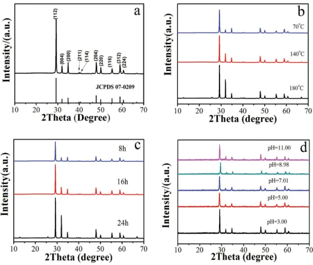
Figure 1 shows the XRD patterns of CdMoO4 microcrystals.
Figure 1b-1d show the XRD patterns of various CdMoO4
products prepared at diferent hydrothermal temperatures, times, and pH values, respectively. All difraction peaks of the XRD patterns (Figure 1b-1d) can be ascribed to
tetragonal-phase CdMoO4 (JCPDS no: 07-0209). The peaks at 29.2°,
31.9°, 34.8°, 47.9° and 59.0° show an excellent match with the (112), (004), (200), (204), (312) crystal planes of tetragonal-phase CdMoO4, indicating that all samples were
tetragonal-phase CdMoO4 (Figure 1a). Figure 1b shows that
the difraction peak intensity strengthens and sharpens with increased temperature, indicating the crystallinity increases with
increased temperature and time24,25. Moreover, the difraction peak intensity ratios (I004/I200) of the (004) and (200) crystal
planes are calculated to be 0.7, 1.9, and 3.6 for the sample prepared at 70, 140, and 180 °C, respectively. Therefore, the microcrystals undergo special anisotropic growth along the c-axis with increased hydrothermal temperature from 70 °C
to 180 °C26,27. Figure 1c shows that the crystallinity increases
with increased time, indicating special anisotropic growth along the c-axis with prolonged hydrothermal time from 8 h to 24 h. Figure 1d shows that the crystals’ orientation does not signiicantly change when the pH is changed, but pH has an important efect on morphology of CdMoO4 crystals
16, as
also can be investigated in Section 3.2. Thus, crystal-growth orientation, morphology and crystallinity can be controlled by hydrothermal temperature, reaction time, and pH.
3.2. CdMoO
4morphologies analyses
The SEM images of CdMoO4 products obtained at
diferent hydrothermal temperatures for 24 h are shown in Figure 2. The hydrothermal temperature signiicantly afects the morphology of products. CdMoO4 microparticles derived
at 50 °C with an irregular shape can be observed in Figure 2a. The panoramic view in Figure 2b clearly illustrates that
CdMoO4 hydrothermally derived at 70 °C mostly consists of relatively uniform microspheres with diameters of 2-8
μm, coexisting with many irregular nanoparticles. One
individual microsphere, as can be clearly observed from the
inset in Figure 2b, has a well-deined spherical shape with a diameter of ca. 7 μm. Figure 2c shows that the microspheres derived at 80 °C possibly consist of aggregations containing nanoparticles and nanosheets. Similar rod-like morphologies
of the products derived at 100 and 140 °C can be clearly
Figure 1: XRD pattern of the as-prepared CdMoO4 products. (a) JCPDS 07-0209, (b) temperature series at pH=5 for 24 h, (c) time series
at pH=5 and 180 oC, and (d) pH series at 70 oC for 24 h.
Figure 2: SEM images of CdMoO4 products obtained at pH=5 for 24 h under diferent hydrothermal temperatures. (a) 50 oC, (b) 70 oC,
39 CdMoO4 Micro-ellipsoids: Controllable Synthesis, Growth Mechanism, and Photocatalytic Activity
Many researchers reported that CdMoO4 materials have been synthesized at a low temperature16,21,22. To obtain CdMoO
4
materials with high crystallinity degree at a relatively benign
synthetic condition, we synthetized CdMoO4 particles at 180
°C. Moreover, the growth process of CdMoO4 at 180 °C was
investigated in this work, SEM images of CdMoO4 products
obtained at diferent growth stages at 180 °C are shown in Figure 3. After 1 h of hydrothermal reaction, products with square pieces are observed (Figure 3a), and the map inset is a well-deined square piece with a side length of ~3.6 μm and a thickness of ~500 nm. Apparently, the most exposed surface of the square piece is the crystal facet. As the reaction proceeded, a round pie shape with an average thickness of ~1.5 μm and diameters ranging within 2-3 μm is observed (Figure 3b). When the reaction time is prolonged to 8 h, the morphology type is still round pie shape but with an average thickness of ~1.2 μm and an average diameter of 3.3 μm (Figure 3c). When the reaction time is 1-8 h, morphological
evolution of the crystal indicates that CdMoO4 grows
layer-by-layer from the sheet structure to round pie shape along the c-axis. Therefore, CdMoO4 crystals grow along the c-axis perpendicular to the crystal facet, as can be observed from
the SEM images (Figure 3a-3c). Finally, when the reaction time is further extended to 16, 24, even 48 h, regular crystal structures began to collapse or even seriously agglomerate, as shown in Figure 3d-3f.
CdMoO4 microcrystals obtained at diferent pH values after hydrothermal treatment at 180 °C for 24 h were carefully
examined by SEM. At pH 1.73, irregular nanoscale shapes are observed (Figure 4a). At pH 3.00, many nanospheres exist on the surface of block structures (Figure 4b). At pH 5.00, the as-prepared CdMoO4 crystallites are well-deined
micro-ellipsoids 3-5 μm in size (Figure 4c). At pH 7.00, the
morphology is ellipsoid but many nanoparticles exist on their surface (Figure 4d). At pH 9.00, irregular lumps can be observed (Figure 4e). “Floccule”-like CdMoO4 crystallites
appear at pH 11.00 (Figure 4f).
3.3. Optical properties, photocatalytic activities
and photostability
Optical absorption of the CdMoO4 microparticles
was measured by using an UV-vis spectrometer. Figure 5 presents a typical UV-vis difuse relectance absorption spectra (DRS) of CdMoO4 products. The optical absorption of the CdMoO4 microcrystals was nearly the same except the
sample obtained at pH=11.00 (Figure 5c). All samples show a great increase in absorbance with wavelengths lower than
ca. 380 nm, which indicated that the absorption was not due to the transition from the impurity level but was due to the
band-gap transition28. The sample prepared at pH=11.00 have
two steep shape, one was the bad-gap transition of CdMoO4,
the other may be the band-gap transition of Cd(OH)2
29.
The photocatalytic activities of the samples were
evaluated by RhB degradation. The photodegradation eiciencies of RhB as a function of irradiation time for
CdMoO4 microcrystals under UV light illumination are
depicted in Figure 6. At pH=5.00, the product derived at
180 °C for 24 h showed the best photocatalytic performance
which can efectively degrade 89% RhB within 30 min.
Moreover, the photocatalytic activities of CdMoO4 products
changed obviously with changing reaction temperature and time. However, the changes in the photocatalytic activities
of CdMoO4 products prepared under diferent pH are not
signiicant. It is well-known that the photocatalytic activity
of catalyst is related to its phase compositions, structures, and
Figure 3: SEM images of CdMoO4 products obtained at pH=5 and 180 oC for diferent hydrothermal times at 180 oC. (a) 1 h, (b) 4 h, (c)
Figure 4: SEM images of CdMoO4 products obtained for diferent pH for 24 h at 180 oC. (a) pH=1.73, (b) pH=3.00, (c) pH=5.00, (d)
pH=7.00, (e) pH=9.00, and (f) pH=11.00.
41 CdMoO4 Micro-ellipsoids: Controllable Synthesis, Growth Mechanism, and Photocatalytic Activity
Figure 6: Photodegradation eiciencies of RhB as a function of irradiation time over CdMoO4 products synthesized under various reaction
conditions. (a) temperature series at pH=5 for 24 h, (b) time series at pH=5 and 180 oC, (c) pH series at 180 oC for 24 h, and (d) repeated
experiment of RhB photodegradation over CdMoO4 products.
morphologies, which are greatly inluenced by the synthesis
conditions30-33. It can be noticed that the photocatalytic activities of CdMoO4 products are highly related to its crystallinity
(Figure 1b-1d) except for the CdMoO4 products prepared
under diferent pH. Crystallinity has a certain efect on the photocatalytic activity. The higher the crystallinity, the fewer the bulk defects, and the higher the photocatalytic activity is. Therefore, the CdMoO4 products with high crystallinity
demonstrated excellent photocatalytic activity. On the other hand, the morphologies and exposed facets of photocatalyst
are also important to its photocatalytic activity34-36. Based on the results of this study, the photocatalytic activity of CdMoO4 is closely related to not only the crystallinity but
also the facet.
The stability of a photocatalyst is also important
for its applications. In this work, circulating runs in the
photodegradation of RhB by CdMoO4 micro-ellipsoids
are examined under UV irradiation without washing the photocatalyst. The photoactivity of CdMoO4 remains at
78% after 10 recycles of RhB photodegradation, as shown in Figure 6d, indicating that the prepared CdMoO4 materials
have moderate photostability.
3.4. Formation mechanism of CdMoO
4micro-ellipsoids
The typical SEM images and XRD patterns of CdMoO4
micro-ellipsoids prepared at 180 °C and pH 5.00 for 24 h are shown in Figure 7. The low-magniication SEM images in Figure 7a show that the product contains numerous micro-ellipsoids with a long axis of about 2-6 μm, indicating that micro-ellipsoids can be prepared on a large scale by the proposed easy method. One individual micro-ellipsoid (Figure 7b) is shown to have a well-deined ellipsoid 6 μm in size, and this ellipsoid is assembled by a nanotablet ~200 nm in size. The high-magniication SEM images further
reveal that these micro-ellipsoids are actually composed
of many plate-like microcrystals (Figure 7c). The XRD pattern of the micro-ellipsoids is displayed in Figure 7d. All difraction peaks can be ascribed to scheelite-type tetragonal
CdMoO4 (JCPDS No. 07-0209), and no impurity peak can
be observed, indicating the formation of pure scheelite-type tetragonal CdMoO4.
To reveal the growth process of CdMoO4
Figure 7: Typical SEM images with diferent magniications (a-c) and XRD patterns (d) of CdMoO4 products obtained at pH=5.00, 180 oC for 24 h.
reaction, and then their morphologies were investigated by SEM. The evolutionary stages are shown in Figure 8. After
1 h of hydrothermal reaction, products with microspheres
and several nanoparticles are observed (Figure 8a). With prolonged reaction time to 4 h, only nanoparticles exist (Figure 8b); at 8 h, smaller nanoparticles are observed (Figure 8c). Further increased reaction time leads to the formation of bulk structures (Figure 8d), and then micro-ellipsoids are observed on the bulks (Figure 8e). Finally, when the reaction time is extended to 24 h, micro-ellipsoids were appearing (Figure 8f). However, when the reaction time is keeping
for 48 h, microspheres and several nanoparticles are both
observed (Figure 8g).
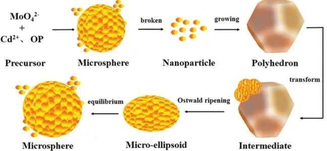
Based on the above observations, a schematic of the
formation mechanism of CdMoO4 ellipsoids is shown in
Figure 9. In the irst step, a cadmium molybdate crystal
nucleus forms from molybdate ion and cadmium ions, and
then an unstable globular structure forms. In the second step, this globular structure breaks into nanoparticles. In the third step, an irregular polyhedron structure forms at the expense of the small particles. In the fourth step, elliptic spherical structures grow on the irregular polyhedron, so the number of irregular polyhedrons decrease and the number of elliptic spherical structures increase. Finally micro-ellipsoids form. After a long time, stable spheres inally appear. The entire process that the CdMoO4
crystals undergo is the Ostwald ripening process37-39. This
spontaneous process occurs because larger particles are more energetically favored than smaller particles. The
formation of many small particles is kinetically favored
(i.e., they are more easily to nucleate). However, large
particles are thermodynamically favored because small
particles have a larger ratio of surface area to volume than large particles and are thus easier to produce. The
competition between thermodynamics and kinetics
drives the crystal-transformation process. Molecules on the surface are energetically less stable than the ones already well ordered and packed in the interior. Large particles, with their greater ratio of volume to surface area, thus have a lower energy state. Hence, many small particles attain a lower energy state if transformed into large particles. Finally the spheres and particles reach the equilibrium state.
4. Conclusions
CdMoO4 micro-ellipsoids were synthesized at an
initial hydrothermal pH of 5.00 and temperature of 180 °C for 24 h in the presence of Triton X-100. Comparative analysis of diferent series of CdMoO4 materials reveals
43 CdMoO4 Micro-ellipsoids: Controllable Synthesis, Growth Mechanism, and Photocatalytic Activity
Figure 8: SEM images of CdMoO4 products obtained at diferent hydrothermal times at pH=5.00, 180
oC. (a) 1 h, (b) 4 h, (c) 8 h, (d) 16
h, (e) 20 h, (f) 24 h, and (g) 48 h.
Figure 9: Schematic illustration of the growth process of CdMoO4 micro-ellipsoids.
The crystal facet and crystallinity of CdMoO4 play a key
part in its photocatalytic activity. Moreover, the formation
mechanism of CdMoO4 micro-ellipsoids was investigated,
which is related to an Ostwald ripening process. Notably, the
CdMoO4 micro-ellipsoids exhibit excellent photocatalytic
5. Acknowledgments
The research was inancially supported by the National Natural Science Foundation of China (51572101) and the
Fundamental Research Funds for Central Universities
(2662015PY047) of China.
6. References
1. Dong M, Lin Q, Sun H, Chen D, Zhang T, Wu Q, et al. Synthesis of Cerium Molybdate Hierarchical Architectures and Their Novel Photocatalytic and Adsorption Performances. Crystal Growth & Design. 2011;11(11):5002-5009.
2. Zhang N, Liu S, Xu YJ. Recent progress on metal core@ semiconductor shell nanocomposites as a promising type of photocatalyst. Nanoscale. 2012;4(7):2227-2238.
3. Zhang L, Zhu Y. A review of controllable synthesis and enhancement of performances of bismuth tungstate visible-light-driven photocatalysts.Catalysis Science & Technology. 2012;2(4):694-706.
4. Li Y, Mi Y, Jiang J, Huang Z. Room-temperature Synthesis of
CdMoO4 Nanooctahedra in the Hemline Length of 30 nm.
Chemistry Letters. 2010;39(7):760-761.
5. Zhang Q, Wang W, Goebl J, Yin Y. Self-templated synthesis of hollow nanostructures. Nanotoday. 2009;4(6):494-507. 6. Lin G, Zheng J, Xu R. Template-Free Synthesis of Uniform
CdS Hollow Nanospheres and Their Photocatalytic Activities. Journal of Physical Chemistry C. 2008;112(19):7363-7370. 7. Sczancoski JC, Bomio MDR, Cavalcante LS, Joya MR, Pizani
PS, Varela JA, et al. Morphology and Blue Photoluminescence Emission of PbMoO4 Processed in Conventional Hydrothermal.
Journal of Physical Chemistry C. 2009;113(14):5812-5822.
8. Adhikari R, Gyawali G, Kim TH, Sekino T, Lee SW. Synthesis of Er3+ loaded barium molybdate nanoparticles: A new approach
for harvesting solar energy. Materials Letters. 2013;91:294-297. 9. Carlsson PA, Jing D, Skoglundh M. Controlling Selectivity in
Direct Conversion of Methane into Formaldehyde/Methanol over Iron Molybdate via Periodic Operation Conditions. Energy & Fuels. 2012;26(3):1984-1987.
10. Zhang M, Shao C, Zhang P, Su C, Zhang X, Liang P, et al. Bi2MoO6 microtubes: Controlled fabrication by using electrospun
polyacrylonitrile microibers as template and their enhanced visible light photocatalytic activity. Journal of Hazardous Materials. 2012;225-226:155-163.
11. Shen M, Zhang X, Dai K, Chen H, Peng T. Hierarchical
PbMoO4 microspheres: hydrothermal synthesis, formation
mechanism and photocatalytic properties. CrystEngComm. 2013;15:(6):1146-1152.
12. Mikhailik VB, Kraus H, Wahl D, Mykhaylyk MS. Studies of electronic excitations in MgMoO4, CaMoO4 and CdMoO4
crystals using VUV synchrotron radiation. Physica Status Solidi (B). 2005;242(2):R17-R19.
13. Jayaraman A, Wang SY, Sharma SK. High-pressure Raman investigation on CdMoO4 and pressure-induced phase
transformations. Physical Review B. 1995;52(14):9886-9889.
14. Adhikari R, Malla S, Gyawali G, Sekino T, Lee SW. Synthesis,
characterization and evaluation of the photocatalytic performance
of Ag-CdMoO4 solar light driven plasmonic photocatalyst.
Materials Research Bulletin. 2013;48(9):3367-3373.
15. Madhusudan P, Zhang J, Cheng B, Yu J. Fabrication of CdMoO4@
CdS core–shell hollow superstructures as high performance visible-light driven photocatalysts. Physical Chemistry Chemical Physics. 2015;17(23):15339-15347.
16. Ren Y, Ma J, Wang Y, Zhu X, Lin B, Liu J, et al. Shape-Tailored Hydrothermal Synthesis of CdMoO4 Crystallites on Varying
pH Conditions. Journal of the American Ceramic Society. 2007;90(4):1251-1254.
17. Wang WS, Zhen L, Xu CY, Shao WZ, Chen ZL. Formation
of CdMoO4 porous hollow nanospheres via a self-assembly
accompanied with Ostwald ripening process and their photocatalytic performance. CrystEngComm. 2013;15(39):8014-8021.
18. Khademolhoseini S, Zakeri M, Rahnamaeiyan S, Nasiri M, Talebi R. A simple sonochemical approach for synthesis of cadmium molybdate nanoparticles and investigation of its photocatalyst application. Journal of Materials Science: Materials in Electronics. 2015;26(10):7303-7308.
19. Wang W, Cui J, Wang P, Zhen L, Shao W, Chen Z.
Self-supported construction of 3D CdMoO4 hierarchical structures
from nanoplates with enhanced photocatalytic properties.RSC Advances. 2014;4(73):38527-38534.
20. Li D, Zhu Y. Synthesis of CdMoO4 microspheres by
self-assembly and photocatalytic performances.CrystEngComm. 2012;14(3):1128-1134.
21. Gong Q, Li G, Qian X, Cao H, Du W, Ma X. Synthesis of single
crystal CdMoO4 octahedral microparticles via
microemulsion-mediated route. Journal of Colloid and Interface Science. 2006;304(2):408-412.
22. Wang WS, Zhen L, Xu CY, Shao WZ. Room Temperature Synthesis, Growth Mechanism, Photocatalytic and Photoluminescence Properties of Cadmium Molybdate Core−Shell Microspheres. Crystal Growth & Design. 2009;9(3):1558-1568.
23. Liu H, Tan L. Synthesis, structure, and electrochemical properties
of CdMoO4 nanorods. Ionics. 2010;16(1):57-60.
24. Yu J, Wang B. Efect of calcination temperature on morphology
and photoelectrochemical properties of anodized titanium
dioxide nanotube arrays. Applied Catalysis B: Environmental. 2010;94(3-4):295-302.
25. Shamaila S, Sajjad AKL, Chen F, Zhang J. Mesoporous titania with high crystallinity during synthesis by dual template system as an eicient photocatalyst. Catalysis Today. 2011;175(1):568-575. 26. McLaren A, Valdes-Solis T, Li G, Tsang SC. Shape and size
efects of ZnO nanocrystals on photocatalytic activity. Journal of the American Chemical Society. 2009;131(35):12540-12541. 27. Umar A, Ribeiro C, Al-Hajry A, Masuda Y, Hahn YB. Growth
of highly c-axis-oriented ZnO nanorods on ZnO/glass substrate: growth mechanism, structural, and optical properties. Journal of Physical Chemistry C. 2009;113(33):14715-14720.
28. Kudo A, Tsuji I, Kato H. AgInZn7S9 solid solution photocatalyst
for H2 evolution from aqueous solutions under visible light
45 CdMoO4 Micro-ellipsoids: Controllable Synthesis, Growth Mechanism, and Photocatalytic Activity
29. Gujar TP, Shinde VR, Kim WY, Jung KD, Lokhande CD, Joo OS. Formation of CdO ilms from chemically deposited Cd(OH)2 ilms as a precursor. Applied Surface Science.
2008;254(13):3813-3818.
30. Dai K, Yao Y, Liu H, Mohamed I, Chen H, Huang Q. Enhancing the photocatalytic activity of lead molybdate by modifying with fullerene. Journal of Molecular Catalysis A: Chemical.
2013;374-375:111-117.
31. Zhu H, Wang J, Wu D. Fast Synthesis, Formation Mechanism, and Control of Shell Thickness of CuS Hollow Spheres. Inorganic Chemistry. 2009;48(15):7099-7104.
32. Shen M, Zhang Q, Chen H, Peng T. Hydrothermal fabrication
of PbMoO4 microcrystals with exposed (001) facets and
its enhanced photocatalytic properties. CrystEngComm. 2011;13(7):2785-2791.
33. D’Arienzo M, Carbajo J, Bahamonde A, Crippa M, Polizzi S, Scotti R, et al. Photogenerated Defects in Shape-Controlled TiO2
Anatase Nanocrystals: A Probe To Evaluate the Role of Crystal Facets in Photocatalytic Processes. Journal of the American Chemical Society. 2011;133(44):17652-17661.
34. Pan J, Liu G, Lu GQ, Cheng HM. On the True Photoreactivity
Order of {001}, {010}, and {101} Facets of Anatase TiO2 Crystals.
Angewandte Chemie International Edition. 2011;50(9):2133-2137. 35. Han X, Kuang Q, Jin M, Xie Z, Zheng L. Synthesis of titania
nanosheets with a high percentage of exposed (001) facets and related photocatalytic properties. Journal of the American Chemical Society. 2009;131(9):3152-3153.
36. Yang HG, Sun CH, Qiao SZ, Zou J, Liu G, Smith SC, et al.
Anatase TiO2 single crystals with a large percentage of reactive
facets. Nature. 2008;453:638-641.
37. Bastús NG, Comenge J, Puntes V. Kinetically controlled seeded growth synthesis of citrate-stabilized gold nanoparticles of up to 200 nm: size focusing versus Ostwald ripening. Langmuir. 2011;27(17):11098-11105.
38. Mahesh S, Gopal A, Thirumalai R, Ajayaghosh A. Light-induced Ostwald ripening of organic nanodots to rods. Journal of the American Chemical Society. 2012;134(17):7227-7230. 39. Johnson NJJ, Korinek A, Dong C, van Veggel FCJM.