Cop
yright
© ABE&M t
odos os dir
eit
os r
eser
vados
.
Cop
yright
© ABE&M t
odos os dir
eit
os r
eser
vados
.
Bone disease in hypoparathyroidism
Doença óssea no hipoparatiroidismo
Bart L. Clarke1
ABSTRACT
Hypoparathyroidism is a rare disorder that may be acquired or inherited. Postsurgical hypo-parathyroidism is responsible for the majority of acquired hypohypo-parathyroidism. Bone disease occurs in hypoparathyroidism due to markedly reduced bone remodeling due to the absence or low levels of parathyroid hormone. Chronically reduced bone turnover in patients with hy-poparathyroidism typically leads to higher bone mass than in age- and sex-matched controls. Whether this increased bone density reduces fracture risk is less certain, because while incre-ased bone mineralization may be associated with increincre-ased brittleness of bone, this does not appear to be the case in hypoparathyroidism. Treatment of hypoparathyroidism with recombi-nant parathyroid hormone may reduce bone mineral density but simultaneously strengthen the mechanical properties of bone. Arq Bras Endocrinol Metab. 2014;58(5):545-52
Keywords
Hypoparathyroidism; bone disease; bone mineral density; bone histomorphometry; fractures; skeleton
RESUMO
O hipoparatireoidismo é uma enfermidade rara que pode ser adquirida ou herdada. Dentre as causas adquiridas dessa doença, destaca-se como maior responsável o hipoparatireoidismo pós-cirúrgico. As manifestações ósseas dessa patologia decorrem devido a uma diminuição marcada no remodelamento ósseo causada pela diminuição ou ausência do hormônio da pa-ratireoide. Esse remodelamento ósseo cronicamente reduzido leva a um aumento da massa óssea, evidenciado quando indivíduos com hipoparatireoidismo são comparados a controles de mesma idade e sexo. Entretanto, não se sabe se esse aumento de massa óssea reduz o risco de fratura. Apesar de o aumento da massa óssea estar associado a um aumento da fra-gilidade óssea em algumas situações, este não parece ser o caso no hipoparatireoidismo. O tratamento com hormônio recombinante da paratireoide pode reduzir a densidade mineral óssea ao mesmo tempo em que melhora as propriedades mecânicas do osso. Arq Bras Endocrinol Metab. 2014;58(5):545-52
Descritores
Hipoparatiroidismo; doença óssea; densidade mineral óssea; histomorfometria óssea; fraturas; esqueleto
1 Department of Internal Medicine
and the Division of Endocrinology, Diabetes, Metabolism, and Nutrition, Mayo Clinic College of Medicine, Rochester, Minnesota, United States
Correspondence to:
Bart L. Clarke
Division of Endocrinology, Diabetes, Metabolism, and Nutrition, Mayo Clinic East 18-A,
200 First Street SW, 55905 – Rochester, Minnesota, United States
Received on Apr/3/2014 Accepted on May/14/2014
DOI: 10.1590/0004-2730000003399
INTRODUCTION
H
ypoparathyroidism is a rare disorder characteri-zed by the presence of decreased serum calcium and absent or inappropriately decreased serum para-thyroid hormone (PTH). This condition is most often acquired, but may also be inherited (1). The acquired form is most often due to removal of, or damage to, the parathyroid glands or their blood supply at the time of neck surgery for thyroid disease, head and neck cancer, or parathyroid disease. Postsurgical hypoparathyroi-dism is responsible for about 75% of acquired cases (2). The next most common cause in adults is thought tobe autoimmune disease, affecting either only the para-thyroid glands, or the parapara-thyroid glands in addition to multiple other endocrine organs. Remaining cases are due to a variety of rare iniltrative disorders, metastatic disease, iron or copper overload, ionizing radiation ex-posure, or rare genetic disorders.
incre-Cop
yright
© ABE&M t
odos os dir
eit
os r
eser
vados
.
ased bone density reduces fracture risk is less certain, because increased bone mineralization may be associa-ted with increased brittleness of bone, although this does not appear to be the case in hypoparathyroidism. Treatment of hypoparathyroidism with recombinant parathyroid hormone may reduce bone mineral density (BMD) but concomitantly strengthen bone.
BONE MINERAL DENSITY
Bone mineral density is usually upper-normal or incre-ased in patients with chronic hypoparathyroidism. One of the irst studies that showed this used single photon absorptiometry to measure skeletal mass in the pro-ximal femur, lumbar spine, and distal radius in 19 fe-males with hypoparathyroidism after surgery for either thyroid carcinoma or hyperparathyroidism (6). Healthy subjects, as well as normocalcemic patients who had undergone the same surgical procedure without de-veloping hypoparathyroidism, were used as controls. Skeletal mass was measured after a mean postoperative interval of 13 years in patients operated on for thyroid carcinoma, and 10 years in patients operated on for hyperparathyroidism. Bone mass was 10-32% greater in hypoparathyroid patients than in controls. In patients who retained parathyroid function after total thyroi-dectomy or surgical treatment of hyperparathyroidism, bone mass did not differ from that in age-matched healthy controls. Long term L-thyroxine treatment using doses that suppressed endogenous TSH produc-tion was not associated with decreased bone mass in this study, although other studies have not found this (12,13). Reduced PTH production, vitamin D treat-ment, and calcium supplementation were all thought to have contributed to the increased bone mass found in the patients with postsurgical hypoparathyroidism.
Greater insight into the architectural basis of the in-crease in bone mass in hypoparathyroidism has been obtained by using peripheral quantitative computed tomography (pQCT). Using this technique, Chen and cols. (14) compared volumetric bone mineral density (vBMD) and geometry of the distal radius and mid-ra-dius among postmenopausal women with postoperati-ve or idiopathic hypoparathyroidism, primary hyperpa-rathyroidism, and normal control individuals. At the 4% distal radius site, which is enriched in cancellous bone, trabecular vBMD was higher in the patients with hy-poparathyroidism, lower in controls, and lowest in pa-tients with primary hyperparathyroidism. At the 20%
mid-radius site, cortical vBMD also was greater in hypo-parathyroidism, lower in controls, and lowest in prima-ry hyperparathyroidism. The BMD differences among these three groups could be explained by differences in bone geometry. At both radial sites, total bone area and both periosteal and endosteal surfaces were greater in primary hyperparathyroidism than in patients with hypoparathyroidism or controls, and cortical thickness and area were higher in patients with hypoparathyroi-dism compared to controls and patients with primary hyperparathyroidism. Increased cancellous bone vo-lume has been shown by high-resolution pQCT, and increased bone mechanical strength has been suggested by microinite element analysis (15).
Cop
yright
© ABE&M t
odos os dir
eit
os r
eser
vados
.
BONE HISTOMORPHOMETRY
Although only two studies have evaluated bone histo-morphometry in patients with hypoparathyroidism, the most comprehensive information on the effects of hy-poparathyroidism on the skeleton has come from histo-morphometric analysis of iliac crest bone biopsies.
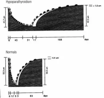
In the irst of these studies, Langdahl and cols. (3) obtained iliac crest bone biopsies from 4 men and 8 women with vitamin D-treated hypoparathyroidism and 13 age- and gender-matched control individuals. Nine of the subjects had postoperative hypoparathyroidism for 2 to 53 years duration, and 3 had idiopathic hypoparathyroidism for their entire lifetime. Ten of the patients were treated with 1-α-hydroxylated vitamin D (0.5 to 3.0 mcg/d), and two received calciferol oil (1600 to 3520 mcg/d). There was a nonsigniicant trend toward an increase in cancellous bone volume in the hypoparathyroid subjects, but the structural indexes, marrow star volume, trabecular star volume, and trabecular thickness were not different from those in control individuals. Bone forming surface and bone-formation rate were reduced signiicantly by 58% and 80%, respectively, in the hypoparathyroid subjects, and remodeling activation frequency in the hypoparathyroid patients was reduced to 0.13 per year, compared with 0.6 per year in control individuals. Initial mineral apposition rate also was lower by a factor of 5 in the hypoparathyroid subjects, but this difference was not statistically signiicant. The total resorption period was prolonged from 26 to 80 days in the hypoparathyroid subjects, and the resorption depth was reduced. The reconstructed remodeling cycles derived from these data are shown in igure 1. The balance between the resorption depth and wall thickness of cancellous bone packets was slightly positive by approximately 5 mm in the hypoparathyroid subjects compared with the control individuals.
Rubin and cols. (16) performed a more recent, larger histomorphometric study involving 24 women and 9 men with hypoparathyroidism treated with vitamin D and 33 age- and gender-matched control subjects. The etiologies of the hypoparathyroid state were post-thyroid surgery (n = 18), autoimmune (n = 13), and DiGeorge syndrome (n = 2), and the mean duration of their disease was 17 ± 13 (SD) years. Vitamin D intake varied between 400 and 100,000 IU/d, calcitriol intake between 0 and 3 mcg/day, and calcium supplementation between 0 and 9 g/d. Ten of the 33 hypoparathyroid subjects received thiazide diuretics. In contrast to the earlier smaller Langdahl
Figure 1. Reconstructed remodeling cycles in cancellous bone in
hypoparathyroid (upper) and normal (lower) subjects. Note that all phases of the remodeling cycle are prolonged in hypoparathyroidism. Reproduced with permission from ref. 3.
Hypoparathyroidism
Normals
Cop
yright
© ABE&M t
odos os dir
eit
os r
eser
vados
.
Figure 2. Low-magniication view of iliac crest bone biopsies obtained
from a normal control subject (left) and a hypoparathyroid patient (right). Biopsies were stained with Goldner-Masson trichrome stain. Note that cancellous bone volume and cortical thickness are increased in the hypoparathyroid patient. Reproduced with permission from ref. 16.
Figure 3. Cancellous and cortical bone measurements obtained by bone histomorphometry in patients with hypoparathyroidism (hatched bars) and
normal controls (open bars). Values are mean ± SD. Drawn from data from Rubin and cols. Reproduced with permission from ref. 2.
The effects of PTH deiciency on cancellous and cortical bone mass, which were observed initially by noninvasive imaging and by 2D histomorphometry, were conirmed recently by the 3D analytical capabi-lity afforded by micro-computed tomography. Results from the study by Rubin and cols. (17) conirmed the increase in cancellous bone volume and trabecular thi-ckness in hypoparathyroid subjects, and demonstrated higher trabecular number and trabecular connectivity in comparison with matched control subjects. In
addi-tion, the structural model index was lower in hypopara-thyroidism, indicating that the trabecular structure was more platelike than rodlike (Figure 5).
Cop
yright
© ABE&M t
odos os dir
eit
os r
eser
vados
.
Figure 5. Micro-CT images of cancellous bone from a patient with
hypoparathyroidism (left) and a normal control subject (right). The cancellous bone in the patient with hypoparathyroidism appears heavier and denser. Reproduced with permission from ref. 17.
Figure 4. High-power magniication views of tetracycline labels in a
hypoparathyroid patient (left) and normal control subject (right). Note that tetracycline labelling was markedly reduced in the patient with hypoparathyroidism, relecting low bone turnover. Reproduced with permission from ref. 16.
Hypoparathyroidism Control 1 mm
MARKERS OF BONE TURNOVER
Previous studies have shown that markers of bone tur-nover are lower in patients with hypoparathyroidism than in healthy age- and sex-matched controls. Markers of bone formation are decreased relative to those in healthy controls, and markers of bone resorption are similarly decreased. These indings generally conirm the bone histomorphometry indings reported in the preceding section, with speciic results discussed in tre-atment trials discussed in the following section on res-ponse to treatment with PTH.
FRACTURES
The mechanism behind PTH activation of bone remo-deling is intimately dependent on the time of exposure of bone cells to parathyroid hormone levels. Sustained high PTH levels trigger catabolism, while transitory elevations induce anabolism. Mendonça and cols. (19) recently performed a small study assessing the impact of hypoparathyroidism on BMD, frequency of subcli-nical vertebral fracture, and mandible morphometry. This study evaluated 33 postmenopausal women, 16 of whom had hypoparathyroidism, and 17 of whom were healthy controls matched for age, weight and height.
BMD of the lumbar spine, total hip, and 1/3 distal ra-dius, radiographic evaluation of vertebral morphome-try, panoramic radiography of the mandible, and bio-chemical evaluation of mineral metabolism and bone remodeling were assessed in both groups. There were no signiicant differences in lumbar spine or total hip BMD between the groups. There was marked hetero-geneity of lumbar spine BMD in the hypoparathyroid patients, with increased BMD in four, normal BMD in nine, osteopenia in one, and osteoporosis in two pa-tients. BMD was decreased in the 1/3 distal radius in the hypoparathyroid patients (P < 0.005). The hypopa-rathyroid group had increased morphometric vertebral fractures and decreased mandible cortical thickness. The study concluded that vertebral fragility fractures occurred in hypoparathyroid patients despite normal or increased BMD.
Underbjerg and cols. (20) recently showed that post-surgical hypoparathyroidism was associated with decreased upper extremity fracture risk. The study eva-luated data on fractures and other outcomes in patients with postsurgical hypoparathyroidism due to nonma-lignant causes in the Danish National Patient Registry, with information conirmed by review of individual hospital records. Individual cases were matched with three age- (± 2 years) and gender-matched controls from the general background population. Compared to controls, patients did not have increased risk of any fracture, whereas risk of fracture of the upper extremi-ties was signiicantly decreased. The authors concluded that postsurgical hypoparathyroidism is associated with protection against upper extremity fractures. Whether these indings apply to patients with hypoparathyroi-dism due to other causes is not yet clear. No other pu-blished studies to date have formally assessed fracture incidence in patients with hypoparathyroidism.
RESPONSE TO TREATMENT WITH PTH
forma-Cop
yright
© ABE&M t
odos os dir
eit
os r
eser
vados
.
tion favors the latter because, over time, bone mass in-creases in patients with hypoparathyroidism. This effect is seen in both cancellous and cortical bone compart-ments (6,7,14,16,21).
To assess effects of PTH therapy on 3-dimensional bone structure, Sikjaer and cols. (22) randomized 62 patients with hypoparathyroidism to 24 weeks of treat-ment with either PTH(1-84) 100 µg/day subcutaneou-sly or similar placebo as add-on therapy to calcium and vitamin D supplementation. Microcomputed tomogra-phy was performed on 44 iliac crest bone biopsies, with 23 biopsies taken from patients on PTH treatment, obtained after 24 weeks of treatment. Compared with placebo, PTH treatment caused a 27% lower trabecu-lar thickness (p < 0.01) and 4% lower trabecutrabecu-lar bone tissue density (p < 0.01), whereas connectivity density was 34% higher (p < 0.05). Trabecular tunneling was evident in 11 (48%) of the biopsies from the PTH tre-atment group. Patients with tunneling had signiicantly higher levels of biochemical markers of bone resorption and formation. In cortical bone, the number of Haver-sian canals per area was 139% higher (p = 0.01) in the PTH treatment group, causing a trend toward increa-sed cortical porosity (p = 0.09). At different subregions of the hip, areal BMD (aBMD) and volumetric BMD (vBMD), as assessed by dual-energy X-ray absorptio-metry (DXA) and quantitative computed tomography (QCT), decreased signiicantly by 1% to 4% in the PTH treatment group. However, at the lumbar spine, aBMD decreased by 1.8% (p < 0.05), whereas vBMD increased by 12.8% (p = 0.02) in the PTH treatment group com-pared with the placebo group.
Rubin and cols. (23) hypothesized that parathyroid hormone(1-84) [PTH(1-84)] treatment would res-tore skeletal properties toward normal in hypopara-thyroidism. This study included 64 subjects with hy-poparathyroidism who were treated with PTH(1-84) for 2 years. All subjects underwent histomorphometric assessment with percutaneous iliac crest bone biopsies at baseline and at 1 or 2 years. Another group of sub-jects had a single biopsy at 3 months, having received tetracycline before beginning PTH(1-84) and prior to the biopsy, using a quadruple-label protocol. Bioche-mical bone turnover markers were measured. Structu-ral changes after PTH(1-84) treatment for two years included reduced trabecular width (144 ± 34 µm to 128 ± 34 µm, p = 0.03) and increased trabecular num-ber (1.74 ± 0.34/mm to 2.07 ± 0.50/mm, p = 0.02). Cortical porosity increased after two years of treatment
from 7.4% ± 3.2% to 9.2% ± 2.4% (p = 0.03). Histomor-phometrically measured dynamic parameters, including mineralizing surface, increased signiicantly at three months, peaked at one year (0.7% ± 0.6% to 7.1% ± 6.0%, p = 0.001), and persisted at two years. Biochemical me-asurements of bone turnover increased signiicantly, peaking after ive to nine months of therapy, and persis-ting for 24 months. The study concluded that PTH(1-84) treatment of hypoparathyroidism is associated with increases in histomorphometric and biochemical indi-ces of skeletal dynamics. Structural changes were felt to be consistent with an increased remodeling rate in both trabecular and cortical compartments, with tun-neling resorption in cortical bone. These changes were interpreted as suggesting that PTH(1-84) improves abnormal skeletal properties in hypoparathyroidism, and restores bone metabolism toward normal eupara-thyroid levels.
Cop
yright
© ABE&M t
odos os dir
eit
os r
eser
vados
.
with hPTH 1-34 in hypoparathyroid adults and adoles-cents has varying effects in the different skeletal com-partments, leading to an increase in trabecular bone and an apparent trabecularization of cortical bone.
Neer and cols. (25) showed that compared with ba-seline in postmenopausal osteoporotic women, BMD increased at the lumbar spine by 2.9% ± 4% (p < 0.05) from 1.24 ± 0.3 to 1.27 ± 0.3 g/cm(2) (T-score 1.7 ± 2.0 to 1.9 ± 2.0) after treatment with teriparatide (PTH 1-34) over 21 months. The lumbar spine is a site that is enriched in cancellous bone. Because PTH is known to be anabolic for cancellous bone, this ob-servation was interpreted to indicate that new, younger bone was being formed as a result of PTH treatment. A more detailed examination of skeletal features using high resolution imaging or bone biopsy will be necessa-ry to elucidate which changes in microarchitectural pa-rameters contribute to the increase in trabecular BMD. Such results also would be of great interest in terms of a comparison between the effects of PTH(1–84) as a therapy for osteoporosis or as replacement therapy for hypoparathyroidism. Along with the increase in lum-bar spine BMD, a decrease in the distal 1/3 radius, a site enriched in cortical bone, was observed, decreasing by 2.4% ± 4.0% (p < 0.05) from 0.73 ± 0.1 to 0.70 ± 0.1 g/cm(2) (T-score, -0.03 ± 2.0 to -0.26 ± 1.0). These results suggest that teriparatide causes endos-teal resorption. These data do not imply that bone is wea kened because salutary effects on microarchitec-ture and bone size could well provide biomechanical advantages despite the reduction in BMD. More detai-led skeletal assessment will be required to answer this question. Overall, these changes in trabecular and cor-tical skeletal compartments recall the pattern seen with PTH treatment of osteoporosis in individuals who do not have hypoparathyroidism (26).
Taken together, these studies suggest that treatment of patients with hypoparathyroidism with PTH of different forms reduces BMD toward normal and improves bone microarchitecture, resulting in improved bone strength. These changes should also lead to reduced fracture risk, but prospective randomized controlled trials have not yet been conducted to demonstrate this.
CONCLUSION
Bone disease occurs in hypoparathyroidism due to the absence or relative deiciency of parathyroid hormo-ne. Lack of parathyroid hormone results in low bone turnover and increased bone mineral density with
as-sociated increased bone mineralization. Whether these changes affect risk of fracture is not yet clear. Never-theless, treatment with parathyroid hormone appears to improve the structural quality of bone.
Financial support: none.
Disclosure: no potential conlict of interest relevant to this article was reported.
REFERENCES
1. Shoback D. Clinical practice. Hypoparathyroidism. N Engl J Med. 2008;359:391-403.
2. Bilezikian JP, Khan A, Potts JT Jr, Brandi ML, Clarke BL, Shoback D, et al. Hypoparathyroidism in the adult: epidemiology, diagnosis, pathophysiology, target-organ involvement, treatment, and chal-lenges for future research. J Bone Miner Res. 2011;26(10):2317-37. 3. Langdahl BL, Mortensen L, Vesterby A, Eriksen EF, Charles P. Bone
histomorphometry in hypoparathyroid patients treated with vita-min D. Bone. 1996;18(2):103-8.
4. Kruse K, Kracht U, Wohlfart K, Kruse U. Biochemical markers of bone turnover, intact serum parathyroid hormone and renal cal-cium excretion in patients with pseudohypoparathyroidism and hypoparathyroidism before and during vitamin D treatment. Eur J Pediatr. 1989;148(6):535-9.
5. Mizunashi K, Furukawa Y, Miura R, Yumita S, Sohn HE, Yoshinaga K. Effects of active vitamin D3 and parathyroid hormone on the serum osteocalcin in idiopathic hypoparathyroidism and pseudo-hypoparathyroidism. J Clin Invest. 1988;82(3):861-5.
6. Abugassa S, Nordenström J, Eriksson S, Sjödén G. Bone mineral density in patients with chronic hypoparathyroidism. J Clin Endo-crinol Metab. 1993;76(6):1617-21.
7. Fujiyama K, Kiriyama T, Ito M, Nakata K, Yamashita S, Yokoyama N, et al. Attenuation of postmenopausal high turnover bone loss in patients with hypoparathyroidism. J Clin Endocrinol Metab. 1995;80(7):2135-8.
8. Seeman E, Wahner HW, Offord KP, Kumar R, Johnson WJ, Riggs BL. Differential effects of endocrine dysfunction on the axial and the appendicular skeleton. J Clin Invest. 1982;69(6):1302-9. 9. Touliatos JS, Sebes JI, Hinton A, McCommon D, Karas JG,
Palm-ieri GM. Hypoparathyroidism counteracts risk factors for osteo-porosis. Am J Med Sci. 1995;310(2):56-60.
10. Sikjaer T, Rejnmark L, Thomsen JS, Tietze A, Bruel A, Andersen G, et al. Changes in 3-dimensional bone structure indices in hypo-parathyroid patients treated with PTH(1-84): a randomized con-trolled study. J Bone Miner Res. 2012;27(4):781-8.
11. Takamura Y, Miyauchi A, Yabuta T, Kihara M, Ito Y, Miya A. Attenu-ation of postmenopausal bone loss in patients with transient hypoparathyroidism after total thyroidectomy. World J Surg. 2013;37(12):2860-5.
12. Sugitani I, Fujimoto Y. Effect of postoperative thyrotropin sup-pressive therapy on bone mineral density in patients with papil-lary thyroid carcinoma: a prospective controlled study. Surgery. 2011;150(6):1250-7.
13. Biondi B, Cooper DS. Beneits of thyrotropin suppression versus the risks of adverse effects in differentiated thyroid cancer. Thy-roid. 2010;20(2):135-46.
Cop
yright
© ABE&M t
odos os dir
eit
os r
eser
vados
.
15. Cohen A, Dempster DW, Müller R, Guo XE, Nickolas TL, Liu XS, et al. Assessment of trabecular and cortical architecture and mechanical competence of bone by high-resolution peripheral computed tomography: comparison with transiliac bone biopsy. Osteoporos Int. 2010;21(2):263-73.
16. Rubin MR, Dempster DW, Zhou H, Shane E, Nickolas T, Sliney J Jr, et al. Dynamic and structural properties of the skeleton in hypo-parathyroidism. J Bone Miner Res. 2008;23(12):2018-24. 17. Rubin MR, Dempster DW, Kohler T, Stauber M, Zhou H, Shane E,
et al. Three dimensional cancellous bone structure in hypopara-thyroidism. Bone. 2010;46(1):190-5.
18. Christen P, Ito K, Müller R, Rubin MR, Dempster DW, Bilezikian JP, et al. Patient-speciic bone modelling and remodelling simula-tion of hypoparathyroidism based on human iliac crest biopsies. J Biomech. 2012;45(14):2411-6.
19. Mendonça ML, Pereira FA, Nogueira-Barbosa MH, Monsignore LM, Teixeira SR, Watanabe PC, et al. Increased vertebral morphometric fracture in patients with postsurgical hypoparathyroidism despite normal bone mineral density. BMC Endocr Disord. 2013;13:1. 20. Underbjerg L, Sikjaer T, Mosekilde L, Rejnmark L. Post-surgical
hypoparathyroidism - risk of fractures, psychiatric diseases,
can-cer, cataract, and infections. J Bone Miner Res. 2014 May 7 [Epub ahead of print].
21. Duan Y, De Luca V, Seeman E. Parathyroid hormone deiciency and excess: similar effects on trabecular bone but differing ef-fects on cortical bone. J Clin Endocrinol Metab. 1999;84(2):718-22.
22. Sikjaer T, Rejnmark L, Thomsen JS, Tietze A, Brüel A, Andersen G, et al. Changes in 3-dimensional bone structure indices in hypo-parathyroid patients treated with PTH(1-84): a randomized con-trolled study. J Bone Miner Res. 2012;27(4):781-8.
23. Rubin MR, Dempster DW, Sliney J Jr, Zhou H, Nickolas TL, Stein EM, et al. PTH(1-84) administration reverses abnormal bone-re-modeling dynamics and structure in hypoparathyroidism. J Bone Miner Res. 2011;26(11):2727-36.
24. Gafni RI, Brahim JS, Andreopoulou P, Bhattacharyya N, Kelly MH, Brillante BA, et al. Daily parathyroid hormone 1-34 replacement therapy for hypoparathyroidism induces marked changes in bone turnover and structure. J Bone Miner Res. 2012;27(8):1811-20. 25. Rubin MR, Sliney J Jr, McMahon DJ, Silverberg SJ, Bilezikian JP.