Cop
yright
© ABE&M t
odos os dir
eit
os r
eser
vados
.
Cop
yright
© ABE&M t
odos os dir
eit
os r
eser
vados
.
The multiple effects of thyroid
disorders on bone and
mineral metabolism
Os múltiplos efeitos das disfunções tireoidianas sobre o metabolismo osteomineral
Ludmilla F. Cardoso1, Léa M. Z. Maciel1, Francisco J. A. de Paula1
ABSTRACT
Differently from most hormones, which commonly are specialized molecules able to inluen-ce other inluen-cells, tissues and systems, thyroid hormones (TH) are pleiotropic peptides, whose primordial function is dificult to identify. The complex action of TH on human economy can be easily witnessed by examining the diverse consequences of TH excess and deiciency du-ring development and after maturity. In particular, different manifestations in bone modeling and remodeling relect the circumstantial consequences of thyroid disturbances, which are age dependent. While hyperthyroidism during childhood enhances bone mineralization and acce-lerates epiphyseal maturation, in adults it induces bone loss by predominant activation of oste-oclast activity. Furthermore, the syndrome of TH resistance is a multifaceted condition in which different sites exhibit signs of hormone excess or deiciency depending on the coniguration of the TH receptor isoform. The investigation of the impact of TH resistance on the skeleton still remains to be elucidated. We present here a thorough review of the action of TH on bone and of the impact of thyroid disorders, including hyper- and hypothyroidism and the syndrome of TH resistance, on the skeleton. Arq Bras Endocrinol Metab. 2014;58(5):452-63
Keywords
Thyroid hormones; osteoporosis; thyrotoxicosis; hypothyroidism; thyroid hormone resistance
RESUMO
Diferentemente da maioria dos hormônios, que usualmente são moléculas especializadas ca-pazes de inluenciar outras células, tecidos e sistemas, os hormônios da tireoide (HT) são pep-tídeos pleiotrópicos, cuja função primordial é difícil de identiicar. A ação complexa dos HT na isiologia humana pode ser facilmente reconhecida ao observar as diversas consequências do excesso e da deiciência de HT durante e após o pleno desenvolvimento. Em particular as diferentes manifestações na modelação e remodelação óssea reletem que as consequências esqueléticas das disfunções tireoidianas dependem das circunstâncias e variam com a idade. Enquanto o hipertireoidismo durante a infância aumenta a mineralização óssea e acelera a maturação epiisária, em adultos induz a perda óssea pela ativação predominante da ação osteoclástica. Além disso, a síndrome de resistência ao HT é uma condição multifacetada na qual diferentes tecidos apresentam sinais de excesso ou deiciência hormonal, dependendo da predominância da expressão das diversas isoformas do receptor de HT. O impacto da re-sistência ao HT sobre o esqueleto ainda é motivo de investigação. Apresentamos aqui uma revisão abrangente sobre as ações ósseas dos HT e o impacto no esqueleto dos distúrbios da tireoide, incluindo hipo e hipertireoidismo e síndrome de resistência ao HT. Arq Bras Endocrinol Metab. 2014;58(5):452-63
Descritores
Hormônios tireoidianos; osteoporose; tireotoxicose; hipotireoidismo; resistência ao hormônio tireoidiano 1 Department of Internal Medicine,
Ribeirão Preto Medical School, University of São Paulo (FMRP-USP), Ribeirão Preto, SP, Brazil
Correspondence to:
Francisco J. A. de Paula Av. Bandeirantes, 3900
14049-900 – Ribeirão Preto, SP, Brazil [email protected]
Received on Feb/25/2014 Accepted on May/12/2014
Cop
yright
© ABE&M t
odos os dir
eit
os r
eser
vados
.
INTRODUCTION
T
hyroid hormones (TH), vitamin D (VD) and steroids belong to a special group of endocrine molecules which produce their effects by signaling in nuclear receptors (NR) (1). Typically they are pleiotro-pic hormones, affecting a signiicant range of cell types in most tissues and in various systems. In common TH, steroids and VD also have intense effect on bone strength and on mineral and energy metabolism. While glucocorticoids and gonadal steroids, respectively, lead to a loss and an increase in bone mass, TH have a more complex effect depending on age, circumstances.The TH 3,5,3’,5’-tetraiodo-L-thyronine (thyroxin or T4) and 3,5,3’-triiodothyronine (T3) are produced and secreted by thyroid follicles which, under TSH stimulation, capture iodide through the membrane protein NIS (a iodide-sodium cotransporter) and uti-lize it for TH synthesis. T4 is considered to be a pro-hormone whose main function is to serve as a substrate for the production of T3 through deiodination by the type 1 (D1) and type 2 (D2) deiodinase enzymes. T3 is the main hormone responsible for the genomic actions of TH, modulating the expression of target genes after binding to the TH receptor (TR), while T4, reverse T3 (rT3) and 3,5-diiodothyronine (T2) seem to be more involved in non-genomic TH actions, which oc-cur more rapidly and usually in consonance with their genomic actions. These last hormones are also funda-mental for the regulation of basal cell activity and are important for rapid adjustments of cell homeostasis (1).
Once released into the blood circulation, TH are transported to the intracellular medium by TH transport-ers (THT), which have 12 transmembrane domains and can be divided into two subfamilies: organic anion trans-porters (NTCP and OATP) and amino acid transtrans-porters (L type – LAT1 and LAT2, and T type – MCT8 and MCT10). The only THT speciic for TH is MCT8, while all the others are denoted secondary transporters (2).
Inside the cell, T3 binds to TR, which belongs to the superfamily of NR and is usually found in the nucleus as a heterodimer with the retinoid X receptor (RXR), modulating gene transcription. Thus, NR are transcription factors activated by their ligands, proteins that bind to the promoter region of target genes in or-der to activate or repress transcription. In the absence of T3, TR recruits corepressor complexes (CoR) which compact chromatin by means of histone acetylation and prevent the action of the transcription machinery. T3
binding to TR changes its tridimensional conforma-tion, which then shows greater afinity for coactivator complexes (CoA). As a consequence, CoR dissociation occurs, leading to histone acetylation and chromatin loosening, with the subsequent transcription of the tar-get gene (1). The biological genomic activity of TH depends on the intracellular concentrations of T3, on D1 and D2 activity, and also on the activity of type 3 deiodinase (D3), which inactivates T3.
TR are coded by the THRA (c-erbAα) and THRB
(c-erbAβ) genes, respectively located on chromosomes 17 and 3, which code by alternative splicing various iso-forms distributed in the organism depending on tissue and on age. While TRβ predominates in the hypothala-mus-pituitary region, the liver and kidneys, TRα is the primary effector of the actions of T3 on the heart, cen-tral nervous system, intestine, skeletal muscle, and bone (3). Different isoforms have diverse effects, as can be observed in experimental models using knockout mice, in which the deletion of different types of TR leads to diverse manifestations.
MATERIALS AND METHODS
The platform Medline-PubMed database, followed by the Embase and Scientiic Electronic Library Online/ Latin American and Caribbean Health Science Litera-ture (SciELO/Lilacs) databases were used to search articles. The search terms used were thyroid hormone and bone in association with one of the following: os-teoporosis, calcium, phosphorus, thyroid hormone re-sistance and growth.
Molecular actions of TH in the skeleton
The ample interface between TH and bone metabolism is demonstrated by the variety of molecules related to the metabolism and action of TH expressed in skeletal tissue, such as TRs, THTs and deiodinases (4-6).
li-Cop
yright
© ABE&M t
odos os dir
eit
os r
eser
vados
.
neage such as interleukin 6 and prostaglandin E2 (5). Moreover, T3 acts in a synergistic manner with osteo-clastogenic hormones such as parathyroid hormone (PTH) (9) and VD (10). It has also been demonstrated that T3 increases the expression of mRNA of the ligand of receptor activator of nuclear factor-κβ (RANKL) in the osteoblast, which activates RANK present in osteo-clast precursors a key step in the osteoosteo-clastogenesis (7).
The TRβ1 and TRα1 isoforms are expressed in the stromal cells of bone marrow, chondrocytes, os-teoblasts and osteoclasts. Quantitative RT-PCR stu-dies have indicated that TRα1 expression is at least ten times higher than TRβ1 expression, indicating that TRα1 is the predominant mediator of the actions of T3 on the skeleton. Studies on adult female rats trea-ted with 3,5-dimethyl-4-(4-hydroxy-3-isopropylbenzyl) phe noxy acetic acid (GC-1), a TRβ-selective analogue, have shown that chronic treatment with GC-1 does not change bone mass. Conversely, treatment with a supra-physiological dose of T3 equimolar to the GC-1 dose causes a generalized bone loss. Taken together, these indings support the concept that the osteopenic effects of T3 are mediated by TRα (11).
Studies using animal models genetically modiied for TR and D2 and with congenital hypothyroidism have made important contributions to the understand-ing of osteomineral effects of TH. Table 1 lists vari-ous representative studies in this line of investigation. In general, these studies demonstrated that mice with mutation of TRα or of both α and β receptors nearly show the same phenotype of retarded growth and bone maturation, with delayed ossiication, reduced postna-tal growth and bone mineralization, delayed closure of the cranial sutures in early life and increased mineraliza-tion during adult life, as observed in hypothyroidism, although with less severity. In contrast, mice with muta-tion in TRβ gene show a thyrotoxic skeletal phenotype, with increased mineralization and advanced ossiication which result in short stature and craniosynostosis in childhood and reduced mineralization and low bone mass in adulthood (12). However, the lower severity of the skeletal phenotype of TR mutants compared to mice with TH deicit or excess actually suggests a sub-stantial overlap in the action of these two receptors re-garding the control of bone development. Thus, up to a point, TRβ and TRα are interchangeable, although the compensatory effects are incomplete, with some ef-fects being isoform-speciic. In addition, TRα1 mutants exhibit reduced expression and signaling of GH and
IGF-1 receptors during the growth, suggesting that these receptors are targets of the physiological action of T3 on bone in vivo. In support to this point, there are studies demonstrating that IGF-I is stimulated by T3 in clones of cells of the osteoblast lineage (13).
Cop
yright
© ABE&M t
odos os dir
eit
os r
eser
vados
.
Table 1. Skeletal phenotypes of genetic modiied mice showing congenital hypothyroidism, TSH resistance, mutation in the type 2 deiodinase and thyroid hormone resistance alpha and/or beta
Model Genotype Hormonal status Young skeleton Adult skeleton Skeletal
phenotype References
Pax8 Lack of essencial transcriptor factor Pax8 for thyroid
development
No thyroid. Undetectable TH; elevated TSH 2000
fold
Serious and persistent linear growth retardation, delayed
endochondral ossiication, reduced cortical bone, reduced
bone mineralization
NR Hypothyroid Mansouri and cols. (61); Friedrichsen and cols. (62)
TSHR TSHR deleted Thyroid hypoplasia. Undetectable TH;
elevated TSH
Death after weaning if not treated with TH
NR NR Marians and cols. (63)
Hyt/hyt Loss of function mutation in TSHR gene
Low TH; elevated TSH 2000 fold
Linear growth retardation, delayed
endochondral ossiication, reduced cortical bone, reduced
mineralization
NR Hypothyroid Beamer and cols. (64); Gu and cols. (65)
D2 Type 2 deiodinase deleted
Elevated TSH and T4; normal T3
Normal growth and development
NR NR Bassett and cols. (5);
Fraichard and cols. (66) TRα0/0 TRα deleted;
TRβ preserved
Euthyroid; normal GH and IGF-I
Transient growth retardation; delayed
endochondral ossiication, reduced
mineralization
Osteosclerosis; increased trabecular volume, reduction of osteoclastic absorption
Hypothyroid Gauthier and cols. (67); Bassett and cols. (5)
TRα1PV/+ TRα mutation with heterozygous dominant negative
TRα receptor
Mild thyroid failure, normal GH, reduced
IGF-I
Serious and persistent growth retardation;
delayed intramembranous and
endochondral ossiication, reduced
mineralization
NR Hypothyroid Kaneshige and cols. (68); O’Shea and cols. (69)
TRβ-/- TRβ deleted; TRα preserved
RTH with elevated TH and goiter
Persistent short stature; advanced ossiication, increased
mineralization
Osteoporosis, reduced mineralization, increased osteoclastic
resorption
Thyrotoxic Forrest and cols. (69); Gauthier and cols. (67);
Bassett and cols. (5)
TRβPV/PV TRβ mutantion with homozigous dominant negative TRβ receptor
RTH serious and goiter; reduced GH
Accelerated prenatal growth; retarded persistent postnatal
growth; advanced ossiication; increased
mineralization
NR Thyrotoxic Kaneshige and cols. (70); O’Shea and cols. (12);
Bassett and cols. (5)
TRα-/-TRβ-/- TRα and TRβ deleted RTH and small goiter Growth retardation, delayed ossiication, reduced mineralization
Death near the weaning
NR Gauthier and cols. (67); Bassett and cols. (5)
TH: thyroid hormone; TSHR: thyroid stimulating hormone receptor; D2: type 2 deiodinase; TRα: thyroid hormone receptor alpha; TRβ: thyroid hormone receptor beta; RTH: resistance to thyroid hormone; NR: not reported.
Consequences for development
Childhood and adolescence are periods of great ske-letal changes, when more than 90% of the bone mass is acquired. During the prepubertal period, bone growth and mineralization are more rapid in the limbs than in the spine (17). Spine growth accelerates during early and intermediate puberty and both axial an
Cop
yright
© ABE&M t
odos os dir
eit
os r
eser
vados
.
are relatively less mineralized during the peripubertal period, contributing to the risk of fractures during this phase. Between six and 16 years of age, bone mass in-creases 2.5 and 3 times in girls and boys, respectively. The peak of bone mass reached at the beginning of adult life is a major determinant of osteoporosis emer-gence after reproductive age (17). Although genetic factors determine 60 to 80% of the variability of skeletal bone mass, other factors related to health and life style can signiicantly inluence bone growth and remodel-ing. Among these factors are various hormonessuch as estrogens (18), androgens and GH (19), with anabolic effects, glucocorticoids with catabolic actions (20,21) and TH with a variable effect depending on the phase of development. T3 is known to be a potent regulator of skeletal growth and maturation, with its actions on bone varying according to developmental stage, with predominance of anabolism during development and of catabolism after bone maturation (22).
Hypothyroidism and thyrotoxicosis
In children, TH deiciency leads to delayed maturation, short stature and changes in the epiphyseal growth plates, while TH excess results in advanced bone age, premature fusion of the epiphyses, and consequent lim-itation of inal stature (23,24).
The relative expression of the two TR genes also varies, not only among tissues, but also during the different phases of development. It has been demon-strated that the TRα1 and TRβ1 isoforms are function-ally present at varying proportions in three osteosar-coma cell lines which express ibroblast, preosteoblast and mature osteoblast phenotypes (ROS 25/1, UMR 1O6 and ROS 17/2.8, respectively), suggesting a pos-sible change in TH action during bone development (25). Indeed, these cells show different responses to T3 treatment.
Together with GH, IGFs, glucocorticoids, estro-gens and androestro-gens, TH represent important systemic factors that inluence bone growth and maturation, es-pecially after the beginning of puberty. Under normal conditions, TH activates both synthesis and degrada-tion of bone matrix, having relevant role for skeletal integrity maintenance (8). One of the mechanisms by which TH produce these effects is an indirect action increasing GH and IGF secretion (26), as reported in classical studies on thyroidectomized rats treated with TH. After thyroidectomy, the animals show a reduction
of pituitary and serum circulatory levels of GH, which is reversed after TH replacement. Additionally, there is evidence that TH can act directly on the epiphyseal plate independently of GH (26), as demonstrated in studies with cultures of epiphyseal plate cells submitted to a T3-rich environment. Within this context, there is inhibition of the clonal expansion of immature chon-drocytes in parallel to an increased differentiation of hypertrophic chondrocytes, the latter representing the end phase of life of these cells, when they can no longer promote growth.
The inal height can be impaired by both lack and excess of TH. Spontaneous nocturnal secretion of GH is low in hypothyroidism and hyperthyroidism (27,28). The rate and the amount of GH released are reduced in adolescents with untreated thyrotoxicosis compared with normal controls. It has been proposed that the reduced GH release during GHRH stimulation in hy-perthyroidism may be explained in part by an increase in hypothalamic somatostatin tone with a concomitant decrease in GHRH (28). While in the presence of thy-rotoxicosis the impairment of height occurs in a pro-portionate manner, congenital hypothyroidism is the only cause of secondary disproportionate short stature, mainly affecting the lower segment of the body.
Resistance to thyroid hormones
Cop
yright
© ABE&M t
odos os dir
eit
os r
eser
vados
.
not known whether the delay in bone age occurring in some of these individuals with RTHβ is primarily due to TRβ dysfunction per se or to the negative dominant effect that mutants may exert on normal TRα1, pre-venting their full physiological role.
Consequences for bone mass
The bone remodeling process is an ingenious mecha-nism that simultaneously contributes to calcium ho-meostasis and bone resistance. Under ideal conditions, the inal product of bone remodeling is the mainte-nance of bone integrity (8). Conversely, imbalance in bone formation and resorption leads to bone loss and deterioration of bone microarchitecture, with conse-quent emergence of bone fragility and an increased risk of fractures (9).
Hypothyroidism and thyrotoxicosis
The intracellular TH metabolism performed by de-iodinases D2 (activator) and D3 (inactivator) in the osteoblasts is a sophisticated mechanism for the main-tenance of intracellular T3 concentrations in a man-ner partially independent of its serum concentrations. Thus, D2 activity is maximum in hypothyroidism and minimum in thyrotoxicosis, in order to maintain con-stant intracellular T3 concentrations. These adaptations may play an important role by preserving bone minera-lization and strength during the phase of installation of thyroid disorders. However, when hypothyroidism and thyrotoxicosis become established, this local feedback mechanism is unable to maintain T3 in bone microen-vironment, with the occurrence of increased
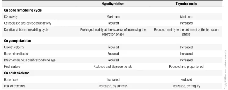
(hypothy-roidism) or reduced (hyperthy(hypothy-roidism) mineralization, and repercussions on bone mass and strength. Thus, an increased risk of fractures occurs in hypothyroidism due to stiffness and instability, or due to fragility in thyro-toxicosis. Expectedly, the risk of fracture according to thyroid status, follows a “U”-shaped curve, with the risk being lower in euthyroidism and increasing with both the lack and the excess of TH (7,8). The main changes and consequences of TH excess or deiciency are summarized in table 2.
Hypothyroidism leads to a low bone turnover, with a reduction of osteoclast bone reabsorption and of os-teoblast formation, slowing the remodeling process and increasing the time taken in the remodeling cycle, mainly due to the prolongation of the mineralization phase. A slight increase in bone mass may even occur, which unfor-tunately does not result in a reduced risk of fracture. On the contrary, the risk of fracture arises before bone mass reaches levels compatible with osteoporosis. Although the mechanisms are not fully understood, impairment of bone quality is believed to occur in association with the reduction of mechanical stimuli due to lower physical ac-tivity and with an increased risk of falls (36-38).
In contrast, in thyrotoxicosis there is an increase in bone turnover, with a reduction of the cycle mainly im-pairing the phase of bone formation, with decoupling of the remodeling process in favor of reabsorption. As a consequence bone loss occurs; indeed hyperthyroidism is a major cause of secondary osteoporotic fractures. The catabolic effects of TH in bone were described more than one century ago, and the clinical reper-cussion in BMD and risk of fractures have been fully characterized more recently in untreated hyperthyroid
Table 2. Main effects of thyroid hormone deiciency or excess on bone turnover in young adult skeleton
Hypothyroidism Thyrotoxicosis
On bone remodeling cycle
D2 activity Maximum Minimum
Osteoblastic and osteoclastic activity Reduced Increased
Duration of bone remodeling cycle Prolonged, mainly at the expense of increasing the resorption phase
Reduced, mainly to the detriment of the formation phase
On young skeleton
Growth velocity Reduced Increased
Bone mineralization Reduced Increased
Intramembranous ossiication/Bone age Reduced Increased
Final stature Reduced and disproportionate Reduced and proportioned
On adult skeleton
Bone mass Increased Reduced
Cop
yright
© ABE&M t
odos os dir
eit
os r
eser
vados
.
subjects. Histomorphometry studies in thyrotoxicosis have demonstrated increased osteoblast and osteoclast activity (11). The evaluation of bone histomorphom-etry on humans and animals have shown that, in the presence of excess TH, osteoblast and osteoclast activ-ity is increased, with a predominance of the latter. As a result, bone metabolism is accelerated, favoring resorp-tion and a negative calcium balance and as a result bone loss (37,38). The ensuing structural changes involve reduction of the thickness and number of trabeculae, implying in a reduction of trabecular bone volume. There is also increased porosity and reduced thickness of cortical bone. Bone fragility has been reported as an early adverse effect that may arise even in subclinical hyperthyroidism (5).
An important clinical aspect that emphasizes the negative impact of hyperthyroidism on the skeleton is the bone recomposition that follows the reestablish-ment of normal TH production. However, there is still controversy about whether this recovery is total or par-tial (39,40). A 2003 meta-analysis study has suggested that treatment of hyperthyroidism alone restores bone mass to normal levels after one to four years, even when no other measure is taken to increase bone mass (41).
Although all skeletal sites are responsive to TH, they exhibit different response to TH stimuli. The long-term administration of high LT4 doses to adult male rats for a period of 3 to 20 weeks resulted in reduced bone min-eral density (BMD) in the femur but not in vertebrae. There was also a signiicant increase in the expression of osteocalcin, osteopontin, alkaline phosphatase and tartrate-resistant acid phosphatase in the femur, but not in the spine specimen. (42,43). In clinical investiga-tion, hyperthyroidism seems to affect cortical bone to a greater extent than trabecular bone, demonstrated by predominant bone loss in the forearm (44). The vari-ability in response pattern is probably related to the dif-ferential expression not only of TRs, but also of other factors that modulate bone remodeling (45).
It is also possible that TH may interfere with the bioavailability and action of estradiol and/or androgens, since both hypo- and hyperthyroidism can cause signii-cant changes in the bioavailability of these steroids (46). These changes range from elevation of sex hormone binding globulin (SHBG), estradiol and testosterone, in the case of thyrotoxicosis, to reduction of these parameters in hypothyroidism. Both TRα and TRβ are expressed in the testicles and TRα1 is the isoform most likely associated with testicular development and
function (47). The ovaries also express TRs, inclusive mRNA of TRα1, TRβ1, TRβ2 and c-erb-TRΔα2 hav-ing been detected in human oocytes (48). These data indicate the existence of a strict interaction between the gonadal and thyroid axis. Certainly, this is one of the multiple mechanisms involved with the emergence of bone disease in patients showing thyroid disorders.
Resistance to thyroid hormone
Animal models of congenital hypothyroidism, thyro-toxicosis and RTH due to THRA and THRB genes deletions or mutations have greatly contributed to the understanding of osteomineral metabolism in different thryoid conditions. The models of TH deiciency, usu-ally due to PAX8 or TSHR mutation, as well as TRα mutants, present a skeletal phenotype of hypothyroi-dism, with the animals showing reduced mineraliza-tion, growth retardation and delayed ossiication du-ring development, but with increased mineralization after maturation (5,12). In human beings, an especially curious clinical model of primary hypothyroidism is congenital hypothyroidism due to thyroid dysgenesis. In addition to low serum TH levels, affected indivi-duals show reduced circulating levels of calcitonin. Previous studies have shown that adult patients with thyroid dysgenesis who started TH replacement during different phases of childhood have a normal bone mass despite the concomitant chronic calcitonin deiciency.
In contrast, experimental models of TH excess usu-ally involve treatment with T3 and TRβ mutants. The thyrotoxic skeletal phenotype varies from increased mineralization and advanced ossiication during de-velopment to reduced mineralization after maturation (5,12). Other experimental models, such as mutants for D2 and dual oxidase (Duox2), the enzyme gen-erating hydrogen peroxide which permits the action of thyroperoxidase, are more complex. In the former, they show normal skeletal growth and development al-though with stiff bones due to increased bone miner-alization. In the last, the bone phenotype still is to be characterized (42).
Cop
yright
© ABE&M t
odos os dir
eit
os r
eser
vados
.
BONE RESORPTION
OSTEOCLASTS (OC)
May express TRs T3 may have direct actions in OC and/or
T3 may stimulate OC through the stimulation of OB synthesis of RANKL and other cytokines involved in osteoclastogenesis
ACTIONSOF T3
STIMULATESEXPRESSIONOF
IL-6, IL-8 PGE2 RANKL ACTSINASYNERGISTICMANNERWITH
PTH VD
Figure 1. Interface between TH and bone metabolism. Bone remodeling on the surface of trabecular bone is illustrated here. T3 stimulates osteoblast differentiation and activity by complex direct actions on TRs and indirect mechanisms, involving diverses growth factors and cytokines. T3 also stimulates osteoclast differentiation and activity, but it still remains unclear whether the effects of T3 on the promotion of bone resorption result from direct actions in osteoclasts or indirectly through effects on osteoblasts. TR, thyroid hormone receptor; IL-6, interleukin 6; IL-8, interleukin 8; PGE2, prostaglandin E2; RANKL, ligand of receptor activator of nuclear factor-κβ; PTH, parathyroid hormone; VD, vitamin D; D2, type 2 deiodinase; D3, type 3 deiodinase; THT, thyroid hormone transporters; MCT8, monocarboxilate transporter 8; LAT: L-type amino acid transporter; TSHR, TSH receptor; IGF-I, insulin-like growth factor I; IGF-IR, IGF-I receptor.
Consequences for osteomineral metabolism
Over the last two decades there has been a signiicant advance in the understanding of bone physiology. It has been unequivocally determined that the skeleton has a marked metabolic activity, utilizing substrates in order to provide the energy used in the bone remodel-ing process. In addition, bone is involved in the en-docrine modulation not only of mineral metabolism, but also of energy metabolism. The ibroblast growth
factor 23 (FGF-23), originally produced by osteocytes, shares with PTH an important role on the modulation of circulatory levels of phosphate. In parallel, FGF-23 inhibits the production of both PTH and calcitriol [1,25(OH)2D] (49). The anatomical structure enables trabecular bone to have greater metabolic activity than its cortical counterpart (49). Bone remodeling is regu-lated by a variety of systemic hormones and local fac-tors that act on osteoblast and/or osteoclast cell lines,
BONEFORMATION
OSTEOBLASTS (OB)
Express TRs, mainly TRα1, but also TRα2 and TRβ1 Express D2 and D3
Express THTs MTC8, LAT1 and LAT2 Express TSHR
ACTIONSOF T3
STIMULATESEXPRESSIONOF
IGF-I / IGF-IR Osteocalcin Type I collagen Alkaline phosphatase Metalloproteases 9 and 13
Cop
yright
© ABE&M t
odos os dir
eit
os r
eser
vados
.
exerting effects on the proliferation of undifferentiated cells and on cell recruitment, differentiation and/or ac-tivation (49). Among the main systemic hormones that participate in the modulation of bone metabolism are the so-called calciotrophic hormones, which include PTH, 1,25(OH)2D and calcitonin, the last one of less importance. In addition, TH, together with insulin, growth hormone, glucocorticoids and sex steroids, have a signiicant inluence on the development and maintenance of bone mass (8).
Hypothyroidism and thyrotoxicosis
Hyperthyroidism causes important changes in calcium and phosphorus metabolism which may lead to an in-crease of as much as three times of the urinary and fe-cal excretion of fe-calcium. The action of TH on renal phosphorus reabsorption starts early, during develop-ment. For instance, it was observed that TH promote the maturation of tubular phosphate transporters in rats. Although TH increase the total urinary excretion of phosphorus, they also increase the renal reabsorp-tion of phosphorus by activating phosphorus channels dependent on the sodium gradient (50-52).
The hypercalcemia of thyrotoxicosis may result from an abnormal calcium eflux from the skeleton, kidneys and gastrointestinal tract to the extracellular luid. The trend to hypercalcemia in thyrotoxicosis has been documented and its incidence varies from 8 to 22%. It is usually mild, but occasionally it is severe enough to be symptomatic and rarely is associated with signs and symptoms of acute hypercalcemia. The mean serum calcium concentrations of hyperthyroid patients are higher than those of control subjects (53-55). Pre-vious studies have shown that hypercalcemia in thyro-toxicosis is mainly due to increased mineral bone mo-bilization. This in turn is caused by a direct stimulation of bone cells by the high TH concentrations, with a consequent increase in bone reabsorption. It has been demonstrated that T3 increased the mRNA expression of the RANKL in preosteoblastic cells. The RANKL is a key molecule for osteoclastogenesis and osteoclast acti-vation. It is synthesized and secreted by the osteoblasts and binds to its receptor, RANK, expressed on osteo-clast precursors and mature osteoosteo-clasts, thus activating osteoclastogenesis and osteoclast activity (7,8). Indeed, in patients with thyrotoxicosis, bone histomorphom-etry is consistent with increased osteoclast reabsorption in cortical bone, leading to increased cortical porosity,
while in cancelous bone these changes are less evident. The biochemical markers of bone formation and reab-sorption, such as osteocalcin (OC), alkaline phospha-tase (AP), bone-speciic AP, urinary pyridinoline and deoxypyridinoline, are increased in patients with hy-perthyroidism, indicating enhanced bone remodeling activity.
The increased mineral bone mobilization and con-sequent hypercalcemia, may lead to the suppression of PTH release in patients with thyrotoxicosis. It has also been reported that hyperphosphatemia, hypercal-ciuria and hyperphosphaturia frequently occur in these patients (53,54). The calcium balance in patients with untreated hyperthyroidism is negative and the losses do not seem to be due to VD deiciency, since they are not reduced by VD treatment (55). Some authors have also proposed that hypercalcemia in thyrotoxicosis may be due to increased adrenergic tonus. In support to this hypothesis there are studies showing that thyrotoxic patients with hypercalcemia may become normocal-cemic after therapy with propranolol alone. Moreover the serum levels of bone turnover markers (e.g., alkaline phosphate and osteocalcin) are elevated in hyperthy-roidism, and remain high for months during its treat-ment, despite normalization of serum TH (54).
Regarding phosphorus metabolism in hyperthy-roidism, the indings are more conlicting. Some au-thors have reported that thyrotoxicosis courses with hyperphosphatemia, while others detected normal phosphorus concentrations, and hypophosphatemia (50,53,54). Hyperphosphatemia in hyperthyroidism appears to be caused by: increased bone reabsorption and tubular phosphate reabsorption; a direct action of TH on the renal N/Pi transporters and by PTH sup-pression induced by hypercalcemia. In the kidneys, studies using immunocytochemistry and speciic anti-bodies against TRα and TRβ have demonstrated the presence of both isoforms in all segments of the proxi-mal tubules. Hyperphosphatemia due to excess TH also seems to trigger compensatory mechanisms for the increase in phosphaturia. Recent studies reported that patients with Graves disease exhibit high serum levels of FGF-23, however this occurrence is exclusive of pa-tients with hyperphosphatemia (56). In other words, the increase in FGF-23 secretion appears to be a conse-quence of high phosphorus concentrations and not an event secon dary to the direct action of TH.
Cop
yright
© ABE&M t
odos os dir
eit
os r
eser
vados
.
Some studies have demonstrated that PTH concentra-tions are unchanged in the presence of hyperthyroid-ism (57-59), others have shown an increase in PTH, while most investigations have indicate a phenomenon of counterregulation between PTH and TH (9,20,41). It has been demonstrated that treatment with T3 in-creases the number of PTH receptors in osteoblastic osteosarcoma cells in a time- and dose-dependent man-ner. Also it has been shown that treatment with PTH increases the number of TRs, suggesting a synergism between these two hormones in the regulation of bone metabolism (9).
There is evidence that T3 and VD act synergisti-cally on osteoclastogenesis. It has been demonstrated that the α and β TR isoforms and vitamin D receptors (VDR), in addition to estrogen and androgen receptors, are coexpressed in the bone marrow of mice, suggesting that this is a prerequisite for this synergism for osteo-clastogenesis (10). In another study on co-cultures of pre-osteoblasts with bone marrow cells, T3 increased the osteoclast formation induced by VD. T4 had the same effects, suggesting its local metabolization to T3, which later led to the identiication of the expression of D2 mRNA in pre-osteoblastic cells. It has also been demonstrated that VD increases D2 expression in these cells in a time- and dose-dependent manner, indicating the presence of an additional synergistic mechanism be-tween VD and T3 in osteoclastogenesis (10).
Resistance to thyroid hormone
No studies have evaluated osteomineral metabolism in RTHα. In RTHβ low serum phosphorus concentra-tions and renal threshold for phosphate loss have been observed (35,60). Additionally it was observed higher serum levels of FGF-23 and calcium concentrations in these patients compared to control subjects (35). These data represent a challenge for new studies to unveil the control of calcium and phosphorus homeostasis and fracture risk in these patients.
CONCLUSIONS
The molecular mechanisms of the actions of TH on the skeleton are complex and only partially understood, occurring in a direct and indirect manner. The pheno-types developed by a wide variety of mutant mice sug-gest that a complex interaction of different TR isoforms with their target genes mediates the effects of TH on
bone tissue, indicating that the skeleton is an important target of T3. TH play a fundamental role in endochon-dral ossiication, in skeletal development and growth, and in the maintenance of bone mass, predominantly through the action of TRα1.
Acknowledgements: writing this review and clinical investigation was enabled by support from National Council for Scientiic and Technological Development (CNPq), (FJAP 306027/2011-9) and FAEPA.
Disclosure: no potential conlict of interest relevant to this article was reported.
REFERENCES
1. Williams GR, Harvey CB. Mechanism of thyroid hormone action. Thyroid. 2002;12(6):441-6.
2. Kinne A, Schülein R, Krause G. Primary and secondary thyroid hormone transporters. Thyroid Res. 2011;3(4)Suppl 1:S7. 3. Lazar MA. Thyroid hormone receptors: multiple forms, multiple
possibilities. Endocr Rev. 1993;14:184-93.
4. Capelo LP, Beber EH, Fonseca TL, Gouveia CH. The monocarbo-xylate transporter 8 and L-type amino acid transporters 1 and 2 are expressed in mouse skeletons and in osteoblastic MC3T3-E1 cells. Thyroid. 2009;19:171-80.
5. Bassett JH, Williams GR. The skeletal phenotypes of TRalpha and TRbeta mutant mice. J Mol Endocrinol. 2009;42:269-82.
6. Abe E, Marians RC, Yu W, Hu XB, Ando T, Li Y, et al. TSH is a ne-gative regulator of skeletal remodeling. Cell. 2003;115(2):151-62. 7. Nicholls JJ, Brassill NJ, Williams GR, Bassett JHD. The skeletal
consequences of thyrotoxicosis. J Endocrinol. 2012;213:209-21. 8. Gouveia CH. O efeito molecular e estrutural do hormônio
tireoi-diano no esqueleto. Arq Bras Endocrinol Metab. 2004;48/1:183-95. 9. Gu WX, Stern PH, Madison LD, Du GG. Mutual up-regulation of
thyroid hormone and parathyroid hormone receptors in rat osteo-blastic osteosarcoma 17/2.8 cells. Endocrinology. 2001;142:157-64. 10. Gruber R, Czerwenka K, Wolf F, Ho GM, Willheim M, Peterlik M.
Expression of vitamin D receptor, of estrogen and thyroid hormo-ne receptor alpha- and beta-isoforms, and the androgen receptor in cultures of native mouse bone marrow and of stromal/osteo-blastic cells. Bone. 1999;24:465-73.
11. Freitas FR, Moriscot AS, Jorgetti V, Soares AG, Passarelli M, Scanlan TS, et al. Spared bone mass in rats treated with thyroid hormone receptor TRβ-selective compound GC-1. Am J Physiol Endocrinol. 2003;285:E1135-41.
12. O’Shea PJ, Basset JHD, Sriskantharajah S, Ying H, Cheng S, Williams GR. Contrasting skeletal phenotypes in mice with an identical mutation targeted to thyroid hormone receptor alpha1 ou beta. Mol Endocrinol. 2005;19(12):3045-59.
13. Varga F, Rumpler M, Klaushofer K. Thyroid hormone increase insulin-like growth factor mNRA levels in the clonal osteoblastic cell line MC3T3-E1. FEBS Lett. 1994;345:67-70.
14. Gouveia CH, Christoffolete MA, Zaitune CR, Dora JM, Harney JW, Maia AL, et al. Type 2 iodothyronine selenodeiodinase is expres-sed throughout the mouse skeleton and in the MC3T3-E1 mou-se osteoblastic cell line during differentiation. Endocrinology. 2005;146:195-200.
Cop
yright
© ABE&M t
odos os dir
eit
os r
eser
vados
.
thyroid stimulating hormone or thyroid hormone. Minerva Endo-crinol. 2005;30:237-46.
16. Ma R, Morshed S, Latif R, Zaidi M, Davies TF. The inluence of thyroid-stimulating hormone and thyroid-stimulating hor-mone receptor antibodies on osteoclastogenesis. Thyroid. 2011;21(8):897-906.
17. Bachrach LK. Skeletal development in childhood and adolescen-ce. In: Rosen C (ed.). Primer on the metabolic bone diseases and disorders of mineral metabolism. Seventh Edition. An oficial pu-blication of The American Society for Bone and Mineral Research. 2008;14:74-80.
18. Alessandri SB, Pereira Fde A, Villela RA, Antonini SR, Elias PC, Martinelli CE Jr, et al. Bone mineral density and body composi-tion in girls with idiopathic central precocious puberty before and after treatment with a gonadotropin-releasing hormone agonist. Clinics (Sao Paulo). 2012;67(6):591-6.
19. Gois MB Jr, Salvatori R, Aguiar-Oliveira MH, Pereira FA, Oliveira CR, Oliveira-Neto LA, et al. The consequences of growth hormo-ne-releasing hormone receptor haploinsuficiency for bone quali-ty and insulin resistance. Clin Endocrinol (Oxf). 2012;77(3):379-84. 20. Lanna CM, Paula FJ, Montenegro RM Jr, Moreira AC, Foss MC.
Parathyroid hormone secretion in chronic human endogenous hypercortisolism. Braz J Med Biol Res. 2002;35(2):229-36. 21. Lanna CM, Paula FJ, Montenegro RM Jr. Fisiopatologia da
osteo-porose induzida por glicocorticóide. Arq Bras Endocrinol Metab. 2003;47(1):9-18.
22. Waung JA, Bassett JHD, Williams GR. Thyroid hormone metabo-lism in skeletal development and adult bone maintenance. Trends Endocrinol Metab. 2011;23(4):155-62.
23. Boersma B, Otten BJ, Stoelinga GB, Wit JM. Catch-up growth af-ter prolonged hypothyroidism. Eur J Pediatr. 1996;155:362-7. 24. Segni M, Gorman CA. The aftermath of childhood
hyperthyroi-dism. J Pediatr Endocrinol Metab. 2001;14:1277-82.
25. Williams GR, Bland R, Sheppard MC. Characterization if thyroid hormone (T3) receptors in three osteosarcoma cell lines of dis-tinct osteoblast phenotype: interactions among T3, vitamin D3, and retinoid signaling. Endocrinology. 1994;136:4304-14. 26. Robson H, Siebler T, Stevens DA, Shalet SM, Williams GR. Thyroid
hormone acts directly on growth plate chondrocytes to promote hypertrophic differentiation and inhibit clonal expansion and cell proliferation. Endocrinology. 2000;141:3887-97.
27. Katz HP, Youlton R, Kaplan SL, Grumbach MM. Growth and gro-wth hormone release in children with primary hypothyroidism and thyrotoxicosis. J Clin Endocrinol Metab. 1969;29:346-8. 28. Chernausek SD, Turner R. Attenuation of spontaneous, nocturnal
growth hormone secretion in children with hypothyroidism and its correlation with plasma insulin-like growth factor-I concentra-tion. J Pediatr. 1989;114:968-72.
29. Bochukova E, Schoenmakers N, Agostini M, Schoenmakers E, Ra-janayagam O, Keogh JM, et al. A mutation in the thyroid hormo-ne receptor alpha gehormo-ne. N Engl J Med. 2012;366(3):243-9. 30. Moran C, Schoenmakers N, Agostini M, Schoenmakers E, Ofiah
A, Kydd A, et al. An adult female with resistance to thyroid hor-mone mediated by defective thyroid horhor-mone receptor alpha. J Clin Endocrinol Metab. 2013;98(11):4254-61.
31. Brucker-Davis F, Skarulis MC, Grace MB, Benichou J, Hauser P, Wiggs E, et al. Genetic and clinical features of 42 kindreds with resistance to thyroid hormone. The National Institutes of Health Prospective Study. Ann Intern Med. 1995;123(8):572-83.
32. Weiss RE, Refetoff S. Effect of thyroid hormone on growth. Les-sons from the syndrome of resistance to thyroid hormone. Endo-crinol Metab Clin North Am. 1996;25(3):719-30.
33. Refetoff S, DeWind LT, DeGroot LJ. Familial syndrome combining deaf-mutism, stippled epiphyses, goiter, and abnormally high
PBI: possible target organ refractoriness to thyroid hormone. J Clin Endocrinol Metab. 1967;27:279-94.
34. Dumitrescu AM, Refetoff S. The syndromes of reduced sensitivity to thyroid hormone. Biochim Biophys Acta. 2013;1830(7):3987-4003.
35. Cardoso LF, de Paula FJA, Maciel LMZ. Evaluation of bone and mineral metabolism in patients with the syndrome of resistance to thyroid hormone. XV Latin American Thryoid Congress LATS. Florianópolis, Brazil, March 20-23/2013:130.
36. Mundy GRaC D, Oyajobi BO. Bone remodeling. In: Favus MJ, ed. Primer on the metabolic bone diseases and disorders of mineral metabolism. 5th ed. Washington, DC: American Society for Bone and Mineral Research. 2003. p. 28-31.
37. Bassett JH, Boyde A, Howell PG, Bassett RH, Galliford TM, Ar-chanco M, et al. Optimal bone strength and mineralization requi-res the type 2 iodothyronine deiodinase in osteoblasts. Proc Natl Acad Sci USA. 2010;107(16):7604-9.
38. Vestergaard P, Bassett L. Fractures in patients with hyperthyroi-dism and hypothyroihyperthyroi-dism: a nationwide follow-up study in 16,249 patients. Thyroid. 2002;12:411-19.
39. Gorka J, Taylor-Gjevre RM, Arnason T. Metabolic and clinical con-sequences of hyperthyroidism on bone density. Int J Endocrinol. 2013;2013:638727. doi: 10.1155/2013/638727. Epub 2013 Jul 22. 40. Allain TJ, Thomas MR, McGregor AM, Salisbury JR. A
histomor-phometric study of bone changes in thyroid dysfunction in rats. Bone. 1995;16:505-9.
41. Mosekilde L, Melsen F, Christensen MS. Interrelationships be-tween bone morphometry, thyroid function tests and serum parathyroid hormone in hyperthyroidism. Calcif Tissue Res. 1977;22:229-35.
42. Vestergaard P, Mosekilde L. Hyperthyroidism, bone mineral and fracture risk – a meta-analysis. Thyroid. 2003;13:585-93.
43. Johnson KR, Marden CC, Ward-Bailey P, Gagnon LH, Bronson RT, Donahue LR. Congenital hypothyroidism, dwarism, and hearing impairment caused by a missense mutation in the mouse dual oxidase 2 gene, Duox2. Mol Endocrinol. 2007;21:1593-602. 44. Gouveia CH, Jorgetti V, Bianco AC. Effects of thyroid hormone
administration and estrogen deiciency on bone mass of female rats. J Bone Miner Res. 1997;12:2098-107.
45. Suwanwalaikorn S, Ongphiphadhanakul B, Braverman LE, Baran DT. Differential responses of femoral and vertebral bones to long--term excessive L-thyroxine administration in adult rats. Eur J En-docrinol. 1996;134:655-9.
46. Krassas GE, Poppe K, Glinoer D. Thyroid function and human re-productive health. Endocrine Reviews. 2010;31(5):702-55. 47. Buzzard JJ, Morrison JR, O’Bryan MK, Song Q, Wreford NG.
De-velopmental expression of thyroid hormone receptors in the rat testis. Biol Reprod. 2000;62:664-9.
48. Zhang SS, Carrillo AJ, Darling DS. Expression of multiple thyroid hormone receptor mRNAs in human oocytes, cumulus cells, and granulosa cells. Mol Hum Reprod. 1997;3(7):555-62.
49. de Lima, Nobrega LHC, de Mendonça RP. Introdução ao meta-bolismo ósseo e mineral. In: Bandeira F, Graf H, Griz L, Faria M, Lazaretti-Castro M (eds.) Endocrinologia e Diabetes, 2.ed., Rio de Janeiro: Medbook – Editora Cientíica Ltda. 2009;363-8.
50. Persons V, Anderson J. The maximum renal tubular reabsor-tive rate for inorganic phosphate in thyrotoxicosis. Cli Sci. 1964;27:313-8.
51. Espinosa RE, Keller MJ, Yusui ANK, Dousa TP. Effect of thyroxi-ne administration on phosphate transport across renal cortical brush border membrane. Am J Physiol. 1984;246(15):F133-9. 52. Euzet S, Lelievre-Pegorier M, Merlet-Benichou C. Maturation of
Cop
yright
© ABE&M t
odos os dir
eit
os r
eser
vados
.
53. Mosekilde L, Eriksen EF, Charles P. Effects of thyroid hormones on bone and mineral metabolism. Endocrinol Metab Clin North Am. 1990;19(1):35-63.
54. Pantazi H, Papapetrou PD. Changes in parameters of bone and mineral metabolism during therapy for hyperthyroidism. J Clin Endocrinol Metab. 2000;85(3):1099-106.
55. Veríssimo JMT, de Oliveira HL, Mazzoncini M, Lomonaco DA. Estudo do metabolismo do cálcio na tireotoxicose humana. Rev Assoc Med Brasil. 1965;11:11-23.
56. Yamashita H, Yamazaki Y, Hasegawa H, Yamashita T, Fukumoto S, Shigematsu T, et al. Fibroblast growth factor-23 in patients with Graves’ disease before and after antithyroid therapy: its impor-tant role in serum phosphate regulation. J Clin Endocrinol Metab. 2005;90(7):4211-5.
57. Langdahl BL, Loft AGR, Eriksen EF, Mokesilde L, Charles P. Bone mass, bone turnover and body composition in former hypo-thyroid patients receiving replacement therapy. Eur J Endocrinol. 1996;134:702-9.
58. Kumeda Y, Inaba M, Tahara H, Kurioka Y, Ishikawa T, Morii H, et al. Persistent increase in bone turnover in Graves’ patients with sub-clinical hyperthyroidism. J Clin Endocrinol Metab. 2000;85:4157-61. 59. Marcocci G, Golia F, Vignali E, Pinchera A. A skeletal integrity in
men chronically treated with suppressive doses of thyroxine. J Bone Miner Res. 1997;12:72-7.
60. Dare GLR, Magalhães PKR, Castro M, Maciel LMZ. Peripheral parameters of thyroid hormone action in resistance to thyroid hormone syndrome: a focus on mineral metabolism. Thyroid. 2009;19(7):785-7.
61. Mansouri A, Chowdhury K, Gruss P. Follicular cells of the thyroid gland require Pax8 gene function. Nat Genet. 1998;19:87-90. 62. Friedrichsen S, Christ S, Heuer H, Schafer MK, Mansouri A, Bauer
K, et al. Regulation of iodothyronine deiodinases in the
Pax8-/- mouse model of congenital hypothyroidism. Endocrinology. 2003;144:777-84.
63. Marians RC, Ng L, Blair HC, Unger P, Graves PN, Davies TF. De-ining thyrotropin-dependent and –independent steps of thyroid hormone synthesis by using thyrotropin receptor-null mice. Proc Natl Acad Sci USA. 2002;99:15776-81.
64. Beamer WG, Cresswell LA. Defective thyroid ontogenesis in fetal hypothyroid (hyt/hyt) mice. Anat Rec. 1982;202:387-93.
65. Gu WX, Du GG, Kopp P, Rentoumis A, Albanese C, Kohn LD, et al. The thyrotropin (TSH) receptor transmembran domain mutation (Pro556--Leu) in the hypothyroid hyt/hyt mouse models in plasma membrane targeting but defective TSH biding. Endocrinology. 1995;136:3146-53. 66. Fraichard A, Chassande O, Plateroti M, Roux JP, Trouillas J, Dehay
C, et al. The T3R alpha gene encoding a thyroid hormone receptor is essential for post-natal development and thyroid hormone pro-duction. EMBO J. 1997;16:4412-20.
67. Gauthier K, Chassande O, Plateroti M, Roux JP, Legrand C, Rous-set B, et al. Different functions for the thyroid hormones recep-tors TRα and TRβ in the control of thyroid hormone production and post-natal development. Embo J. 1999;18:623-31.
68. Kaneshige M, Suzuki H, Kaneshige K, Cheng J, Wimbrow H, Barlow C, et al. A targeted dominant negative mutation of the thyroid hormone alpha 1 receptor causes increased mortali-ty, infertilimortali-ty, and dwarism in mice. Proc Natl Acad Sci U S A. 2001;98(26):15095-100.
69. Forrest D, Sjoberg M, Vennstrom B. Contrasting developmental and tissue-speciic expression of alpha and beta thyroid hormone receptor genes. EMBO J. 1990;9:1519-28.