Ultrasonographic mapping of the extracranial carotid artery
bifurcation for surgical planning: gender diferences
Mapeamento ecográfico da bifurcação das artérias carótidas extracranianas para
planejamento cirúrgico: diferenças baseadas no gênero do paciente
Sandra Maria Pontes1, Fanilda Souto Barros2, Leonard Hermann Roelke2, Maria Alice Taylor Almeida2, João Luiz Sandri3, Cláudio de Melo Jacques3, Daniela Pontes Nofal3, Sérgio X. Salles Cunha4
Abstract
Context: Doppler ultrasonography is an established method for diagnosis, preoperative imaging and follow-up of extracranial carotid artery disease.
Objetive: he evaluation of gender diferences in carotid artery bifurcation Doppler ultrasonography mapping.
Methods: High resolution Doppler ultrasonography of 500 carotid bifurcations was performed in 192 women and 308 men before surgical treatment. Gender diferences were analyzed based on B-mode, color-low, duplex doppler transverse and longitudinal images. Diameter percent stenoses, plaque length, distal internal and common carotid artery diameters, and distance from the carotid bifurcation to the ear lobe were compared. Mean, standard deviation, minimum and maximum values were described. Statistical comparisons were performed based on Student’s t and χ2 tests.
Results: Carotid stenoses averaged 70±11% (30-95%) in women and 72±12% (40-98%) in men (p=0.013). he prevalence of 90-99% stenosis was greater in men, 14.3 vs 7.8% (p=0.029). Carotid plaques were longer in men, 2.3±0.8 vs 1.9 ±0.6 cm (p<0.001). Mean diameters of the distal internal carotid artery, 4.9±0.9 vs 4.6 ±0.8 mm, and of the common carotid artery, 7.6±1.3 vs 7.1±1.4 mm, were greater in men (p=0.001). h e distance from the ear lobe to the bifurcation was also greater in men, 5.9±1.1 vs 5.3±0.9 cm (p<0.001).
Conclusions: Doppler ultrasonography preoperative mapping demonstrated that the parameters measured were greater in men than in women. Detailed planning of carotid plaque treatment must take into consideration individual diferences such as those associated with the patient’s gender.
Keywords: ultrasonography; carotid stenosis; surgery.
Resumo
Contexto: A ecograia das artérias carótidas extracranianas já se estabeleceu como método diagnóstico de imagem pré-operatória, e para seguimento de pacientes.
Objetivo: Avaliar diferenças do mapeamento ecográico em função do gênero masculino ou feminino dos pacientes.
Métodos: Ultrassonograia de alta resolução foi realizada antes do tratamento cirúrgico de 500 bifurcações carotídeas em 192 mulheres e 308 homens. Análise de diferenças baseadas no gênero foi feita em imagens modo B e luxo a cor, transversal e longitudinal, e medidas duplex doppler de velocidades. Porcentual de estenose expressa em redução de diâmetro, comprimento de placa, diâmetros das artérias carótida interna distal e comum, e distância da bifurcação ao lóbulo da orelha foram comparados. Média, desvio padrão, mínimo e máximo foram descritos. Comparações estatísticas foram baseadas em testes t de Student e do χ2.
Resultados: Estenoses carotídeas mediram 70±11% (30-95%) em mulheres e 72±12% (40-98%) em homens (p=0,013). Prevalência de estenoses no intervalo 90-99% foi mais alta em homens, 14,3 vs 7,8% (p=0,029). As placas foram mais extensas nos homens, 2,3±0,8 vs 1,9±0,6 cm (p<0,001). O diâmetro médio foi maior nos homens, tanto da carótida interna distal, 4,9±0,9 vs 4,6±0,8 mm, como da carótida comum, 7,6±1,3 vs 7,1±1,4 mm (p=0.001). A distância da bifurcação ao lóbulo da orelha foi maior nos homens, 5,9±1,1 vs 5,3±0,9 cm (p<0,001).
Conclusões: O mapeamento ecográico demonstrou que as medidas analisadas foram maiores em pacientes do gênero masculino. O planejamento detalhado do tratamento da placa carotídea deve considerar diferenças individuais como as associadas ao gênero do paciente.
Palavras-chave: ultrassonograia; estenose das carótidas; cirurgia.
Study carried out at Angiolab – Vascular Laboratory – Vitória (ES), Brazil.
1 PhD in Medicine from Fundação Técnica e Educacional Souza Marques – Rio de Janeiro (RJ), Brazil. 2 PhD in Medicine from Universidade Federal do Espírito Santo – Vitória (ES), Brazil.
3 PhD in Medicine from Escola Superior de Ciências da Santa Casa de Misericórdia de Vitória – Vitória (ES), Brasil. 4 PhD in Biomedical Engeneering from Marquette University – Milwaukee (Wisconsin), United States.
Introduction
Echography has been established as a fundamental feature in the diagnosis, treatment, preoperative mapping, and follow-up of extracranial carotid artery stenosis1-10. Initially, in the
1980’s, color-duplex echography (or Doppler ultrasonography – DUS) was used to select patients for contrast angiography, the method traditionally used for carotid endarterectomy planning. Alternative diagnostic protocols were introduced when scientiic studies showed Doppler ultrasonography – a non-invasive, low-cost method - to be an alternative to contrast angiography, which is an invasive and expensive method, besides causing complica-tions in some patients. Noninvansive ultrasonography includes additional advantages over luminography – the study of vessel lumen by longitudinal imaging provided by the radiographic method of contrast angiography. Doppler ultrasonography provides allows cross-sectional luminography, measurements of low velocity and direct visualization of the atheromatous plaques. hus, the combination of DUS and contrast angiogra-phy or, more recently, computed tomograangiogra-phy (CT) angiograangiogra-phy and magnetic resonance angiography (MRA)11 has created a
philosophy of double exams as the irst and second opinion on the carotid treatment planning.
An unusual alternative has been a protocol involving two independent echographic examinations: the irst a di-agnostic test and the second a preoperative mapping1. Such
preoperative mapping collects detailed information that helps in the surgical treatment, but is not necessary to the diagnosis. his paper analyzes data obtained by ultrasono-graphic mapping of the carotid artery of patients with in-dication for endarterectomy due to symptoms, risk factors and/or previous diagnostic ultrasonography. he values of measurements that are helpful in planning carotid surgery vary between individuals, justifying preoperative mapping.
Surgical practice takes into consideration individual diferences. including those related to gender, which have always been focus of attention. he objectives of this study were to analyze descriptive statistical variables related to DUS indings and to determine diferences related to gen-der. he null hypotheses tested refer to equalities and po-tential inequalities of preoperative DUS indings between genders.
Methods
Inclusion and exclusion criteria, sample characteristics, echographic methods used for extracranial carotid bifurca-tion mapping, and statistical analysis are described in this section. he ethical principles of patients’ anonymity were
rigorously followed in clinical data report. Principles of eth-ical physician-patient relationship were in compliance with our non-invasive laboratory protocol.
Since we did not have access to previous data to esti-mate an appropriate sample size, the number of cases of our sample was based on data from relevant international clini-cal trials12,13. Consecutive cases were analyzed to determine
the adequate number of cases to be included. Statistical analysis ater the inclusion of 500 cases showed signiicant diferences, which justiies the publication of our indings without additional further recruitment of patients.
Inclusion criteria
Patients were randomly selected as they were referred to the Angiolab Vascular Laboratory, Vitória (ES), Brazil, which medical staf has over ten years of experience with vascular ultrasonography. Data were collected consecu-tively, based on the appointment date. Only one carotid bi-furcation per patient was included in the study, in order to avoid the bias of including unilateral data in some patients and bilateral data in others. In cases of bilateral mapping, the bifurcation with higher-grade stenosis was selected for inclusion in the study.
Exclusion criteria
In cases of bilateral mapping, the bifurcation with the lowest estimate for stenosis was excluded of the analysis. Incomplete data were also excluded: a) diiculty in estimate the appropriate percentage of stenosis or arterial diameters at arterial wall calciication with acoustic shadowing; b) conlicting data in single measurements of carotid plaque longitudinal extension.
Sample characteristics
his study included 500 patients, that is, 500 carotid bi-furcations of 192 females and 308 males. Mean age of both groups were 72±9 years, ranging from 45 to 95 and from 41 to 95 for females and males, respectively (p=0.91; un-paired Student’s t test). Right-to-let bifurcation ratio was similar: 55%/45% (n=105/89) and 49%/51% (n=152/156) for females and males, respectively (p=0.35; χ2 test).
Carotid ultrasonography
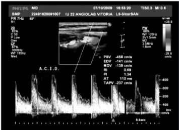
Ultrasound and HDI-5000), using 5 to 13 MHz transduc-ers. The protocol included: a) B-mode gray-scale or col-or-coded images with several anatomical measurements (Figure 1 and 2); b) duplex Doppler ultrasound imag-ing, with measurements of flow velocity in the internal, external, and common carotid arteries and at point of maximal stenosis (Figure 3); c) Doppler color flow map-ping; d) longitudinal and cross-sectional graphs for ca-rotid bifurcations. The report also included qualitative descriptions of the plaque as homogeneous or heteroge-neous, calcified or non-calcified and irregular or regular surface.
he measurements analyzed in the study were: a) esti-mates of carotid stenosis based on anatomical and function-al anfunction-alysis of Doppler low velocity; b) plaque extension; c) distal diameter of the internal carotid artery; d) diameter of the common carotid artery; e) level of carotid bifurcation in relation to the ear lobe.
Statistics
Data obtained from men and women were analyzed separately. Statistical tests were performed in Excel® for windows. Stenosis prevalence was divided into intervals: 90-99%, 80-89%, 70-79%, 60-69%, 50-59%, and <50%. Gender diferences of continuum variables (stenosis per-centage, plaque extension, bifurcation diameter and level) were identiied by unpaired Student’s t test. Diferences re-garding interval prevalence between genres were compared by the χ2 test.
Figure 1. Longitudinal color echography image showing distal and internal carotid arteries. Note the estimates of plaque extension and distal common carotid artery, distal internal carotid artery and minimum residual lumen. The distance between the plaque and the ear lobe is estimated by the position of the transcutaneous transducer.
Figure 2. Cross-sectional echographic imaging of the internal carotid artery in minor lumen region. Estimates of low axis of the elliptical lu-men (more common) or diameter of a circular lulu-men and original ar-tery diameter at the point of highest stenosis.
Figure 3. Duplex Doppler imaging showing high velocities at the point of peak stenosis of the proximal internal carotid that conirm anatomi-cal indings.
Results
Carotid stenosis
he mean degree of carotid stenosis was 70±11% (30 to 95%) for women and 72±12% (40 to 98%) for men. Despite the similarity of results between genres, the diference was statistically signiicant (p=0.013). Table 1 depicts the preva-lence of stenosis for each of the aforementioned intervals. Only the prevalence in the 90-99% interval, which was higher in males, was statistically signiicant.
Plaque extension
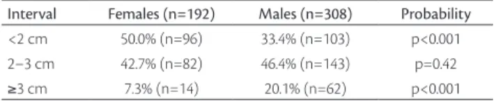
p<0.001). Table 2 shows the prevalence of plaque extension for three intervals: minor, median, and major. Comparison between genders showed higher prevalence of plaques less than 2 cm long in women and plaques more than 3 cm in men. he vast majority of plaques were less than 3 cm in both genders.
Distal diameter of the internal carotid artery
he diameter of the internal carotid artery distal to the bulb was signiicantly greater in men (4.9±0.9 mm – 2.0 to 9.0 mm) than in women (4.6±0.8 mm – 2.0 to 8.0 mm; p=0.001). Table 3 shows that the comparison of speciic di-ameters found large or small carotid arteries carotid in both men and women. However, prevalence of distal internal ca-rotid diameter between 4 and 5 mm was higher in females and from 5 to 6 mm in males.
Diameter of the common carotid artery
he common carotid artery diameter was signiicant-ly higher in men (7.6±1.3 mm – 3.7 to 11.8 mm) than in women (7.1±1.4 mm – 4.0 to 10.0 mm), with p<0.001. Table 4 shows a high prevalence of diameter less than 7 mm in females, and more than 8.5 mm in males. Most common carotid arteries in both genders were in the 7.0 to 8.5 mm interval.
Carotid bifurcation level
he distance from the ear lobe to carotid bifurcation was signiicantly greater in men (5.9±1.1 cm – 2.5 to 10.0 cm) as compared to women (5.3±0.9 cm – 3.0 to 8.0 cm), with p<0.001. Table 5 shows that even though the prevalence was low, women presented higher bifurcations more frequently, which could be a contraindication for surgical treatment. Most bifurcations in women were located 4 to 6 cm from the ear lobe, while most bifurcations in men were located at a distance of more than 6.0 cm.
Discussion
his section emphasizes: a) addresses the value of the carotid ultrasonographic mapping as an essential exam for open or endovascular extracranial catorid treatment plan-ning; b) selection of ultrasonographic variables evaluated; c) comparative analysis between male and female genders. he results of the comparative statistical analyses justify the discussion on the importance of a second DUS exami-nation and on the variables chosen for analysis, besides
emphasizing that individual variations should be taken into account in treatment planning.
Preoperative ultrasonographic mapping is a comple-mentary method to the initial diagnosis and restricted to patients with indication for surgery. It is performed strictly on the side that is being treated. A complementary and speciic mapping should not be included in diagnos-tic ultrasonography in order to avoid high costs, for the majority of patients evaluated in non-invasive vascular laboratories are candidates to medical rather than surgical treatment.
Doppler ultrasonography has a well-established role in the evaluation of patients referred to surgery. In a reference service, almost 90% of carotid bifurcation operations were preceded by an ultrasonographic study10.
Interval Females (n=192) Males (n=308) Probability 90–99% 7.8% (n=15) 14.3% (n=44) p=0.029 80–89% 20.8% (n=40) 24.4% (n=75) p=0.36 70–79% 38.0% (n=73) 34.1% (n=105) p=0.37 60–69% 25.5% (n=49) 20.1% (n=62) p=0.16 50–59% 4.7% (n=9) 6.2% (n=19) p=0.48 <50% 3.1% (n=6) 1.0% (n=3) p=0.078
Table 1. Carotid bifurcation stenosis.
Interval Females (n=192) Males (n=308) Probability <2 cm 50.0% (n=96) 33.4% (n=103) p<0.001 2–3 cm 42.7% (n=82) 46.4% (n=143) p=0.42
≥3 cm 7.3% (n=14) 20.1% (n=62) p<0.001
Table 2. Carotid plaque extension.
Interval Females (n=192) Males (n=308) Probability <4 mm 12.0% (n=23) 10.1% (n=31) p=0.50 4.0–4.9 mm 49.0% (n=94) 36.7% (n=113) p=0.007 5.0–5.9 mm 30.2% (n=58) 39.6% (n=122) p=0.033
≥6.0 mm 8.9% (n=17) 13.6% (n=42) p=0.11
Table 3. Diameters from distal internal carotid artery to carotid bulb.
Interval Females (n=192) Males (n=308) Probability <7 mm 36.5% (n=70) 23.4% (n=72) p=0.002 7.0–8.4 mm 47.9% (n=92) 50.6% (n=156) p=0.55
≥8.5 mm 15.6% (n=30) 26.0% (n=80) p=0.007
Table 4. Diameters representing the common carotid artery.
Interval Females (n=192) Males (n=308) Probability <4 cm 5.7% (n=11) 1.3% (n=4) p=0.005 4.0–5.9 cm 63.5% (n=122) 37.3% (n=115) p<0.001
≥6.0 cm 30.7% (n=59) 61.4% (n=189) p=0.001
Doppler ultrasonography has also been regarded as an essential method in the perioperative evaluation of carotid endarterectomies2,5. The location and length of
the incision have been guided by ultrasonography5, but
ultrasonography is so sensitive that is has allowed en-darterectomy in carotid previously diagnosed with oc-clusion by other radiographic or RMI techniques14. Some
reference centers already use it as a guide technique in endovascular procedures4,7.
The importance of ultrasonographic evaluation of the extracranial carotid arteries based on correlations between anatomic measurements and flow velocities has been emphasized in the literature3,15. Particularly, DUS
exams showing high degree stenosis (more than 80%) and non-significant stenosis (less than 50%) have high positive predictive values, when compared to “lumino-graphic” methods, such as contrast angiography. For mid-range stenosis (60 to 80%), DUS positive predictive values have been increased when high flow velocity has been added to the measurements.
he term arteriography, popularly associated with ra-diologic luminography has been misused. Such technique is not actually “arteriographic”, for it does not yield direct information on the arterial wall or on atheromatous le-sions. Besides this technical failure, radiological luminog-raphy has shown severe complications rates of 0.4 to 1.2% in studies of carotid endarterectomy16,17. In medical
prac-tice, the increase in the number of asymptomatic patients currently treated has minimized the need for detailed in-formation about intracranial arteries such as the aortic arch. Ultrasonography has been regarded as a reliable im-aging technique for the assessment of patients examined immediately ater symptoms onset18.
Echography, on the other hand, is a true arteriogra-phy of the carotid bifurcation wall, showing the degree of stenosis, plaque extension, the characteristics of the atheromatous plaque, including ulceration, the level of the carotid bifurcation and residual lumen diameters. Studies of echographic characterization have shown that unstable plaques are associated with higher incidence of symptoms on plaque follow-up and at endovascular treatment19,20. Cross-sectional ultrasonographic images
explain why radiologic luminography may underesti-mate the degree of stenosis in the presence of elliptic lumens (the majority of cases) 11 or even in cases of
over-lapping images of the internal and external carotid arter-ies. Besides that, color Doppler mapping allows indirect evaluation of severe anomalies of the aortic arch or in-tracranial arteries.
It should be emphasized however that the measure-ments of flow velocity are influenced by the heart rate and rhythm, by carotid stenosis, tortuosities or con-tralateral occlusion, or by the patients’ gender. In such instances, the images, allow for correction of stenosis estimates. In our service we have moved on from a di-agnostic ultrasonographic exam followed by the contrast radiologic examination to confirm the diagnosis to an algorithm of a double ultrasonographic investigation1.
This way, contrast arteriography is not used or used very selectively in our patients. The diagnostic ultrasonogra-phy is performed by examiner 1, based on clinical indi-cations. If a stenosis less than 50% is found, the case is discussed and usually referred to medical management and long-term follow-up. In patients with indication for open or endovascular carotid surgery, a second preoper-ative DUS mapping is performed by examiner 2. In case of contradictions between the two examinations, the pa-tient is again examined by the first examiner or another imaging method (magnetic resonance or CT scan) is ordered. Our surgical team also performs echographic mapping as a means of confirming DUS performed in vascular laboratories elsewhere. Out of the nine cases (<2%) reported as less than 50% carotid stenosis by DUS mapping, six had the initial examination performed else-where, two had ulceration of the carotid plaque and one patient had carotid dissection, with a flap.
An algorithm based in two ultrasonographic studies – one for diagnosis and other for preoperative mapping – reduces costs in 70 to 90%, compared to radiologic or magnetic resonance imaging. he latter methods are more expensive and sometimes less reliable for planning the de-tails of the surgical procedure. he approach, the length and location of the incision for endarterectomy may be ef-iciently planned using ultrasonographic mapping alone5.
In summary, ultrasonography is fundamental for the diagnosis of carotid stenosis. CT scan or MRI may be per-formed as a means of conirming degree of stenosis grade or for a thorough evaluation of a symptomatic patient21,22.
Our experience shows that ultrasonographic mapping may replace these expensive methods and provide consis-tent data for open or endovascular surgical planning.
One of the aims of this study is to avoid conjoined anal-ysis of subgroups that are better analyzed separately. For in-stance, gender subgroups should be evaluated separately.
Data from the literature have shown that even though the prevalence of signiicant stenosis did not depend on the patient’s gender8, restenosis ater endarterectomy
females8. Greater diameters among men were expected
and conirmed23,24. he length of stay in the hospital was
shorter in men compared to women12. Blood low velocity
in the carotid artery may also difer between genders13.
Justiication for the selection of variables was as fol-lows: carotid stenosis diameter percentage may not be a strictly scientiic variable, for a carotid stenosis with a circular lumen is rare. his variable, however, is used in medical practice, and it has been used in the classic ran-domized trials that have conirmed the value of surgical carotid treatment. Plaque extension helps planning the incision for endarterectomy and selecting the appropriate devices for endovascular treatment. Previous knowledge of the common and internal carotid diameters prepare the surgical team for problems associated with small caliber vessels – specially in women – and helps planning endo-vascular treatment, including placement of ilters and se-lection of balloon catheters and stents. he distance from the carotid plaque to the ear lobe helps in the choice of in-cision site and calls the surgeon’s attention to a potentially high bifurcation, with its attendant diiculties of exposure of the segment to be treated.
his study has shown gender diferences in degree of stenosis (percentage), carotid plaque extension, carotid artery diameter, and anatomic location of the plaque and the carotid bifurcation. Men present a higher prevalence of critical stenosis, plaques longer than 3 cm and greater distance between the plaque and the ear lobe. A carotid bifurcation high up in the neck was particularly important to identify, for it is associated with intraoperative techni-cal diiculties. A suggestion could be made for screening males earlier than usual. Even though gender diferences do exist, , further studies are necessary those men and women that do not it the usual patterns.
We concluded that non invasive ultrasonographic im-aging of the extracranial carotid arteries’ anatomy show signiicant diferences between males and females. hese indings emphasize reinforce that a pre-treatment proto-col should include information on individual anatomic diferences that might contribute to the eiciency of open and endovascular procedures.
References
1. Sandri JL. Endarterectomia carotídea somente com duplex. In: Nectoux Filho JL, Salles Cunha S, Paglioli AS, de Souza GG, Pereira AH (editores). Ultra-sonograia vascular. Rio de Janeiro: Revinter; 2000, p. 71-5.
2. Ascher E, Markevich N, Kallakuri S, et al. Intraoperative carotid artery duplex scanning in a modern series of 650 consecutive primary en-darterectomy procedures. J Vasc Surg. 2004;39(2):416-20.
3. Salles-Cunha SX, Ascher E, Hingorani AP, et al. Efect of ultra-sonography in the assessment of carotid artery stenosis. Vascular. 2005;13(1):28-33.
4. Ascher E, Marks NA, Schutzer RW, et al. Duplex-assisted internal carotid artery balloon angioplasty and stent placement: a novel approach to minimize or eliminate the use of contrast material. J Vasc Surg. 2005;41(3):409-15.
5. Ascher E, Hingorani A, Marks N, et al. Mini skin incision for carotid endarterectomy (CEA): a new and safe alternative to the standard approach. J Vasc Surg. 2005;42(6):1089-93.
6. Menezes FH, Luccas GC, Matsui IA, et al. Avaliação através da ul-tra-sonograia duplex da medida de reestenose da carótida interna dos pacientes submetidos à endarterectomia aberta da bifurcação carotídea, com eversão parcial da carótida interna. J Vasc Bras. 2005;4(1):47-54.
7. Ascher E, Hingorani AP, Marks N. Duplex-assisted internal carotid artery balloon angioplasty and stent placement. Perspect Vasc Surg Endovasc her. 2007;19(1):41-7.
8. Freitas P, Piccinato CE, Martins WP, et al. Aterosclerose carotídea avaliada pelo eco-dopppler: associação com fatores de risco e doenças arteriais sistêmicas. J Vasc Bras. 2008;7(4):298-307.
9. Blackshear Junior WM, Connar RG. Carotid endarterectomy with-out angiography. J Cardiovasc Surg (Torino). 1982; 23(6):477-82.
10. Chiesa R, Melissano G, Castellano R, et al. Carotid endarterectomy: experience in 5425 cases. Ann Vasc Surg. 2004;18(5):527-34.
11. Pan XM, Saloner D, Reilly IM, et al. Assessment of carotid artery stenosis by ultrasonography, conventional angiography, and mag-netic resonance angiography: correlation with ex vivo measure-ment of plaque stenosis. J Vasc Surg. 1995;21(1):82-9.
12. Hernandez N, Salles-Cunha SX, Daoud YA, et al. Factors re-lated to short length of stay after carotid endarterectomy. Vasc Endovascular Surg. 2002;36(6):425-37.
13. Comerota AJ, Salles-Cunha SX, Daoud Y, et al. Gender difer-ences in blood velocities across carotid stenoses. J Vasc Surg. 2004;40(5):939-44.
14. Ascher E, Markevich N, Hingorani A, et al. Pseudo-occlusions of the internal carotid artery: a rationale for treatment on the basis of a modiied carotid duplex scan protocol. J Vasc Surg. 2002;35(2):340-5.
15. Beebe HG, Salles-Cunha SX, Scissons RP, et al. Carotid arterial ultra-sound scan imaging: A direct approach to stenosis measurement. J Vasc Surg. 1999;29(5):838-44.
16. Endarterectomy for asymptomatic carotid artery stenosis. JAMA. 1995;273(18):1421-8.
17. Hobson RW 2nd, Weiss DG. Fields WS, et al. Eicacy of carotid endarterectomy for asymptomatic carotid stenosis. he Veterans Afairs Cooperative Study Group. N Engl J Med. 1993; 328:221-7.
18. Wardlaw JM, Stevenson MD, Chappell F, et al. Carotid ar-tery imaging for secondary stroke prevention: both imaging modality and rapid access to imaging are important. Stroke. 2009;40(11):3511-7.
20. Biasi G, Froio A, Dietrich EB et al. Carotid plaque echolucency increases the risk of stroke in carotid stenting: the Imaging in Carotid Angioplasty and Risk of Stroke (ICAROS) study. Circulation 2004;110(6):756-62.
21. Roi M, Lüshcer TF. Management of patients with carotid artery stenosis. Herz. 2008;33(7):490-7.
22. Lanzino G, Tallarita T, Rabinstein AA. Internal carotid artery stenosis: natural history and management. Semin Neurol. 2010;30(5):518-27
23. Kreiza J, Arkuszewski M, Kasner SE, et al. Carotid artery diameter in men and women and the relation to body and neck size. Stroke. 2006;37(4):1103-5.
24. den Hartog AG, Algra A, Moll FL, et al. Mechanisms of gender-related outcome diferences after carotid endarterectomy. J Vasc Surg. 2010;52(4):1062-71.
Correspondence Sandra Maria Pontes Rua José Teixeira, 290 – Praia do Canto CEP 29055-310 – Vitória (ES), Brasil E-mail: [email protected]
Author’s contribution Conception and design: SMP, FSB, JLS, CMJ Analysis and interpretation: SMP, FSB, LHR, MATA, JLS, CMJ, DPN, SXSC Data collection: SMP, FSB, LHR, MATA, JLS, CMJ, DPN Writing the article: SMP, FSB, SXSC Critical revision of the article: SMP, SXSC Final approval of the article*: SMP, FSB, LHR, MATA, JLS, CMJ, DPN, SXSC Statistical analysis: SXSC Overall responsibility: SMP *All authors have read and approved of the inal version of the article