Detection of high risk human
papillomavirus by hybrid
capture II
®
according cytological
indings in women treated for
squamous intraepithelial lesions
of the cervix, period 2006/2010
Detección del virus del papiloma
humano de alto riesgo por captura
híbrida II
®
según hallazgos
citológicos en mujeres tratadas por
lesiones escamosas intraepiteliales
de cuello uterino, período 2006/2010
Pamela Mongelós
Malvina Páez
Isabel Rodriguez-Riveros
Graciela Giménez
Amalia Castro
Laura Mendoza
Health Sciences Research Institute, National University of Asunción, Asunción, Paraguay.
Conlicts of interest: Authors declared there were no conlicts of interest
Corresponding author: Laura Mendoza. Departamento de Salud Pública y Epidemiología, Institu-to de Investigaciones en Ciencias de la Salud, Universidad Nacional de Asunción, Iturbe 1184, P.O. Box 2017, Asunción, Paraguay. E-mail: [email protected]
Abstract
Objective: To determinate the frequency of high risk human papillomavirus (HR-HPV) by hybrid capture II ® (CH II®), according
cytology results in women treated for squa-mous intraepithelial lesions of the cervix (SIL). Material and Methods: A descriptive cross-sectional study of a series of cases that included 122 women treated, 79 (75%) for low grade SIL (LSIL) and 43 (35%) for high grade SIL (HSIL) attending at the HPV Laboratory at the Health Sciences Research Institute (IICS), National University of Asunción (UNA), for post-treatment control during period 2006/2010. Results: A total of 28% (34/122) of women treated for SIL were positive for HR-HPV, detecting viral infection in 20% of women with no SIL (NSIL) (22/108), in 83% of women with LSIL (10/12) and in 100% of women with HSIL (2/2). Of 34 women positive for HR-HPV, 10 women (29%) had high values (100 pg / mL or more) of relative viral load, detecting an increase of positive cases with severity of the lesion (28% NSIL, 30% LSIL, 50% HSIL). Conclusion: HR-HPV detection by CH II®
and high relative viral load values especially in women with NSIL could help to identify treated women at risk of developing recur-rence, thereby contributing to strengthening the cervical cancer prevention program.
Keywords: HPV. Post-treatment control. Hybrid Capture II ®. Paraguayan women.
Resumen
Objetivo: Determinar la frecuencia del virus de papiloma humano de alto riesgo onco-génico (HR-HPV) por captura híbrida II ®
(CH II®) según hallazgos citológicos en
mu-jeres tratadas por lesiones escamosas intra-epiteliales (SIL) de cuello uterino. Material y Método: Estudio descriptivo de corte transverso de una serie de casos, en donde se incluyeron 122 mujeres tratadas, 79 (65%) por SIL de bajo grado (LSIL) y 43 (35%) por SIL de alto grado (HSIL) que concurrieron al Laboratorio de HPV del Instituto de Investigaciones en Ciencias de la Salud, Universidad Nacional de Asunción, para realizarse un control post-tratamiento, pe-riodo 2006/2010. Resultados: Se observó un total del 28% (34/122) de mujeres tratadas por SIL positivas para HR-HPV, detectándo-se infección viral en un 20% de las mujeres con ausencia de SIL (NSIL) (22/108), 83% de las mujeres con LSIL (10/12) y 100% de las mujeres con HSIL (2/2). De las 34 mujeres positivas para HR-HPV, 10 mujeres (29%) presentaron valores altos (100 pg/mL o más) de carga viral relativa, detectándose un aumento de casos positivos con la severidad de la lesión (28% NSIL, 30% LSIL, 50% HSIL). Conclusión: La detección de HR-HPV por CH II®, así como los valores de carga viral
relativa altos, en especial en mujeres con NSIL podrían ayudar a identificar mujeres tratadas con riesgo a desarrollar recidivas, contribuyendo así a fortalecer el programa de prevención de cáncer de cuello uterino.
Palabras claves: HPV. Control post tra-tamiento. Captura Híbrida II ®. Mujeres
Paraguayas. Carga viral relativa. Diagnóstico citológico.
Introduction
Cervical cancer is the third most fre-quent type of cancer in women worldwide, including 529,828 new cases and 275,128 deaths in 2008, of which 31,712 occurred in Latin America and the Caribbean. A total of 85% of new cases take place in developing countries. The standard incidence rate by age in South America is 24.1 cases/100,000 women. In Paraguay, the incidence and mortality rates are 35.0 and 16.6/100,000 women, respectively. These rates are sig-nificantly higher than those recorded in neighboring countries such as Argentina, Brazil, Uruguay and Chile1.
The human papillomavirus (HPV) is a factor involved in the development of cer-vical cancer. There are more than 100 types of HPV, of which approximately 40 infect the anogenital mucosa. These are categorized into high-risk viruses (HR-HPV) and low--risk viruses (LR-HPV), according to their oncogenic potential, and the HR-HPV (16, 18, 31, 33, 35, 39, 45, 51, 52, 56, 58, 59, and 66) account for 95% of all cervical cancer cases2-5.
The cytological diagnosis (Papanicolaou test) is a simple low-cost method used to detect the cytopathic effect of this virus, al-though not determining the viral type. As the HPV does not grow in traditional cell cultu-res, molecular methods have been recently used to identify the viral genotype, such as the Hybrid Capture II® test (HC-II®), which
detects 13 types of HR-HPV and enables the viral load to be estimated6,7.
surgical margins show a risk of treatment failure of 2-6%, regardless of the treatment used. The cumulative rate of invasion eight years after treatment is 5.8 cases/1,000 wo-men, which is five times higher than that of the general population8-14.
In addition to considering the com-promised surgical margins as a recurrence factor, there are other factors that promote the persistence or recurrence of squa-mous intraepithelial lesions (SIL) after treatment, such as age, parity, cytological diagnosis and degree of lesion prior to treatment15.
As a result, a continuous and thorough follow-up is important after the treatment. Recent studies suggest that the combined use of cytology and the HC-II® test
increa-ses effectiveness when selecting women at risk of developing residual or recurrent SIL after six months of treatment16-19.
In Paraguay, previous studies have ob-served a high frequency of HR-HPV in wo-men without cervical lesions and cofactors of risk (a high number of sexual partners and multiparity, among others) associated with the development of cervical cancer, com-parable to or higher than those identified in other developing countries, which could partly explain the high incidence of such type of cancer in this country20-23.
In Paraguay, there have been no studies on the frequency of HR-HPV in women treated for SIL. Thus, considering the high--risk sexual behavior of Paraguayan women, which could promote post-treatment viral infection and increase the risk of recurrence, the present study aimed to determine the frequency of HR-HPV using the HC-II® test,
according to cytological results in women treated for cervical SIL, between 2006 and 2010.
Methods
Study population
A descriptive cross-sectional study of a series of cases was conducted with 122 women treated for cervical lesions
who sought the Department of Public Health and Epidemiology of the Instituto de Investigaciones en Ciencias de la Salud
(IICS –Health Sciences Research Institute) of the Universidad Nacional de Asunción
(UNA – National University of Asunción), between 2006 and 2010, with a medical recommendation to have the HC-II® test
performed for HR-HPV detection.
The treated women who went to the IICS-UNA originated from different public and private health centers of the Central Department of Paraguay. The present study included women who had a cytohistologi-cal diagnosis of their lesions prior to the treatment, were treated at least six months before, and had an updated cytological diagnosis performed after the treatment, when the HR-HPV was detected by the HC-II test. Women who sought the HC-IICS, UNA, more than two years after the treatment had been performed were excluded from this study. The treatment applied depen-ded on the medical criteria and was based on the Paraguayan Manual on Norms and Procedures for Cervical Cancer Control and Prevention, developed by the Ministry of Public Health24.
Prior to treatment, of all 122 participa-ting women, 79 had a confirmed histological diagnosis of low-grade SIL (LSIL) and 43 of high-grade SIL (HSIL). Of all women, 97 un-derwent LEEP and 25, cervical conization. All women completed a questionnaire that included demographic, socioeconomic, gyneco-obstetric and sexual behavior cha-racteristics. All information was processed while protecting patients’ confidentiality. This research project was approved by the Research Ethics Committee of the IICS of the
Universidad Nacional de Asunción, under number M07/10.
The minimum sample size of 144 women treated was calculated using the table in the 13E Appendix for a descrip-tive study with a dichotomous variable, considering 30% of expected frequency of HR-HPV after the treatment, an amplitude (ω) of 0.15 and a 95% confidence interval
Detection of cervical lesions in women treated using cytological diagnosis
The cytological analysis was performed in the participating health centers. Samples were obtained by Pap smear and placed on polished microscope slides that were properly identified. These slides were fully immersed in a wide-rimmed container with 96% ethyl alcohol for 15 minutes. Slides were subsequently removed from the container and left to dry for ten minutes. Finally, fixing spray was applied to them. The cytological results were classified according to the 2001 Bethesda system26.
Detection of HR-HPV in women treated using the HC-II® test
The study sample was collected in the IICS, UNA, using a cytobrush, which was introduced into a collection tube provided by its manufacturing company, Qiagen (Germany).
The samples were processed with the HC-II test, which detects 13 types of HR-HPV: 16, 18, 31, 33, 35, 39, 45, 51, 52, 56, 58, 59 and 68. Following the manufacturer’s protocol, the cervical samples were treated with sodium hydroxide, aiming to denature the DNA. The simple-stranded DNA was hy-bridized in a solution with a cocktail of RNA probes from the 13 types of HR-HPV. Each reaction mix including RNA-DNA hybrids was transferred to microplates sensitized with anti-hybrid antibodies, enabling their immobilization. The RNA-DNA hybrids bound to antibodies were put into contact with a second antibody conjugated with alkaline phosphatase. The non-binding material was removed by successively wa-shing it, and a chemiluminescent reactive (Lumi-Phos 530) was subsequently added as substrate for the alkaline phosphatase. The luminescence produced by this reaction was measured with a luminometer. The unit of measurement of light was expressed as rela-tive light units (RLU). Posirela-tive and negarela-tive controls originated from Qiagen were used in triplicate.
The relative viral load was obtained by comparing the sample RLU with those of
the positive control (RLU/PC). Samples with RLU/PC ≥ 1.0 pg/mL were considered as positive. The relative viral load was divided into four categories according to their values in pg/mL: 1 ≤ value < 10 pg/mL (low relative viral load); 10 ≤ value < 100 pg/mL (average relative viral load); 100 ≤ value < 1,000 pg/ mL (high relative viral load); and value ≥ 1,000 pg/mL (very high relative viral load)27.
It should be emphasized that profes-sionals who participated in the sample processing and reporting of results from the HC-II® test were blind to the cytological
results. Consequently, the results of both methods were analyzed independently.
Statistical analysis
Data analysis was performed using descriptive statistics procedures and the Epi Info software, version 3.2 (CDC, Atlanta, USA). Chi-square analysis was performed to identify the association among type of tre-atment, length of time since the treatment and frequency of HR-HPV infection, using the Epi Info software, version 3.2. A p-value <0.05 was considered to be significant in all data analyses performed.
Results
The mean age of the 122 participating women was 34 ± 10 years (95%CI17-67). Information about socioeconomic charac-teristics, obstetric history and sexual beha-vior could be obtained from 86% of the study population (105/122 women) (Table 1).
In all, 28% (34/122) of participants treated for SIL were positive for HR-HPV, and viral infection was detected in 20% of women without SIL (NSIL) (22/108), 83% of those with LSIL (10/12) and 100% of those with HSIL (2/2) (Table 2).
It should be emphasized that there were no significant differences among frequen-cies of HR-HPV according to type of treat-ment, and 27/97 (28%) women treated with LEEP and 7/25 (28%) treated with cervical conization had the viral infection (p = 0.7).
Table 1 -Socioeconomic characteristics, obstetric history and sexual behavior among women treated for cervical squamous intraepithelial lesions.
Tabla 1 - Características socioeconómicas, antecedentes obstétricos y conducta sexual de las mujeres tratadas por lesiones escamosas intraepiteliales de cuello uterino.
Variables* n (%)
Level of education
Complete primary school 32 (30)
Secondary school and higher 73 (70)
Smoking habit
Yes 7 (7)
No 98 (93)
Use of hormonal contraceptive
Yes 35(33)
No 70(67)
Pregnancy
Presence 80 (76)
Absence 25 (24)
Number of pregnancies
>2 34 (32)
<2 46 (44)
Age of irst pregnancy (in years)
≤19 28 (27)
>19 52 (50)
Age of irst sexual intercourse (in years)
<19 57(54)
≥19 48 (46)
Number of sexual partners
>2 40 (38)
≤2 65 (62)
* Information about socioeconomic characteristics, obstetric history and sexual behavior could be obtained from 86% of the study population (105/122 women).
*Fue posible obtener información acerca de las características socioeconómicas, antecedentes obstétricos y conducta sexual del 86% de la población en estudio (105/122 mujeres).
Table 2 -HR-HPV results for CH II® in women treated for cervical squamous intraepithelial
lesions according cytologic diagnosis.
Tabla 2 - Resultado de HR-HPV por CH II® en mujeres tratadas por lesiones escamosas
intraepiteliales según el diagnóstico citológico.
Cytological diagnosis n HR-HPV infection
Positive n (%) Negative n (%)
NSIL 108 22 (20) 86 (80)
LSIL 12 10 (83) 2 (17)
HSIL 2 2 (100) 0 (0)
Total 122 34(28) 88(72)
HR-HPV: high risk human papilloma virus; NSIL: no squamous intraepithelial lesion, LSIL: low grade squamous intraepithelial lesion; HSIL: high grade squamous intraepithelial lesion.
differences in frequencies of HR-HPV be-tween women who had a test to detect viral infection until one year after their treatment (32% 25/77 women) and those who did so after one year (20%, 9/45 women), p = 0.1.
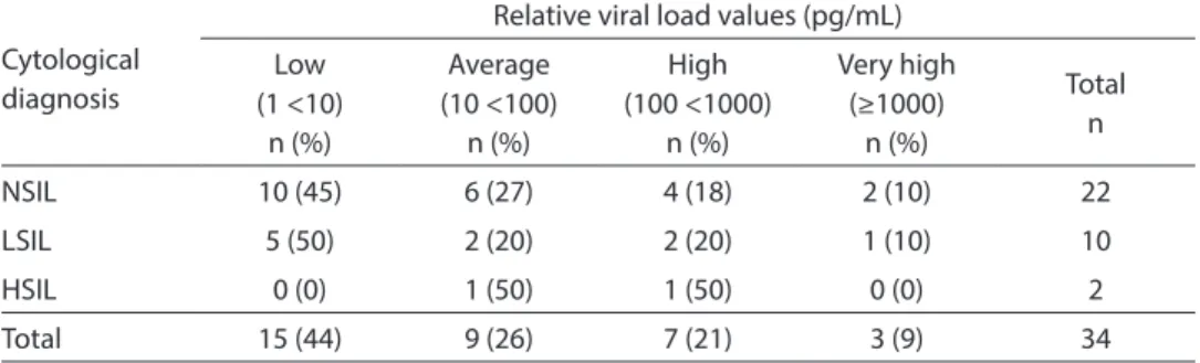
In terms of the relative viral load, of all women positive for HR-HPV, 29% (10/34) had high or very high values. According to the cytological diagnosis, 28% of women with NSIL, 30% with LSIL and 50% with HSIL had high or very high relative viral loads. It should be emphasized that none of the rela-tive viral load values in women with HSIL was low (Table 3).
Discussion
There is a high incidence of pre-neoplas-tic lesions and cervical cancer in Paraguay, which are considered to be a public health problem. The present study showed prelimi-nary results of HR-HPV detection with the HC-II® test, according to the cytological
di-agnosis performed in women treated for SIL. In this study, the frequency of women treated for SIL and positive for HR-HPV (28%) was similar or higher than those found in Brazil (33% or 22/67 women and 22% or 16/74 women), Korea (18% or 44/243 women) and Belgium (29% or 21/72 women), where women positive for HR-HPV had a high frequency (from 18% to 44%) of
recurrence of SIL16,28-30. This suggests that
post-treatment HR-HPV detection can be a useful tool in the follow-up and guidance of treated women.
With regard to the cytological diagnosis, 20% of women with NSIL were positive for HR-HPV. This lack of agreement between the HC-II® test and the cytological
diag-nosis could have been due to the fact that the former enables the viral presence to be detected even when there is no lesion6,22.
Previous studies suggest that women with NSIL and positive for HR-HPV have a higher risk of developing cervical lesions. Koutsky et al. observed that 30% of women with a normal cytological diagnosis and positive for HR-HPV developed intraepi-thelial lesions in the course of two years. It should be emphasized that 80% (86/122) of women with NSIL had negative results for HR-HPV and, considering the high nega-tive predicnega-tive value of the HC-II test, have a reduced risk of developing high-grade lesions14,16-18,31-35.
The results found in the present study suggest that the frequency of HR-HPV in treated women does not vary according to the type of treatment and length of time spent to perform the post-treatment control (until two years), which could be due to other factors playing a role in the persis-tence of post-treatment HR-HPV infection.
Table 3 - Frequency of women treated for cervical squamous intraepithelial lesions positive for HR-HPV by CH II® accoding relative viral load values.
Tabla 3 - Frecuencia de mujeres tratadas por lesiones escamosas intraepiteliales positivas para HR-HPV por CH II® según valores de carga viral relativa.
Cytological diagnosis
Relative viral load values (pg/mL)
Low (1 <10)
n (%)
Average (10 <100)
n (%)
High (100 <1000)
n (%)
Very high (≥1000)
n (%)
Total n
NSIL 10 (45) 6 (27) 4 (18) 2 (10) 22
LSIL 5 (50) 2 (20) 2 (20) 1 (10) 10
HSIL 0 (0) 1 (50) 1 (50) 0 (0) 2
Total 15 (44) 9 (26) 7 (21) 3 (9) 34
NSIL: no squamous intraepithelial lesion; LSIL: low grade squamous intraepithelial lesion; HSIL: high grade squamous intraepi-thelial lesion.
Previous studies observed that women with HSIL infected with HPV-16, 18, 33 and 45 and those with multiple types of HPV prior to LEEP or cervical conization have a sig-nificantly higher frequency of viral infection recurrences after the treatment15,36,37.
With regard to the relative viral load, previous studies suggest that patients with a persistent and high relative viral load have an increased risk of developing HSIL in a short period of time, even in the absence of detectable cytological changes38,39. In terms
of the cytological diagnosis and in agree-ment with the results observed in the pres-ent study, the greater the severity of SIL, the higher the frequency of women with a high or very high relative viral load. It should be emphasized that there were no low relative viral load values among women with HSIL, which indicates that women positive for HR-HPV with a low relative viral load have a lower probability of developing HSIL, as observed in other studies. Thus, the present
results point out that relative viral load values could contribute to post-treatment guidance and control of women, identifying those at a greater risk of developing cervical lesions again38,39.
The present study had certain limita-tions, due to the impossibility of completing the sample comprised of 144 women and the lack of data on women’s relative viral load prior to the treatment. Nonetheless, the results observed suggest that the use of the HC-II® test for HR-HPV detection
and determination of relative viral load as a complement to the cytological diagnosis could contribute to the identification of women treated for pre-neoplastic lesions at a greater risk of having recurrences. The data obtained could help to strengthen the Paraguayan Cervical Cancer Prevention, Detection and Treatment Program, devel-oped by the Ministry of Public Health and aimed at improving the service provided by Primary Health Care units.
References
1. Ferlay J, Shin HR, Bray F, Forman D, Mathers C, Parkin DM. Estimates of worldwide burden of cancer in 2008: GLOBOCAN 2008. Int J Cancer 2010; 127: 2893-917. 2. Carestiato FN, Silva KC, Dimetz T, Oliveira LH,
Cavalcanti SM. Prevalence of human papillomavirus infection in the genital tract determined by hybrid capture assay. Braz J Infect Dis 2006; 10(5): 331-6. 3. zur Hausen H. Papillomavirus infections-a major cause
of human cancers. Biochim Biophys Acta 1996; 1288(2): 55-78.
4. Walboomers JM, Jacobs MV, Manos MM, Bosch FX, Kummer JA, Shah KV et al. Human papillomavirus is a necessary cause of invasive cervical cancer worldwide. J Pathol 1999; 189(1): 12-9.
5. Wright TC Jr, Schiffman M, Solomon D, Cox JT, Garcia F, Goldie S, et al. Interim guidance for the use of human papillomavirus DNA testing as an adjunct to cervical cytology for screening. Obstet Gynecol 2004; 103(2): 304-9.
6. Dôres GB, Taromaru EK, Bonomi CG, Longatto Filho A, Gilli NP, Matsubara S, et al. HPV infection detected by hybrid capture II: correlation with morphological findings. DST – J Bras Doenças Sex Transm 2005; 17(4): 255-8.
7. Carvalho MOO, Almeida RW, Leite FMS. Detection of Human Papillomavirus DNA by the Hybrid Capture Assay. Braz J Infect Dis 2003; 7(2): 121-5.
8. Kucera E, Sliutz G, Czerwenka K, Breitenecker G, Leodolter S, Reinthaller A. Is high-risk human papillomavirus infection associated with cervical intraepithelial neoplasia eliminated after conization by large-loop excision of the transformation zone? Eur J Obstet Gynecol Reprod Biol 2001; 100: 72-6.
9. Nagai Y, Maehama T, Asato T, Kanazawa K. Persistence of human papillomavirus infection after therapeutic conization for CIN 3: is it an alarm for disease recurrence? Gynecol Oncol 2000; 79: 294-9. 10. Bollen LJM, Tjong-A-Hung SP, van der Velden J, Mol
BW, ten Kate FW, ter Schegget J et al. Prediction of recurrent and residual cervical dysplasia by human papillomavirus detection among patients with abnormal cytology. Gynecol Oncol 1999; 72: 924-9.
12. Gardeil F, Barry-Walsh C, Prendiville W, Clinch J, Turner M. Persistent intraepithelial neoplasia after excision for cervical intraepithelial neoplasia grade III. Obstet Gynecol 1997; 89: 419- 22.
13. Mohamed-Noor K, Quinn MA, Tan J. Outcomes after surgical cold knife conization with complete and incomplete excision of abnormal epithelium: a review of 699 cases. Gynecol Oncol 1997; 67: 34-8.
14. Soutter WP, de Barros Lopes A, Fletcher A, Monaghan JM, Duncan ID, Paraskevaidis E et al. Invasive cervical cancer after conservative therapy for cervical intraepithelial neoplasia. Lancet 1997; 349: 978-80. 15. Nam K, Chung S, Kim J, Jeon S, Bae D. Factors associated
with HPV persistence after conization in patients with negative margins. J Gynecol Oncol 2009; 20(2): 91-5. 16. Park JY, Bae J, Lim MC, Lim SY, Lee DO, Kang S et al. Role
of high risk-human papilloma virus test in the follow-up of patients who underwent conization of the cervix for cervical intraepithelial neoplasia. J Gynecol Oncol 2009; 20(2): 86-90.
17. Jeong NH, Lee NW, Kim HJ, Kim T, Lee KW. High-risk human papillomavirus testing for monitoring patients treated for high-grade cervical intraepithelial neoplasia.
J Obstet Gynaecol Res 2009; 35(4): 706-11.
18. Ribaldone R, Boldorini R, Capuano A, Arrigoni S, Di Oto A, Surico N. Role of HPV testing in the follow-up of women treated for cervical dysplasia. Arch Gynecol Obstet 2010; 282(2): 193-7.
19. Aerssens A, Claeys P, Beerens E, Garcia A, Weyers S, Van Renterghem L, et al. Prediction of recurrent disease by cytology and HPV testing after treatment of cervical intraepithelial neoplasia. Cytopathology 2009; 20(1): 27-35.
20. Rolón PA, Smith JS, Muñoz N, Klug SJ, Herrero R, Bosch X et al. Human papillomavirus infection and invasive cervical cancer in Paraguay. Int J Cancer 2000; 85(4): 486-91.
21. Ruoti de García de Zúñiga M, Arrom de Fresco CH, Ruoti Cosp M, Orué E. Conocimientos, actitudes y prácticas sobre el test de Papanicolau (PAP) en mujeres embarazadas consultantes de hospitales públicos del Departamento de Alto Paraná, Paraguay Mem Inst Investig Cienc Salud. 2008; 6(2): 48-58.
22. Mendoza L, Páez M, Insaurralde A, Rodriguez MI, Castro A, Kasamatsu E. Detection of High Risk Human Papillomavirus Cervical Infections by the Hybrid Capture in Asunción, Paraguay. Braz J Infect Dis 2009; 13(3): 207-10
23. WHO/ICO. Information Centre on HPV and Cervical Cancer (HPV Information Centre). Human Papillomavirus and Related Cancers in Paraguay. Summary Report 2010. Available at: www.who.int/ hpvcentre. [Accessed March 2011]
24. Ministerio de Salud Pública y Bienestar Social. Manual Nacional de Normas y Procedimientos para la Prevención y el Control del Cáncer de Cuello Uterino. Asunción: OPS; 2010.
25. Solomon D, Davey D, Kurman R, Moriaty A, O’Connor D, Prey M et al. For the Forum Group Members and the Bethesda 2001 Workshop. The 2001 Bethesda System. Terminology for Reporting Results of Cervical Cytology.
JAMA 2002; 287: 2114-9.
26. Browner WS, Black D, Newman TB, Hulley SB. Estimación del tamaño de muestra y de la potencia. En: Hulley SB Cumming SR. Diseño de la investigación clínica. Un enfoque epidemiológico. Barcelona: Ediciones Doyma S.A.; 1993. pp. 153-65.
27. Lorincz AT, Castle PE, Sherman ME, Scott DR, Glass AG, Wacholder S. Viral load of human papillomavirus and risk of CIN 3 or cervical cancer. Lancet 2002; 360(9328): 228-9.
28. Verguts J, Bronselaer B, Donders G, Arbyn M, Van Eldere J, Drijkoningen M et al. Prediction of recurrence after treatment for high-grade cervical intraepithelial neoplasia: the role of human papillomavirus testing and age at conisation. BJOG 2006; 113(11): 1303-7.
29. Sarian LO, Derchain SF, Pittal Dda R, Andrade LA, Morais SS, Figueiredo PG. Human papillomavirus detection by hybrid capture II and residual or recurrent high-grade squamous cervical intraepithelial neoplasia after large loop excision of the transformation zone (LLETZ).
Tumori 2005; 91(2): 188-92.
30. Figueirêdo PG, Derchain SF, Sarian LO, Gontijo RC, Andrade LA, Campos EA et al. Detecção do DNA do Papilomavírus Humano após Excisão da Zona de Transformação com Alça Diatérmica para Tratamento de Neoplasia Intra-epitelial Cervical. RBGO 2003; 25(1): 9-15.
31. Cuzick J, Clavel C, Petry KU, Meijer CJ, Hoyer H, Ratnam S et al. Overview of the European and North American studies on HPV testing in primary cervical cancer screening. Int J Cancer 2006; 119(5): 1095-101. 32. Castle PE, Wacholder S, Sherman ME, Lorincz AT,
Glass AG, Scott DR et al.. Absolute risk of a subsequent abnormal pap among oncogenic human papillomavirus DNA-positive, cytologically negative women. Cancer
2002; 95(10): 2145-51.
33. Koutsky LA, Holmes KK, Critchlow CW, Stevens CE, Paavonen J, Beckmann AM et al. A cohort study of the risk of cervical neoplasia grade 2 or 3 in relation to papillomavirus infection. N Engl J Med 1992; 327: 1272-8.
34. Mergui JL, Levêque J.What kind of follow-up after surgical treatment for high-grade cervix lesion? Gynecol Obstet Fertil 2008; 36(4): 441-7.
36. Cecchini S, Carozzi F, Confortini M, Zappa M, Ciatto S. Persistent human papilloma virus infection as an indicator of risk of recurrence of high-grade cervical .intraepithelial neoplasia treated by the loop electrosurgical excision procedure. Tumori 2004; 90(2): 225-8.
37. Wu D, Zheng Y, Chen W, Guo C, Yu J, Chen G, Huang Y. Prediction of residual/recurrent disease by HPV genotype after loop excision procedure for high-grade cervical intraepithelial neoplasia with negative margins.
Aust N Z J Obstet Gynaecol 2011; 51(2): 114-8.
38. Dalstein V, Riethmuller D, Prétet JL, Le Bail Carval K, Sautière JL, Carbillet JP et al. Persistence and load of risk HPV are predictors for development of high-grade cervical lesions: a longitudinal French cohort study. Int J Cancer 2003; 106: 396-403.
39. Jeong NH, Lee NW, Kim HJ, Kim T, Lee KW. High-risk human papillomavirus testing for monitoring patients treated for high-grade cervical intraepithelial neoplasia.
J Obstet Gynaecol Res 2009; 35(4): 706-11.