Review
Printed in Brazil - ©2011 Sociedade Brasileira de Química 0103 - 5053 $6.00+0.00*e-mail: [email protected]
Modulation of the Catalytic Activity of Porphyrins by Lipid- and
Surfactant-Containing Nanostructures
Iseli L. Nantes,*,a Nelson Durán,a,b Sandra M. S. Pinto,a Fabiana B. da Silva,a
Juliana S. de Souza,a Noemia Isoda,a Roberto A. S. Luz,a Tiara G. de Oliveiraa and
Vinícius G. Fernandesa
aCentro de Ciências Naturais e Humanas, Universidade Federal do ABC, Rua Santa Adélia, 166,
Bairro Bangu, 09210-170 Santo André-SP, Brazil
bInstituto de Química, Universidade Estadual de Campinas,
CP 6154, 13083-970 Campinas-SP, Brazil
Fatores estruturais que modulam a atividade das poririnas englobam os ligantes equatoriais, o metal central e o número e estrutura dos seus ligantes axiais. A esses fatores se associam o microambiente fornecido por apoproteínas, solventes e membranas. Poririnas são bastante utilizadas para a construção de estruturas supramoleculares com diferentes aplicações. A modulação da atividade das poririnas tem sido freqüentemente obtida pela imitação da natureza, ou seja, fornecimento de diferentes microambientes. A associação das poririnas a nanoestruturas lipídicas pode mudar a atividade destes compostos de tal forma a mimetizar diferentes enzimas como a SOD, citocromo P450, peroxidases e outros. As formas reativas das poririnas são estados de alta valência: metal-oxo-π cátion e metal-oxo produzidos pela reação com peróxidos e perácidos. A modulação da atividade de poririnas por nanoestruturas lipídicas também tem sido obtida para hemeproteínas uma vez que nanoestruturas lipídicas afetam a conformação das proteínas.
The structural factors modulating porphyrin activity encompass pyrrole and equatorial ligands, as well as the central metal and the number and structure of their axial ligands. Of equal importance is the microenvironment provided by apoproteins, solvents and membranes. Porphyrins are often used to construct supramolecular structures with different applications. The modulation of activity of the porphyrins has been frequently achieved by mimicking nature, i.e., by the provision of different microenvironments for these molecules. The association of porphyrins to surfactant- and lipid-containing nanostructures has changed the activity of these compounds to mimic different enzymes such as SOD, cytochrome P450, peroxidases and others. In determined conditions, the reactive forms of the porphyrins are high-valence states of oxo-metal-π cations and oxo-metal produced by the reaction with peroxides and peracids. The modulation of porphyrin activity by surfactant- and lipid-containing nanostructures has also been achieved for hemeproteins, as the lipid nanostructures affect the conformation of proteins.
Keywords: porphyrin, micelles, liposomes, nanostructures
1. Introduction
Porphyrins are aromatic heterocyclic macrocycles derived from a moiety formed by four pyrrole rings interconnected by methine bridges. The pyrrole nitrogen atoms are able to coordinate a transition metal to be able to accept two axial ligands. Porphyrins and their derivatives participate in a diversity of biological processes, including
cell respiration, detoxiication of xenobiotics, oxygen
transport, peroxide cleavage, NO• (nitric oxide) synthesis,
fatty acid oxidation and light harvesting.1,2 In particular,
protoporphyrin IX is a common prosthetic group of hemoglobin and of a diversity of enzymes, such as the antioxidant enzyme catalase and cytochrome P450, which is responsible for the reactions of hydroxylation of saturated carbon, the epoxydation of double bonds, the oxidation of heteroatoms, desalkylation and the oxidation of aromatic
The study of synthetic porphyrins has also gained interest due to their catalytic properties. Different derivatives of synthetic porphyrins have been obtained to
attain long-living species and improved catalysts.4-8
Various factors inluence the activity of metaloporphyrins. The structural factor modulating porphyrin activity encompasses pyrrole and equatorial ligands, as well as the central metal and the number and structure of their axial ligands. Equally important for the activity of porphyrin is the microenvironment provided by apoproteins, solvents
and membranes.2,9
Porphyrins commonly form aggregates in aqueous
media due to π-π interactions and hydrophobicity, and the
association of these compounds to lipid nanostructures is one of the strategies to prevent the formation of
aggregates.10 Furthermore, the microenvironment created
by lipid nanostructures provides an increase of the local substrate concentration and catalyst and, thus, improves
the catalytic activity of the porphyrins.11
Lipid structures constitute an appropriate support for catalysts and present a potential application in biological media. The presence of neutral or charged interfaces and hydrophobic domains allows the association of molecules with different degrees of hydrophobicity and facilitates the
interaction with biological membranes and their mimetics.12
The systems comprised of lipid nanostructures and porphyrins can be prepared by different experimental strategies, according to the desired type of lipid structure
and properties.13 The complexes formed by micelles
and porphyrins can be obtained by using different types of surfactants above the critical micellar concentration (CMC). Table 1 shows different surfactant types with their corresponding CMC values.
Different types and size of liposomes can be obtained
by different techniques.13 Multilamellar vesicles (MLV)
were the irst liposome preparation to be described in
detail and are the most widely used.20 The preparation of
MLV is simple, considering that the bilayer organization of phospholipids and synthetic lipids is a spontaneous process. The lipids are deposited from organic solvents in a thin ilm on the wall of a round bottom lask by rotary evaporation, under reduced pressure, or by purging nitrogen or argon concomitant with manual lask rotation. To obtain liposomes containing a mixture of lipids, the appropriate amount of the components should be mixed in the organic solvent prior to the deposition. Following this, an aqueous buffer is added, and the lipids are hydrated at a temperature above the transition temperature of the lipid or above the transition temperature of the highest melting component in the mixture. A thin ilm of lipid is desirable to facilitate the eficient hydration of the bilayer.
To obtain small unilamelar liposomes (SUV), the usual MLV preparation is subsequently sonicated either with a
bath type sonicator21,22 or a probe sonicator under an inert
atmosphere (usually nitrogen or argon).23-26 SUV can also
be obtained by the extrusion of hydrated lipid ilms.27 With
this method, the liposome size is controlled by the pore ilter. The preparation of phospholipid vesicles can also be achieved by the removal of detergents from detergent-phospholipid mixtures, which results in the formation of unilamellar vesicles. This method has been widely used for
the reconstitution of membranes.28,29 An alternative method
for the preparation of small unilamellar vesicles that avoids both sonication and detergents is the ethanol injection
technique described by Batzri and Korn.30 This technique
consists of the rapid injection of lipids dissolved in ethanol into a buffer solution, where they spontaneously form small unilamellar vesicles. This procedure is simple, rapid, and gentle. Another technique based on the use of organic solvents is the ether infusion technique, introduced by
Deamer and Bangham.31 Large unilamellar vesicles can also
be formed from water-in-oil emulsions of phospholipids and buffer in an excess organic phase, followed by the
removal of the organic phase under reduced pressure.32
2. Protoporphyrin IX and Synthetic Porphyrins
The properties of porphyrins and derivatives, such as redox potential, axial ligation, light gathering capacity and energy transducing, can be modulated by the out-of-plane
distortions of these lexible chromophores.1 In this regard,
the conformation and activity of porphyrins and derivatives can be inluenced by axial ligands and their association
with lipid aggregates, such as micelles and liposomes.1,33 As
models mimicking enzymes and for the development of new catalysts, porphyrins associated with lipid aggregates, such as aqueous and reverse micelles and liposomes, have been the focus of various studies. Metaloporphyrins associated with lipid nanostructures have been described as being able to act as models of peroxidases, superoxide dismutase,
cyclooxigenase and cytochrome P-450.2,34 Generally, the
activation of the porphyrin moiety to a catalytically active species, or the metal-oxo form, is obtained by a reaction
with hydrogen peroxide, organic peroxides and peracids.35-40
The reactive species metal-oxo porphyrin (Figure 1), produced by the reaction with hydrogen peroxide, is
effective as a catalyst of oleine epoxidation.41-47
Data from the literature reports an increase of the reaction speciicity when the porphyrin is associated with lipid structures, since the epoxidation products predominate
in this condition.18 Figure 2 shows the general mechanism
Table 1. Possible main surfactants to be used for the association of porphyrins with micelles
Surfactant Molecular structure *CMC (mmol L-1) **CMC (mmol L-1)
SDS14 8.1 6.5
L-SDP15 3.0
CTAB14 0.9 0.15
Triton X-10016 0.240
Brij 3516 0.060
Tween 8016 0.012
Arachidonic acid17 0.06
DTAO18 0.032
Pluoronic P8519 0.03 wt.%
*Pure water, **10 mmol L-1 salt, when salt is the counterion.
Moreover, the association to micelles leads to an
increase of substrate speciicity.48-50 Apart from the chemical
catalysis, the photoactivation of porphyrin/lipid aggregates
has also been described.18 The photocatalysis process
conducted by porphyrins is based on the generation of excited states of these molecules, followed by electron- or energy-transfer reactions able to trigger the chemical reactions of interest (Figure 3).
In the homogeneous phase, Fe3+ porphyrin complexes
are able to photocatalyze the oxidation of alkanes and alkenes. The general mechanism involves photoexcitation in the wavelength range of the axial ligand to metal charge-transfer bands, which leads to the homolytic cleavage
of the Fe3+-ligand bond followed by the oxidation and
the detachment of the ligand as a radical species and the
oxidizes the alkanes and alkenes leading to, in the presence of oxygen, the formation of peroxy derivatives and radical
propagation.18 The photocurrent generation from porphyrin/
fullerene complexes is an important application and appears
as an innovation in the existing photovoltaic systems.51
Interesting results have been obtained with organized molecular systems, in which porphyrins are associated
to polymers and lipid nanostructures, in a biomimetic approach to induce hydrocarbon oxofunctionalization.
An interesting example is the association of Fe3+TDCPP
(Fe3+meso-tetrakis(2,6-dichlorophenyl)-porphyrin)
to micellar aggregates.18 Aqueous solutions of DTAO
(N,N-dimethyltetradecylamine N-oxide) are able to dissolve
both Fe3+TDCPP (2 × 10⁻⁵ mol L−1) and considerable amounts
of cyclohexene or cyclooctene (up to 5 × 10⁻² mol L−1). The
irradiation of the system in the presence of oxygen leads to the formation of cyclohex-2-en-1-one as a main oxidation product of cyclohexene. The reaction is favored because the location of the porphyrin in the nonpolar region of
the micelles, where the heterolytic cleavage of the Fe3+
porphyrin cyclohexenyl peroxide intermediate occurs, is not favored. The complex substrate and the catalyst undergoes intramolecular decomposition, inciting the
starting Fe3+ porphyrin complex and the corresponding
ketone. Cyclohexene and cyclooctene are also oxidized to the corresponding epoxides, and the system exhibits a selectivity higher than 90% in the case of cyclooctene. One interesting study reported that Triton X-100 micellar solution was able to favor remarkble catalytic activity of manganese pyrochlorophyllide a, an effect similar to that exhibited by PEG (polyethylene glycol). The dramatic effects of lipid aggregates, such as aqueous and reverse micelles and liposomes, on the catalytic activity of porphyrins are associated with the alterations of symmetry and aggregation modes of these molecules, in which many factors are involved, including the concentration of
Table 2. Possible main surfactants to be used to associate liposomes with porphyrins
Surfactant Molecular structure
DMPC
DODAB14
OAna
Figure 1. Schematic representation of the mechanism of preparation of the reactive species M-oxo porphyrin.
Figure 3. General mechanism of photocatalysis conducted by porphyrins.
porphyrin, the structure of porphyrin and the concentration of surfactants.35-39
One example of the effect of micellar aggregates on the structure of porphyrins is provided by the analysis of the UV-Visible (UV-Vis) and magnetic circular dichroism (MCD) spectral changes exhibited by the synthetic porphyrin Fe3+TPFPP (Fe3+
meso-tetrakis-1,2,3,4,5-
pentaluorphenyl-porphyrin) after its association with charged micelles. The UV-Vis and MCD spectroscopy are extremely useful techniques to identify changes in the structure of porphyrins, as the electronic transitions occurring in these compounds are extremely sensible to the molecular structure and symmetry related to changes in the orbital energy. Particularly in the MCD technique, the use of left and right circularly polarized light (lcp and rcp, respectively) associated with the magnetic field allows researchers to obtain refined details about structural changes.
Metaloporphyrins present two types of electronic
transitions resulting from light absorption: π-π* transitions,
occurring at the organic moiety, and π-d transitions,
involving the porphyrin ring π orbitals and the central metal
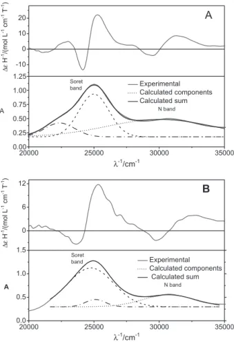
d orbitals. These transitions contribute to the characteristic Q and Soret bands, a ingerprint of porphyrins and can be also evidenced in the corresponding MCD spectra (see band assignments in Figure 4A and 4B). The UV-Vis and
MCD spectra of Fe3+TPFPP, obtained in the respective
anionic and cationic SDS and CTAB micelles and depicted
in Figure 4, give evidence of an interesting effect of the medium on the orbital energy of porphyrins. The UV-Vis
spectrum of Fe3+TPFPP associated with CTAB micelles
presents the typical porphyrinoid Q, Soret and N bands resulting from transitions, respectively, in ascending energies. In the electronic absorption spectra, the N
band appears at around 30000 cm−1 and the Soret band at
25000 cm−1 in CTAB and SDS micelles. Figure 4A and 4B,
light gray lines show the experimental UV-Vis and MCD spectra obtained in the presence of the surfactants CTAB and SDS, respectively. The UV-Vis spectra obtained in both conditions were decomposed to show the contribution of the overlapped transitions contributing to the spectral composite (thick black solid lines). The Soret band of the porphyrin in CTAB micelles is narrower than the spectrum obtained from SDS micelles, as a result of the difference in the energy of the transitions that contributes to the spectra in different media.
The MCD spectra of Fe3+TPFPP in CTAB and SDS
micelles also show signiicant differences according to peculiar structures exhibited by the porphyrin in different environments. The MCD spectroscopy was also useful to study the effect of the microenvironment on the structure of CD silent non-chiral porphyrins. In a non-chiral material
optical activity is absent and the lcp and rcp transitions
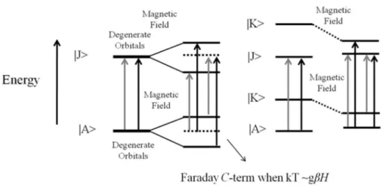
are not differentiated. The application of a magnetic
excited (|J>) states (Zeeman effect) or the combination of
an intermediate state (|K>) with ground or excited states.
Therefore, due to the Zeeman effect, optical activity is induced in the non-chiral material, since it differentiates
the lcp and rcp transitions (Figure 5). MCD transitions,
associated with degenerate orbital splitting, exhibit a derivative band shape, with the zero-crossing coincident with the peak of the corresponding Soret and Q bands
(Faraday A-term). On the other hand, MCD transitions
associated with the mixing of an intermediate state (|K>)
with ground or excited states, exhibit an absorption band shape (Faraday B-term). In a condition in which kT >>gbH
(splitting ∆E), the splitting of a double degenerate ground
state by the magnetic ield results in two spin sublevels that are equally populated. However, at low temperatures,
when kT ca.gbH only one spin sublevel is predominantly
populated resulting in a MCD transition with an absorption
band shape, called as C-term. However, temperature
dependent MCD transitions are not always associated to
degenerate orbital splitting. In a similar way, temperature-dependent MCD transition with absorption band shape can also result from the mixing of |K> with |A> being one sublevel predominantly populated at low temperatures. See
Figure 5, for details about MCD spectroscopy. 6,51
In the case of Fe3+TPFPP, the MCD spectra obtained
both in CTAB and SDS micelles exhibit a non-symmetrical derivative feature, due to the contribution of a Faraday
A-term associated with the π-d transition overlapped with
the positive and negative contribution of Faraday B-term
associated with the π-π* transition. The Soret band peak
and the zero crossing of the Fe3+TPFPP MCD spectrum
obtained in CTAB were the same as those obtained by the association with the anionic SDS micelles. Thus, apparently the charge of the micelle had no inluence on the energy of the ligand to metal transition (LMCT).
However, the association of Fe3+TPFPP with SDS micelles
led to changes in the energy levels of π-π* transitions. SDS
micelles promoted a higher separation of the vibrational levels, resulting in a broader Soret band. The changes in
the energy levels of the Fe3+TPFPP π-π* transitions led to
a higher overlap of the positive envelope of the Faraday
B-term on the negative envelope of the LMCT bands at
the spectral regions of the Q and Soret bands. Therefore, UV-Vis and MCD spectroscopy give evidence that the microenvironment provided by different micelle types have a strong inluence on the structure and, consequently, on the catalytic activity of the porphyrins.
The different microenvironments provided by the lipid nanostructures, as well as the symmetry and structural changes, contribute to modulate the catalytic activity of the pophyrins. Furthermore, data from the literature shows
that the ionization pKa of the water molecule at the sixth
coordination position of the central metal is drastically modiied when the porphyrin is associated with charged interfaces. In the case of positively charged interfaces, the
lowering of the coordinated-water pKa value favors the
peroxidase activity of the porphyrins.52 Another important
aspect to be considered is the impairment of porphyrin aggregation when they are associated with micelles, and this effect is also observed when porphyrins are associated
with lipid bilayers.53-55 Therefore, its association with lipid
nanostructures can give to synthetic porphyrins catalytic
properties similar to those of hemeproteins.56-59
The peroxidase activity of metaloporphyrins associated with lipid nanostructures has been also used for therapeutic applications. Metaloporphyrins associated with liposomes have been used for cancer treatment as inducers of oxidative stress and cell death. The mechanism to induce cell death is the generation of a hydroxyl radical from the peroxide cleavage.60,61
3. Hemepeptides
Microperoxidases are hemepeptides produced by the sequential pepsin- and trypsin-catalyzed hydrolysis of the horse-heart cytochrome c. The resulting structure retains the heme group covalently attached to cysteine residues 14
and 17 and with histidine 18 as the ifth axial ligand.62-64
According to the sequence of native cytochrome c, from which these peptide fragments are derived, it is possible to obtain four microperoxidase types: microperoxidase-6 (CAQCHT), microperoxidase-8 (MP-8) (CAQCHTVE),
microperoxidase-9 (MP-9) (KCAQCHTVE) and microperoxidase-11 (VQKCAQCHTVE). The eficient peroxidase activity of microperoxidases results from the loss of the methionine 80 residue at the sixth
coordination position.62-64 Figure 6 shows the structure of
microperoxidase 9.
These hemepeptides are able to form the same enzyme intermediates during the reaction with peroxides and have been largely used as attractive water-soluble models to study the reaction mechanism of peroxidases. Micelles of CTAB, a cationic surfactant (Table 1), provide a microenvironment
Figure 5. Molecular orbitals, splitting and mixing, promoted by the magnetic ield, resulting in the MCD spectral contribution of Faraday A- and B-terms, respectively. Lcp and rcp are represented by gray and black arrows, respectively. At left, the splitting of degenerate orbitals by the magnetic ield (Zeeman effect) results in the lcp and rcptransitions with different energies. When kT ca.gbH the associated transition is dependent of temperature (Faraday
C-term). At right, the mixing of an intermediate state (|K>) with ground or excited states also results in the lcp and rcp transitions with different energies and the mixing of |K> with |A> can result in a temperature-dependent transition. In both conditions, Zeeman effect and magnetic ield-induced mixing of states, a CD silent molecule exhibits a MCD signal.
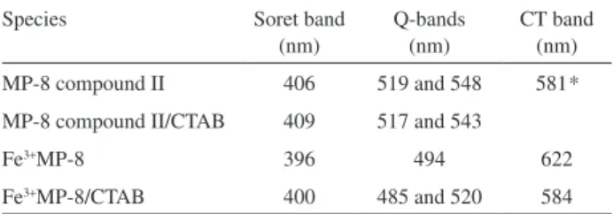
with an alkaline interface and a hydrophobic core that modify
the structure and spectral properties of Fe3+microperoxidases,
as compared to a homogeneous medium (Table 3).56 These
structural modiications have repercussions on the catalytic activity of the hemepeptides.
In a series of studies, we have demonstrated that the
association of Fe3+MPs with CTAB micelles gives to the
hemepeptides the characteristics of a true enzyme, as they
became capable of discriminating substrates,56,65 exhibiting
catalytic cycles without catalyst degradation, and controlling
the route of the reaction with diphenylacetaldehyde.66
The structure of MPs has a net negative charge, due to the presence of heme propionate groups and the carboxy group of the C-terminal oligopeptide chain, which are responsible for the electrostatic afinity for the cationic CTAB micelle surface. However, the hydrophobic groups present in the oligopeptide chain with the hydrophobic heme group also cause a signiicant afinity for the micelle core. Thus, a model for MPs associated with CTAB micelles was proposed, in which the heme propionate groups are associated with the micelle surface and the porphyrin ring and oligopeptide side chains are partially inserted into the micelle.
Microperoxidases associated with CTAB micelles exhibit the red shift of the Soret band that is typical of the
heme group in a hydrophobic microenviroment.67 Changes
in the EPR parameters for the high spin state of MPs suggest that inside the micelle, the hemepeptides are not
coordinated with water but probably instead with OH− or
with the ε-amino group of its N-terminal lysine residue. The
intra-chain coordination with the N-terminal lysine residue
was demonstrated for MP-9 in solution.68
In the complex with the MPs, the micelle constitutes an artiicial enzymatic active site that exhibits a higher afinity for hydrophobic substrates. MP-9/CTAB and MP-8/CTAB
complexes exhibit different afinities for t-BuOOH (tert
-butylhydroperoxide) and HOOH. MP-9/CTAB behaves as a true peroxidase and exhibits a peroxidase catalytic cycle
during its reaction with t-BuOOH. In this way, this heme
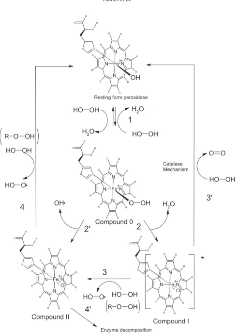
peptide/micelle aggregate behaves as an artiicial enzyme that was named lipoenzyme. The catalytic cycles observed when peroxide was the only available substrate are depicted in Figure 7.69,70
Primus et al.71 described the formation of the MP-8
compound II in the presence of high HOOH concentrations (0.2-1.2 × 10⁻³ mol L−1) in homogeneous media. In this
system, the catalyst was progressively inactivated to a species that exhibited Soret band bleaching. In the presence of CTAB micelles and high peroxide concentrations
(0.2-1.2 × 10⁻³ mol L−1), we have identiied the occurrence of
the same process for MP-8 and MP-9, suggesting that, both in a homogeneous medium and as associated with CTAB micelles, MP-8 and MP-9 compounds I are able to oxidize HOOH to produce compound II.
The occurrence of an oxidative attack and the bleaching of the heme group or a reversible peroxidase cycle without the degradation of the catalyst are dependent on the hemepeptide/micelle ratio and the peroxide concentration. However, whatever the conditions, an association with CTAB micelles favors the formation of the high-valence
intermediate oxoferryl.71 As shown in Figure 7, the
formation of high-valence intermediates of peroxidases is preceded by the coordination of a deprotonated peroxide molecule with the heme iron, leading to a complex that is known as compound 0 (Figure step 1). The deprotonation of the hydroperoxide in compound 0 is a crucial step in the catalytic cycle of peroxidases and cytochrome P450, and consequently, protons act as competitive inhibitors
for the reaction.71 In horseradish peroxidase, the distal
His42 ligand acts as an acid/base catalyst that favors the deprotonation of the hydroperoxide substrate at the enzyme active site and the subsequent heterolytic cleavage
of the peroxide.72-74 Differing from those of horseradish
peroxidase, MPs in an aqueous medium do not exhibit a site pocket with a distal basic residue participating in the peroxide deprotonation and favoring the O–O peroxide bond cleavage. These characteristics should be the cause for the low reactivity of the hemopeptides at low pH values. In an aqueous medium, the alkaline pH leads to the deprotonation of the MP bound water, which then either assists concerted hydrogen peroxide deprotonation and coordination of the hydroperoxo group to the metal center or is directly oxidized by hydrogen peroxide to a
metalhydroperoxo MP.71
In the presence of CTAB micelles, the enzymatic cycle occurs eficiently at pH 7.4. The micellar alkaline interface favors the deprotonation of the water molecule bound to heme iron and subsequent change of the substrate molecule with the formation of a MP compound 0 (Figure 5, step 1). In the following, there are two possible mechanisms for
Table 3. Effect of CTAB on the spectral characteristics of Fe3+microperoxidase-8
Species Soret band
(nm)
Q-bands (nm)
CT band (nm)
MP-8 compound II 406 519 and 548 581*
MP-8 compound II/CTAB 409 517 and 543
Fe3+MP-8 396 494 622
Fe3+MP-8/CTAB 400 485 and 520 584
the cleavage of peroxides (ROOH): a heterolytic cleavage, generating the alcohol derivative (ROH), and compound I (Figure 7, step 2), which reacts with another peroxide molecule, generating compound II and the peroxyl radical
ROO• (Figure 7, step 3) or a homolytic cleavage of ROOH
to form compound II directly (Figure 5, step 3’). In any case,
native Fe3+MP is regenerated after the reaction of compound
II with another ROOH molecule (Figure 7, step 4), and the catalytic cycle is guided to completion. The recycling of MP high-valence species to the resting form in the MP/CTAB micelle complex is evidence that this system behaves as a true peroxidase. As a result, the MP/CTAB
micelle complex is an interesting model to study peroxidase reaction mechanisms and to be applied in nanotechnological devices as a replacement of biological peroxidases.
The complex Fe3+MP-9/CTAB is also able to oxidize
aldehydes such as diphenylacetaldehyde.56 It was
demonstrated that the association of Fe3+MP-9 and
Fe3+MP-11 with CTAB micelles (MP-9/CTAB and
MP11/CTAB) provides a microenvironment that shifts
positively the redox potential of microperoxidases by ca.
100 mV. The addition of diphenylacetaldehyde (DPAA) to the MP-9/CTAB compound II regenerated the native form
of the enzyme, Fe3+MP-9/CTAB, and is what characterizes
the occurrence of a peroxidase cycle. Furthermore,
Fe3+MP-9/CTAB, regenerated during the peroxidase
cycle, reacted with residual DPAA in the medium and was
converted to Fe2+MP-9/CTAB, which indicates that both
Fe3+MP-9/CTAB and its oxyferryl form can use aldehydes
as reducing agents. The reaction of MP-9/CTAB with DPAA produced benzophenone as inal product, and a signiicant difference was observed in the benzophenone yield, according to the micelle/MP-9 molar ratio. The yield of the product reaction of MP-9/CTAB with DPAA, benzophenone, was dependent of the micelle/MP-9 molar ratio. This difference was assigned to the accessibility of the MP-9 amino group to the aldehyde. Spectral analysis showed that the decrease of the micelle/MP ratio favors the association of hemepeptide pairs with the micelle. In a dimeric association with CTAB micelles, Lys13 amino groups could be forced to be located outside of the micelle, where the concentration of DPAA was signiicantly lower due to the partition of the aldehyde, preferentially inside the micelle core. However, in the monomeric form, it is expected that the Lys13 amino groups are to be found inside the micelle core and, thus, are prone to react with DPAA, leading to the formation of Schiff base adducts. Furthermore, in the monomeric form, the peptide chain of MP-9 should be inside the micelle and, thus, is prone to be attacked by intermediate free radicals formed in the course of the reaction. The trapping of the intermediate radicals contributes to the decreasing of the yield of benzophenone without the impairment of the heme iron reduction, a process that precedes the formation of these radicals.
4. Hemeproteins
In cells, the association of apoproteins with the iron protoporphyrin IX, the heme group, is a strategy to modulate its biological properties. The apoproteins provides a variety of microenvironments and axial ligands in such a way that the heme group is able to coordinate
molecular oxygen and NO• for storage as a transport
well and, furthermore, to participate in electron transport, peroxide cleavage, hydroxylation, epoxidation, and
NO• synthesis, among other activities. However, even
in the case of hemeproteins, the association with lipid nanostructures, such as micelles and liposomes, can change the properties of the porphyrin. In this review, we present two hemeproteins whose activity is modulated by their association with micelles and liposomes: horseradish peroxidase (HRP) and cytochrome c.
HRP C is an acidic hemeprotein that exhibits afinity for the cationic CTAB micelles and DODAB liposomes. These surfactants promote structural changes in the HRP
structure, leading to the enhancement of the enzyme activity on the aldeydes: PPA (2-phenylpropionaldehyde)
and DPAA.75 Otherwise, the alkaline hemeprotein,
cytochrome c, exhibits a low afinity to cationic interfaces but is highly susceptible to the effects of acidic surfactants
such as SDS, AOT and cardiolipin.57,76 The association
of cytochrome c with the anionic aqueous and reverse micelles, SDS or AOT/hexane, respectively, leads the heme group of the protein to a less rhombic symmetry that is characterized by electron paramagnetic resonance, UV-Vis, CD, magnetic CD, fluorescence, and Raman
resonance.57 In this condition, the replacement of Met80
by another strong field ligand, probably Lys79, was detected. The cytochrome c species obtained with these anionic surfactants was named as alternative low-spin state cytochrome c (ALSScytc). The ALSScytc retained the capacity to cleave tertbutylhydroperoxide and to be reduced by dithiothreitol and diphenylacetaldehyde, but not by ascorbate. Compatible with a more open heme crevice, ALSScytc exhibited a redox potential that was approximately 200 mV lower than the wild-type protein (+220 mV) and was
more susceptible to the attack of free radicals.57
When associated with negatively charged liposomes, cytochrome c establishes both electrostatic and lipid extended interactions.77 In a systematic study,57 a clear correlation
between the nature of the lipid acyl chain and the spin states
of cytochrome cinteracting with different types of lipid
membranes was shown. Three spin states of cytochrome c were observed in different proportions, according to the lipid acyl chain type and the head group charge present in the
bilayer: a native cytochrome clow-spin state with rhombic
symmetry, a low-spin state with less rhombic symmetry, and a high spin state. The proportion of the spin states of
cytochrome cthat were bound to bilayers was also dependent
on the lipid/protein ratio. This result suggested the existence of two or more protein sites interacting with the lipids,
which was further corroborated.77,78 In the complex with acid
lipids, due to the insertion of a lipid acyl chain inside the cytochrome c structure, this hemeprotein becomes a more eficient peroxidase and can even react with lipid peroxide derivatives.79-82
5. Conclusion
the selectivity of substrate access to the catalytic center. Even for hemepeptides and hemeproteins, micelles and liposomes still have a signiicant inluence on the activity of the porphyrin, as they are able to change peptides, the protein structure and the axial ligands, as well as modulate and select access of substrates. Given these properties, surfactants are attractive devices to construct catalysts with porphyrins and their derivatives.
Acknowledgments
Support from FAPESP, CNPq and CAPES are acknowledged. We are grateful to Professor Otaciro R. Nascimento for the discussions about magnetic circular dichroism theory. Professors ILN and ND are grateful to the students, co-authors of the present article, who participated in the Nanotechnology course of the Graduate Program in Science and Technology/Chemistry of UFABC and who were evaluated by their contributions to this review.
Iseli L. Nantes is a Professor of
Bioenergetics at the Universidade Fe d e ra l d o A B C - U FA B C . S h e received her PhD at the University of São Paulo (USP) working on the enzymatic generation of excited states in model systems, chloroplasts and mitochondria (1995), under the supervision of Professor Giuseppe Cilento. She completed her Postdoctoral research at the University of Campinas (UNICAMP) and USP (1995-1998) and became a Professor of Biochemistry at the University of Mogi das Cruzes (1998-2009). In 2009, she joined UFABC as a Full Professor of Bioenergetics, her present position. Her present research interests are the structure and functions of porphyrins, hemepeptides and hemeproteins. She is investigating the development of porphyrin-containing nanocatalysts as well as the effects of porphyrins and hemeproteins, particularly, cytochrome c and derivates on the mechanisms of cell death. She is the Coordinator of Thematic and a Multi-user Facilities projects, both of them granted by the Fundação de Amparo à Pesquisa do Estado de São Paulo (FAPESP).
Nelson Durán is a Professor
of Chemistry at the University of Campinas (UNICAMP) (Brazil) and a visiting professor at the Universidade Federal do ABC-UFABC. He received his PhD from the University of Puerto Rico (USA) working on the
photolysis and thermolysis of 1,2-dioxolanes (1972). He was an Associate Professor at the Universidad Catolica de Valparaiso, Chile (1973-1975) and carried out a Visiting Professorship at Universidade de São Paulo, Brazil (1975), investigating enzymatic generation of excited states intermediates. In 1978, he joined the Chemistry Institute of UNICAMP (Brazil), working in Biological Chemistry and Biotechnology. His present research interests are nanobiotechnology in cosmetics and in pharmaceuticals, in addition to metallic nanoparticles as antibiotics carriers. He is the Coordinator of the Brazilian Nanobiotechnology Network and a member of the Brazilian Nanocosmetics and Carbon Nanotubes Networks and of the Committee of Brazil-Argentina Nanotechnology Center and belongs to the Consultant Committee for the Nanotechnology area in the Brazilian Science and Technology Ministry.
Sandra Marilia de Souza Pinto
obtained a BSc in Biological Sciences f ro m t h e C e n t ro U n i v e rs i t á r i o Augusto Motta (2005) and a MSc d eg re e f ro m t h e U n i v e rs i d a d e Fe d e ra l d e S ã o Pa u l o ( 2 0 0 9 ) . Currently, she is a PhD student of the Biosystems Graduate Program at the Universidade Federal do ABC (UFABC).
Roberto Alves de Sousa Luz
o b t a i n e d a B S c i n C h e m i s t r y (2007) and a MSc degree from the Universidade Federal do Piauí. Currently, he is a PhD student of the Chemistry Graduate Program at the Universidade Federal do ABC.
Juliana dos Santos de Souza
obtained a BSc in Chemistry (2007) from the Universidade Estadual de São Paulo (2008). Currently, she is an MSc student of the Chemistry Graduate Program at the Universidade Federal do ABC.
Noemia Isoda obtained a BSc
Tiara Gomes de Oliveira obtained a BSc in Food Engineering from the University of São Paulo (2008). She participated in the Nanotechnology course as a special student.
Vi n í c i u s G o d o i Fe r n a n d e s
obtained a BSc in Biomedicine from the Bandeirante University of São Paulo (2009). He participated in the Nanotechnology course as a special student.
Fabiana Braz da Silva obtained a
BSc in Chemistry: Graduate Teacher Programme with Technological Assignment from the Sao Bernardo do Campo College (2007). She participated in the Nanotechnology course as a special student.
References
1. Drain, C. M.; Varotto, A.; Radivojevic, I.; Chem. Rev.2009,
109, 1630.
2. Aida, T.; Inoue, S. In The Porphyrin Handbook; Kadish, K. M. Smith, K. M. Guillard R., eds.; Academic Press: San Diego, 2000, ch. 42.
3. Ponka, P.; Am. J. Med. Sci.1999,318, 241.
4. Da Silva, D. C.; De Freitas-Silva, G.; do Nascimento, E.; Rebouças, J. S.; Barbeira, P. J.; de Carvalho, M. E.; Idemori, Y. M.; J. Inorg. Biochem. 2008,102, 1932.
5. Bochot, C.; Bartoli, J. F.; Frapart, Y.; Dansette, P. M.; Mansuy, D.; Battioni, P.; J. Mol. Catal. A.: Chem. 2007,263, 200. 6. Alkordi, M. H.; Liu, Y. L.; Larsen, R. W.; Eubank, J. F.;
Eddaoudi, M.; J. Am. Chem. Soc. 2008,130, 12639.
7. Suijkerbuijk, B.; Schamhart, D. J.; Kooijman, H.; Spek, A. L.; Van Koten, G.; Gebbink, R.; Dalton Trans. 2010,39, 6198. 8. Groves, J. T.; Nemo, T. E.; J. Am. Chem. Soc. 1983,10, 5786. 9. Drain, C. M.; Smeureanu, G.; Patel, S.; Gong, X. C.; Garno, J.
A.; New. J. Chem. 2006,30, 1834.
10. Wang, Y. T.; Jin, W. J.; Spectrochim. Acta, Part A.2008,70, 871.
11. Komatsu, T.; Moritake, W.; Nakagawa, A.; Tsuchida, E.; Chem. --Eur. J.2002,8, 5469.
12. Nagami, H.; Umakoshi, H.; Shimanouchi, T.; Kuboi, R.;
Biochem. Eng. J.2004,21, 221.
13. Szoka, F.; Papahadjopoulos, D.; Annu. Rev. Biophys. Bioeng.
1980,9, 467.
14. Atkin, R.; Craig, V. S. J.; Wanless, E. J.; Biggs, S.; Adv. Colloid Interface Sci. 2003,103, 219.
15. Grassert, I.; Schinkowski, K.; Vollhardt, D.; Oehme, G.;
Chirality1998,10, 754.
16. Hait, S. K.; Moulik, S. P.; J. Surfactants Deterg.2001,4, 303. 17. Glick, J.; Santoyo, G.; Casey, P. J.; J. Biol. Chem.1996,271,
2949.
18. Maldotti, A.; Andreotti, L.; Molinari, A.; Varani, G.; Cerichelli, G.; Chiarini, M.; Green Chem. 2001,3, 42.
19. Batrakova, E. V.; Kabanov, A. V.; J. Controlled Release2008,
130, 98.
20. Bangham, A. D.; Standish, M. M.; Watkins, J. C.; J. Mol. Biol.
1965,13, 238.
21. Johnson, S. M.; Bangham, A. D.; Hill, M.W.; Korn, E. D.;
Biochim. Biophys. Acta, Biomembr. 1971,233, 820.
22. Papahadj, D.; Watkins, J. C.; Biochim. Biophys. Acta, Biomembr.
1967,135, 639.
23. Abramson, M. B.; Katzman, R.; Gregor, H. P.; J. Biol. Chem.
1964, 239, 70.
24. Hauser, H. O.; Biochem. Biophys. Res. Commun. 1971,45, 1049.
25. Huang, C. H.; Biochemistry1969,8, 344.
26. Saunders, L.; Gammack, D.; Perrin, J.; J. Pharm. Pharmacol.
1962,14, 567.
27. Tien, H. T.; Bilayer Lipid Membranes (BLM): Theory and Practice, 1th ed.; Marcel Dekker: New York, 1974.
28. Razin, S.; Biochim. Biophys. Acta, Biomembr.1972,265, 241. 29. Korenbrot, J. I.; Annu. Rev. Physiol. 1977,39, 19.
30. Batzri, S.; Korn, E. D.; Biochim. Biophys. Acta, Biomembr.
1973,298, 1015.
31. Deamer, D.; Bangham, A. D.; Biochim. Biophys. Acta, Biomembr. 1976,443, 629.
32. Szoka, F.; Papahadjopoulos, D.; Proc. Natl. Acad. Sci. U. S. A.
1978,75, 4194.
33. Mishra, P. P.; Bhatnagar, J.; Datta, A.; J. Phys. Chem. B. 2005,
109, 24225.
34. Pessoto, F. S.; Inada, N. M.; Nepomuceno, M. D.; Ruggiero, A. C.; Nascimento, O. R.; Vercesi, A. E.; Nantes, I. L.; Chem. Biol. Interact.2009,181, 400.
35. Steinbeck, C. A.; Hedin, N.; Chmelka, B. F.; Langmuir2004,
20, 10399.
36. Vanesch, J. H.; Feiters, M. C.; Peters, A. M.; Nolte, R. J. M.;
J. Phys. Chem.1994, 98, 5541.
37. Barber, D. C.; Freitagbeeston, R. A.; Whitten, D. G.; J. Phys. Chem. 1991,95, 4074.
38. Maiti, N. C.; Mazumdar, S.; Periasamy, N. J.; J. Porphyrins Phthalocyanines 1998,2, 369.
41. Zhou, X. T.; Ji, H. B.; Chem. Eng. J. 2010,156, 411. 42. Monnereau, C.; Ramos, P. H.; Deutman, A. B. C.; Elemans, J.;
Nolte, R. J. M.; Rowan, A. E.; J. Am. Chem. Soc.2010,132, 1529.
43. Merlau, M. L.; Grande, W. J.; Nguyen, S. T.; Hupp, J. T.;
J. Mol. Catal. A: Chem. 2000,156, 79.
44. Anzenbacher, P.; Kral, V.; Jursikova, K.; Gunterova, J.; Kasal, A.; J. Mol. Catal. A: Chem. 1997,118, 63.
45. Zhao, Y. C.; Xiang, Y. Z.; Pu, L.; Yang, M.; Yu, X. Q.; Appl. Catal., A2006,301, 176.
46. Zhou, X. T.; Tang, Q. H.; Ji, H. B.; Tetrahedron Lett. 2009,50, 6601.
47. Monfared, H. H.; Aghapoor, V.; Ghorbanloo, M.; Mayer, P.;
Appl. Catal., A2010,372, 209.
48. Heijnen, J. H. M.; de Bruijn, V. G.; Van Den Broeke, L. J. P.; Keurentjes, J. T. F.; Chem. Eng. Process. 2003,42, 223. 49. Santos, A. C.; Luz, R. A. S.; Ferreira L. G. F.; Santos-Júnior, J.
R.; Silva, W. C.; Quim. Nova, 2010,33, 539.
50. Zhou, Y. B.; Ryu, E. H.; Zhao, Y.; Woo, L. K.; Organometallics
2007,26, 358.
51. Nantes, I. L.;Crespilho, F. N.; Mugnol, K. C. U.; Chaves, J. C. A.; Luz, R. A. S.; Nascimento, O. R.; Pinto, S. M. S. In Circular Dichroism: Theory and Spectroscopy; Rodgers, D. S., ed.;Nova Science Publishers: New York, 2010,ch. 8.
52. Travascio, P.; Sen, D.; Bennet, A. J.; Can. J. Chem.2006,84, 613.
53. Omodeo-Sale, F.; Monti, D.; Olliaro, P.; Taramelli, D. P.;
Biochem. Pharmacol. 2001,61, 999.
54. Ishigure, S.; Mitsui, T.; Ito, S.; Kondo, Y.; Kawabe, S.; Kondo, M. Dewa, T.; Mino, H.; Itoh, S.; Nango, M.; Langmuir2010,
26, 7774.
55. Umakoshi, H.; Morimoto, K.; Ohama, Y.; Nagami, H.; Shimanouchi, T.; Kuboi, R.; Langmuir2008,24, 4451. 56. Prieto, T.; Marcon, R. O.; Prado, F. M.; Caires, A. C. F.; Di
Mascio, P.; Brochsztain, S.; Nascimento, O. R.; Nantes, I. L.;
J.Phys.Chem.2006,8, 1963.
57. Mugnol, K. C. U.; Ando, R. A.; Nagayasu, R. Y.; Faljoni-Alario, A.; Brochsztain S.; Santos, P. S. Nascimento, O. R.; Nantes, I. L.; Biophys. J. 2008,94, 4066.
58. Nagatomo, H.; Matsushita, Y.; Sugamoto, K.; Matsui, T.;
Tetrahedron: Asymmetry2003,14, 2339.
59. Cantonetti, V.; Monti, D.; Venanzi, M.; Bombelli, C.; Ceccacci, F.; Mancini, G.; Tetrahedron: Asymmetry2004,15, 1969. 60. Hiraka, K.; Kanehisa, M.; Tamai, M.; Asayama, S.; Nagaoka,
S.; Oyaizu, K.; Yuasa, M.; Kawakami, H.; Colloids Surf., B
2008,67, 54.
61. Yuasa, M.; Oyaizu, K.; Horiuchi, A.; Ogata, A.; Hatsugai, T.; Yamaguchi, A.; Kawakami, H.; Mol. Pharm.2004,1, 387. 62. Aron, J.; Baldwin, D. A.; Marques, H. M.; Pratt, J. M.; Adams,
P. A.; J. Inorg. Biochem. 1986,27, 227.
63. Wang, J. S.; Vanwart, H. E.; J. Phys. Chem.1989,93, 7925. 64. Munro, O. Q.; Marques, H. M.; Inorg. Chem.1996,35, 3752. 65. Prieto, T,; Nascimento, O. R.; Tersariol, I. L. S.; Faljoni-Alario,
A.; Nantes, I. L.; J. Phys. Chem. B2004,108, 11124. 66. Prieto, T.; Nantes, I. L.; Nascimento, O. R.; Prog. Colloid Polym.
Sci.2004,128, 1.
67. Makarska, M.; Radzki, S.; Legendziewicz, J.; J. Alloys Compd.
2002,341, 233.
68. Riposati, A.; Prieto, T.; Shida, C. S.; Nantes, I. L.; Nascimento, O. R.; J. Inorg. Biochem.2006,100, 226.
69. Claiborne, A.; Fridovich, I.; J. Biol. Chem.1979,254, 4245. 70. Hiner, A. N. P.; Ruiz, J. H.; Lopez, J. N. R.; Canovas, F. G.;
Brisset, N. C.; Smith, A. T.; Arnao, M. B.; Acosta, M.; J. Biol. Chem. 2002,277, 26879.
71. Primus, J. L.; Grunenwald, S.; Hagedoorn, P. L.; Albrecht-Gary, A. M.; Mandon, D.; Veeger, C. T.; J. Am. Chem. Soc. 2002,124, 1214.
72. Demontellano, P. R. O.; Annu. Rev. Pharmacol. Toxicol.1992,
32, 89.
73. Savenkova, M. I.; Kuo, J. M.; de Montellano, P. R. O.;
Biochemistry1998,37, 10828.
74. Smith, A. T.; Veitch, N. C.; Curr. Opin. Chem. Biol. 1998, 2, 269.
75. Prieto, T.; Mugnol, K. C. U.; Araujo, J. C.; Sousa, F. L.; Soares,V. A.; Cilento, G.; Nantes, I. L. In Catalysis and Photochemistry in Heterogeneous Media; Nantes, I. L.; Brochesztain, S. eds.; Research Signpost: Kerala, 2007, ch. 1.
76. Zucchi, M. R.; Nascimento, O. R.; Faljoni-Alario, A.; Prieto, T.; Nantes, I. L.; Biochem. J. 2003,370, 671.
77. Kinnunen, P. K. J.; Chem. Phys. Lipids1992,63, 251. 78. Kawai, C.; Prado, F. M.; Nunes, G. L. C.; Di Mascio, P.;
Carmona-Ribeiro, A. M.; Nantes, I. L.; J. Biol. Chem. 2005,
280, 34709.
79. Araujo, J. C.; Prieto, T.; Prado, F. M.; Trindade, F. J.; Nunes, G. L. C.; dos Santos, J. G.; Di Masco, P.; Castro, F. L.; Fernandes, G. J. T.; Valter, J. F.; Araujo, A. S.; Politi, M. J.; Brochsztain, S.; Nascimento, O. R.; Nantes, I. L.; J. Nanosci. Nanotechnol.
2007,7, 3643.
80. Estevam, M. L.; Nascimento, O. R.; Baptista, M. S.; Di Mascio, P.; Prado, F. M.; Faljoni-Alario, A.; Zucchi, M. D.; Nantes, I. L.; J. Biol. Chem. 2004,279, 39214.
81. Kawai, C.; Nantes, I. L.; Baptista, M. D. S.; FEBS J. 2010,277, 224.
82. Nantes, I. L.; Faljoni-Alario, A.; Vercesi, A. E.; Santos, K. E.; Bechara, E. J. H.; Free Radic. Biol. Med.1998,25, 546.
Submitted: January 5, 2011
Published online: May 26, 2011