Arq Neuropsiquiatr 2005;63(1):150-153
Department of Neurology of the Hospital de Clínicas de Porto Alegre RS, Brazil: 1MD, PhD, Neurologist; 2MD, Resident of Neurology. Received 2 April 2004, received in final form 29 July 2004. Accepted 22 September 2004.
Dr. Carlos Rieder - Rua Ramiro Barcelos 2350/2040 - 90035-003 Porto Alegre RS - Brasil. E-mail: [email protected]
HEAD TREMOR AND PROGRESSIVE MULTIFOCAL
LEUKOENCEPHALOPATHY IN AIDS PATIENTS
Report of two cases
Carlos R.M. Rieder
1, Sofia C. Ziomkowski
2ABSTRACT - Progressive multifocal leukoencephalopathy (PML) is caused by replication of JC virus in oligo-dendrocytes of immunocompromised patients. Common manifestations are focal motor and sensory de-ficits, gait abnormalities, speech and language disturbances, cognitive disorders, headache, and visual impair-ment. Although the occurrence of movement disorders is rare in PML, bradykinesia, rigidity, dystonia, myoclonic jerks and myoclonic ataxia have been described. Head tremor associated with PML has not been previous-ly reported. We report two cases of PML in whom head tremor was present.
KEY WORDS: progressive multifocal leukoencephalopathy, tremor, movement disorders, AIDS.
Tremor cefálico e leucoencefalopatia multifocal progressiva em pacientes com SIDA: relato de dois casos
RESUMO - A leucoencefalopatia multifocal progressiva (LMP) é causada pela replicação do vírus JC em oligo-dendrócitos de pacientes imunocomprometidos. As manifestações mais comuns incluem déficits motores e sensitivos, alterações da marcha, da fala e da linguagem, cefaléia e distúrbios visuais e cognitivos. Embora a presença de distúrbios do movimento não seja tão freqüente na LMP, bradicinesia, rigidez, abalos mioclôni-cos e ataxia mioclônica já foram descritos. Nós relatamos dois casos de LMP associados com tremor cefálico.
PALAVRAS-CHAVE: leucoencefalopatia multifocal progressiva, tremor, distúrbios do movimento, SIDA.
Progressive multifocal leukoencephalopathy (PML) is a demyelinatingdisease of the central n e r v-ous system (CNS) caused by the JC virus strain of the papovavirus family. The virus infects and d e s t r o y s oligodendrocytes, causing patchy areas of demyeli-nation in the cerebral white matter. This disease o c-c u r s almost exc-clusively in immunoc-compromised in-dividuals, particularly in patients with acquired im-munodeficiency syndrome (AIDS), lymphoma and chronic and myeloproliferative diseases, and trans-plant recipients. Low CD4 cell count (<100 cells p e r microliter) is postulated to permit reactivation of latent JC virus resulting in the clinical expression of PML1 , 2. PML is present in up to 5% of AIDS p a t i e n t s , causing considerable morbidity and rapid progres-sion to death within a few months3,4. Recently, hi-ghly active antiretroviral therapy (HAART) and o t h-er forms of antiviral treatment have been shown t o increase survival in these patients4,5.
The clinical picture of PML is a consequence of
progressive white matter destruction and varies wi-th lesion location. The presenting manifestations of PML in patients with AIDS do not appear to dif-fer substantially from those in patients with othe r immunosuppressive conditions. Common manifes-tations consist of focal motor and sensory deficits, gait abnormalities, speech and language disturban-ces, cognitive disorders, headache, and visual imp a i r-m e n t6 . Although the basal ganglia circuitry may b e involved in the pathology of PML, movement disor-ders, at least at the onset of PML, are very rare. Bra-dykinesia and rigidity, dystonia, myoclonic jerks and myoclonic ataxia have been described6-8 . Head tre-mor has not been described so far in such patients. We now report two cases of PML in whom head tremor was present.
CASES
Arq Neuropsiquiatr 2005;63(1) 151
sis was observed in the left eyelid. EEG showed a diff u s e increase in slow activity, with no evidence of cortical spi-kes. Lumbar puncture yielded a clear cerebrospinal fluid (CSF) under normal pressure and a positive JC virus PCR ( Table 1). Viral serology was IgG positive for CMV and H S V. Serology for toxoplasma was positive for immunoglobu-lin G (IgG) and negative for IgM. VDRL, HTLV-I and II serol-ogy were negative. Magnetic resonance imaging (MRI) showed hyperintense lesions involving the thalamus, m e-sencephalon, red nuclei, pontine tegmentum and cere-bellum without gadolinium enhancement or mass eff e c t (Fig 1). During the following months the patient develo-ped a spastic tetraparesis, anarthria and dysphagia, a l o n g with deterioration in her general physical condition. S h e Table 1. Cerebrospinal fluid findings.
Case 1 Case 2 Leukocyte count (cells/µL) 6 1
Erythrocyte (cells/µL) 0 4
Cell type lymphocytes NA
Glucose (mg/dL) 78 84
Protein (mg/dL) 118 29
Bacterial culture negative negative
Gram stain negative negative
Mycobacteria culture negative negative
Fungal culture negative NA
Cryptococcus neoformans
Antigen negative NA
Cytomegalovirus IgG R NR
Toxoplasma IgG NA NR
Toxoplasma IgM NA NR
Herpes simplex IgG NR NR
FTA-ABS NR NR
VDRL NR NR
Epstein-Baar virus PCR negative negative
JCV PCR positive positive
Toxoplasma gondii PCR NA negative
Herpes virus type 6 PCR negative negative Herpes simplex type 1 and
2 PCR negative NA
Herpes zoster PCR negative NA
Cytomegalovirus PCR negative negative Mycobacteria PCR negative negative
Enterovirus PCR negative NA
R, reactive; NR, not reactive; NA, not available.
speech disturbance along with blurred vision, which had insidiously developed in the previous four months. She had pulmonary tuberculosis one year before, and was treated successfully with isoniazid, rifampin and pyrazinamide for 9 months. She has been on multiple antiretroviral regimens, the last combination consisting of zidovudine, didanosine and indinavir. At admission
her CD4 cell count was 253 cells/mm3. On the
neurologi-cal examination the patient was alert and fully. Her spe-ech was dysarthric and she was unable to walk because of a severe gait ataxia. Sitting was difficult without sup-port. The patient was quite ataxic on bilateral finger-to-nose and heel-to-knee testing. She had also a high amplitude 4 Hz “no-no” type of head tremor. The tremor persisted when lying down. It was intensified by volunta-ry movements and disappeared during sleep. She had a right-sided weakness and a dystonic posturing of the fin-gers of her right arm, and episodes of irregular distal a r m t r e m o r. Muscle tone was increased in both legs, and she had a right-sided hyperreflexia. Her plantar responses were flexor. Abduction of the right eye was impaired. Upward and downward gaze was impaired. An abduct-ing nystagmus of the left eye was present. Pupils were 2 mm, round, isocoric, and reacted to light. Partial
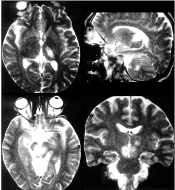
pto-Fig 1. Cranial MRI (T2-weighted) showing hyperintensities in t h e left thalamus (a, b), cerebellum (b) and midbrain (c, d).
152 Arq Neuropsiquiatr 2005;63(1)
have involvement of the posterior fossa, and in 5% to 10% the disease is confined to these structures1 0 -14. Approximately 90% of HIV-infected patients who have PML at their initial evaluation have CD4 lym-phocyte counts of fewer than 200 cells/mm3. Am-plification of the JCV genome by PCR is highly sen-sitive and specific for PML and it has been suggest-ed that this diagnostic test may eliminate the nesuggest-ed for brain biopsy2.
Whilst head tremor is observed in other neurolo-gical conditions such as stroke, multiple sclerosis and head trauma its origin often remains u n c l e a r1 4 , 1 5. We believe that the involvement of structures in the dentate-rubro-thalamo-cortical pathway as d o c-umented by MRI, was responsible for the head tre-mor. Cases of cerebrovascular events due to mid-brain infarction or thalamic lesions leading to head tremor have been published1 5. According to experi-mental studies head tremor may be generated by synchronized bursts of thalamic cells of the ventro-medial nucleus16 , which was not affected in this case. Nevertheless, induction of tremor by lesions of other thalamic nuclei cannot be excluded in o u r first case.
The head tremor presented here resembles Hol-mes tremor. HolHol-mes tremor, or rubral tremor, or midbrain tremor, is characterized by its presence a t rest, enhancement during maintenance of a fixed posture, and increasing amplitude with intention-al voluntary movements. Frequency is slow, usuintention-al- usual-ly between 2-4 Hz1 7. The anatomy of this tremor is complex and incompletely understood, but there has been broad agreement that the tremor follows interruption of the dentate projections pathways, particularly in the region of the superior cerebel-lar peduncle and red nucleus1 8. Since MRI revealed a lesion in the red nucleus, we conclude that the h e a d tremor is most likely a rubral tremor due to PML.
Stereotactic lesions performed in monkeys in the parvicellular red nucleus, brachium conjunctivum decussation and ventromedial mesencephalon ( s u b s-tantia nigra) demonstrated that damage to all t h r e e areas was necessary for sustained tremor19 .These findings confirm clinicopathologic observations in humans. A combination of damage to the red nu-cleus and neighboring cerebello-thalamic, cebello-olivary, and nigrostriatal fibers tracts are re-quired for the development of Holmes tremor (an association of rest, postural and kinetic tremor)19.
In summary, the increasing spectrum of clinical and imaging findings being increasingly reported
became comatose and died from Staphylococcus aureus
pneumonia. No autopsy was performed.
Case 2 – A 51-year-old man presented with a 3-months history of progressive generalized weakness, dysarthria, dysphagia and visual difficulties. There were no sensory or sphincter dysfunction. His past medical his-tory was unremarkable. He denied any hishis-tory of known HIV infection, promiscuous bisexual or homosexual rela-tionship, and exposure to blood or blood products. His physical examination at the time of admission revealed a gait and limb ataxia, and scanning dysarthria. Upward gaze of both eyes was impaired. Superficial and deep sensations were preserved. HIV-1 serology was positive using enzyme-linked immunosorbent assay technique. One week after admission he developed an intense iso-lated “no-no” type head tremor at rest, with increasing amplitude during maintenance of a fixed posture and intentional voluntary movements. During the following month the patient developed a symmetrical spastic tetraparesis, together with deterioration in his general physical condition. MRI showed hyperintense zones in cerebellum and cerebellar peduncle (Fig 2). CSF findings are shown in Table 1 with a positive JC virus PCR. In the subsequent month he developed severe cachexia, became comatose, and finally died of pneumonia.
DISCUSSION
As far as we know, head tremor associated with PML has not been previouly reported. The occurren-ce of movement disorders is uncommon in PML. E v e n in cases of HIV-related PML with basal ganglia in-volvement, focal movement disorders have rarely been reported9. Bradykinesia and rigidity may be detected in a minority of patients with advanced d i s e a s e7. Dystonia and jerky movements of the h a n d have been observed as a consequence of contralate-ral lesions in the basal ganglia6. Myoclonic ataxia in a PML patient was recently described8. We now report two AIDS patients with head tremor in w h o m the appearance of white matter MRI lesions as w e l l as a positive PCR test for JC-virus in the CSF led to the diagnosis of PML. In one case PML was the ini-tial manifestation of AIDS.
Arq Neuropsiquiatr 2005;63(1) 153
in the literature suggest that PML should be con-sidered in the differential diagnosis of all HIV pos-itive patients with uncommon clinical presentations and MRI findings compatible with demyelination.
REFERENCES
1. Simpson D, Tagliati M. Neurologic manifestations of HIV infection. A n n Intern Med 1994;121:769-785.
2. Berger JR, Major EO. Progressive multifocal leukoencephalopathy. Se-min Neurol 1999;19:193-200.
3. Berger JR, Pall L, Lanska D, et al. Progressive multifocal leukoence-phalopathy in patients with HIV infection. J Neurovirol 1998;4:59-68. 4. C l i ff o rd DB, Yiannoutsos C, Glicksman MS, et al. HAART improves pro g-nosis in HIV-associated progressive multifocal leukoencephalopathy. Neurology 1999;52:623-625.
5. De Luca A, Giancola ML, Ammassari A, et al. Potent anti-re t roviral ther-apy with or without cidofovir for AIDS-associated pro g ressive multifo-cal leukoencephalopathy: extended follow-up of an observational stu-dy. J Neurovirol 2001;7:364-368.
6. Stockhammer G, Poewe W, Wissel J, Kiechl U, Maier H, Felber S. Pro-gressive multifocal leukoencephalopathy presenting with an isolated focal movement disorder. Mov Disord 2000;15:1006-1009.
7. Singer C, Berger JR, Bowen BC, et al. Akinetic-rigid syndrome in a 13-y e a r-old female with HIV- related pro g ressive multifocal leukoencepha-lopathy. Mov Disord 1993;8:113-116.
8. Fontoura P, Vale J, Lima C, Scaravilli F, Guimaraes J. Progressive myo-clonic ataxia and JC virus encephalitis in an AIDS patient. J Neurol Neurosurg Psychiatry 2002;72:653-656.
9. Bhatia KP, Morris JH, Frackowiak RS. Primary progressive multifocal
leukoencephalopathy presenting as an extrapyramidal syndrome. J Neurol 1996;243:91-95.
10. Whiteman M, Post MJD, Berger JR, et al. PML in 47 HIV+ patients. Radio-logy 1993;187:233-240.
11. Berger JR, Mucke L. Prolonged survival and partial recovery in AIDS-associated pro g ressive multifocal leukoencephalopathy. Neuro l o g y 1988;38:1060-1065.
1 2 . K a s t rup O, Maschke M, Diener HC, Wanke I. Pro g ressive multifocal leukoencephalopathy limited to the brain stem. Neuro r a d i o l o g y 2002;44:227-229.
13. Rubin DI, Norris S, Flint R. Isolated pontine progressive multifocal leukoencephalopathy: unusual magnetic resonance imaging features. J Neuroimaging 2002;12:63-66.
14. Okuda B, Tachibana H, Sugita M, Maeda Y. Bilateral internuclear oph-thalmoplegia, ataxia, and tremor from a midbrain infarction. Stroke. 1993;24:481-482.
1 5 . Otto S, Buttner T, Schols L, Windmeier DT, Przuntek H. Head tremor due to bilateral thalamic and midbrain infarction. J Neurol 1995;242:608-610. 16. L a m a r re Y, Joff roy AJ. Experimental tremor in monkey: activity of thal-amic and precentral cortical neurons in the absence of peripheral feed-back. Adv Neurol 1957;106:109-163.
17. Deuschl G, Krack P. Tremors: differential diagnosis, neurophysiology, and pharmacology. In Jankovic J, Tolosa E (EDS). Parkinson’s disease and movement disorders, 3rdedition. Baltimore: Williams & Wilkins,
1998:419-452.
18. S h e p h e rd GM, Tauboll E, Bakke SJ, Nyberg-Hansen R. Midbrain tre m o r and hypertrophic olivary degeneration after pontine hemorrhage. Mov Disord 1997;12:432-437.