Arq Neuropsiquiatr 2005;63(1):46-49
Faculdade de Medicina, Universidade de São Paulo (FMUSP), São Paulo, SP, Brazil: 1Department of Neurology, FMUSP and Division of Neurosurgery, Hospital das Clinicas da FMUSP, São Paulo São Paulo SP Brazil; 2Department of Clinical Anatomy, FMUSP. Received 10 May 2004, received in final form 6 August 2004. Accepted 28 September 2004.
Dr. Arthur W. Poetscher - Avenida Albert Einstein 627/1308 - 05651-901 São Paulo SP - Brasil. E-mail: [email protected]
PARA-MUSCULAR AND TRANS-MUSCULAR
APPROACHES TO THE LUMBAR
INTER-VERTEBRAL FORAMEN
An anatomical comparison
Arthur Werner Poetscher
1, Guilherme Carvalhal Ribas
2,
Alexandre Yasuda
2, Koshiro Nishikuni
2ABSTRACT - Foraminal and extra-foraminal disc herniations comprise up to 11.7% of all lumbar disc her-niations. Facetectomy, which had been the classic approach, is now recognized as cause of pain and insta-bility after surgery. Otherwise, posterior lateral approaches through a trans-muscular or a para-muscular tech-nique offer no significant damage to key structures for spinal stability. The surgical anatomy of these approach-es has already been dapproach-escribed, but they were not compared. In order to quantify the angle of vision towards the intervertebral foramen offered by each technique, 12 fresh cadavers were dissected and studied regard-ing these approaches. The angle presented by trans-muscular approach was wider in all studied lumbar leve-ls. Surgery through the trans-muscular approach is performed with a better working angle, requiring a smal-ler resection of surrounding tissues. Therefore, minor surgical trauma can be expected. Our measurements support previously published data that point the trans-muscular approach as the best surgical option. KEY WORDS: intervertebral disc displacement, low back pain, lumbar vertebrae, sciatica, comparative study.
Acessos paramuscular e transmuscular ao forame intervertebral lombar: comparação anatômica
RESUMO - As hérnias de disco lombares apresentam-se como foraminais ou extra-foraminais em até 11,7% dos casos. Seu tratamento cirúrgico através de facetectomias pode causar dor e instabilidade, o que não ocorre com a utilização de acessos cirúrgicos posteriores laterais ao canal central, quer seja por via transmus-cular ou paramustransmus-cular. Nosso objetivo foi comparar o ângulo de trabalho relativo ao forame interverte-bral permitido por cada via e avaliar possíveis implicações cirúrgicas. Dissecções e medidas realizadas em do-ze cadáveres frescos demonstraram que este ângulo é maior na via transmuscular em todos os níveis lom-bares estudados. Dado que um ângulo mais favorável permite uma ressecção menor das estruturas envolvi-das para que se estabeleça o campo de trabalho, a cirurgia por meio da via transmuscular permitirá, então, um trauma cirúrgico menor. Nossos resultados corroboram publicações prévias que apontam a via transmus-cular como a melhor opção cirúrgica.
PALAVRAS-CHAVE: deslocamento do disco intervertebral, dor lombar, vértebras lombares, ciática, estudo com-parativo.
Foraminal and extra-foraminal lumbar disc her-niations have historically been a challenge in spine surgery due to their particular anatomical relation-ship with the nerve roots and the facet joints, com-prising up to 11.7% of all lumbar disc herniations1,2.
Facetectomy used to be the standard technique in treating such herniations for a long time1,3, 4,5.
Des-pite the familiar surgical anatomy and the success in relieving root compression in most of the cases,
this approach has been reviewed and criticized. The incidence of back pain after surgery, related to ins-tability, is significant, as pointed out by several au-thors6-10. Haher also demonstrated that an increase
in axial load on the intervertebral disc may occur in this situation, therefore accelerating degenerati-ve process11. Use of posterior lateral approaches,
Arq Neuropsiquiatr 2005;63(1) 47
option. The posterior lateral approach was initially developed for lumbar arthrodesis, as described by Watkins12and Wiltse13. Nevertheless, only in 1985,
Recoulles-Arche reported his experience with the trans-muscular approach for the surgical treatment of foraminal and extra-foraminal disc herniations14.
Two years later, Reulen8and Fankhauser15published
their own experience with para-muscular and trans-muscular techniques.
The target of both approaches is the lateral as-pect of the inter-vertebral foramen, in between the transverse processes. When using the trans-mus-cular technique, this space is reached by splitting the para-vertebral muscles, whereas, when perform-ing a para-muscular approach, these muscles are detached from the spinous processes and then re-tracted beyond the facet articulations. Although some authors have related their experience with both approaches, these techniques were not specifi-cally compared regarding either the surgical anato-my or patients outcome2,4. Despite the fact that
Reulen8,9and Schlesinger10have suggested that the
trans-muscular technique may provide a wider an-gle towards the intervertebral foramen, no objec-tive measurements have been published yet. A larg-er angle may plarg-ermit a bettlarg-er visualization within the intervertebral foramen, therefore saving bone and ligament resection and diminishing the surgical trauma.
Since the difference between these angles may be important to establish the better surgical option, we report the measurements obtained after dissec-tion of 12 fresh cadavers.
METHOD
The “angle of vision” was defined as the one between the sagittal plane of lateral aspect of the intervertebral
foramen and the line that connects the lateral border of the incision, in the same axial plane, with the interver-tebral foramen.
Since this angle is deeply situated, a direct measure is not feasible. In order to overcome this constraint, we defined a right triangle, as shown in Figure 1. One of the cathetus, called “foramen depth” here of, is the dis-tance from the intervertebral foramen to the coronal pla-ne that crosses the apex of the spinous process. The sec-ond cathetus is the distance from the apex of the spin-ous process, in the same sagital plane, to the point were it intersects the line that begins in the intervertebral fora-men and crosses the most lateral point of the incision, in the same axial plane. This line is the hypotenuse, which represents the “access depth”. Given that cos α= c/h, where c = foramen depth, and h = access depth, the angle of vision’s cosine equals the foramen depth divided by the access depth. This trigonometric relationship allowed us to calculate the angle.
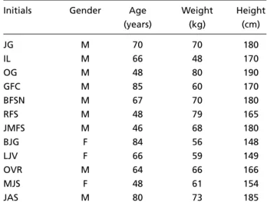
Twelve fresh adult cadavers were dissected and meas-ured. They were randomly selected, having as exclusion criteria either history of spinal disease or findings that suggested so during the dissection. On average, the age was 64 years (range: 46 - 85 years), the height, 169.75 cm. (range: 148 - 190 cm.), and the weight, 56.8 kg (range: 48 - 80 kg). The detailed data are shown in Table 1.
All cadavers were dissected in ventral decubitus, with the arms along the body, in order to maintain the lum-bar lordosis.
Skin was incised at three centimeters from the spin-ous processes, para-median, in a randomly selected side. Once the incision length could be a variable (it changes the muscular retraction), all specimens were incised from L1 to L5. The space between the subcutaneous tis-sue and the toraco-lumbar aponeurosis was dissected to-wards the midline. Para-vertebral muscles were then com-pletely detached and retracted as lateral as possible, allowing us to visualize the transverse processes. A
re-Fig 1. Coronal and sagittal planes, depth of access and depth of foramen.
Table 1. Anthropometric data of dissected cadavers.
Initials Gender Age Weight Height (years) (kg) (cm)
JG M 70 70 180
IL M 66 48 170
OG M 48 80 190
GFC M 85 60 170
BFSN M 67 70 180
RFS M 48 79 165
JMFS M 46 68 180
BJG F 84 56 148
LJV F 66 59 149
OVR M 64 66 166
MJS F 48 61 154
48 Arq Neuropsiquiatr 2005;63(1)
para-muscular: L1-L2: 32˚, L2-L3: 26˚, L3-L4: 25˚, L4-L5: 23˚), reaching a significant difference (t test, p-<0.003).
In both approaches, the angle of vision was smal-ler at the lower lumbar spine (23˚and 36˚at L4-L5, 32˚and 48˚at L1-L2, for para-muscular and trans-muscular, respectively). This difference was also sig-nificant (para-muscular: p<0.03, trans-muscular: p<0.001).
DISCUSSION
The essential divergence between the para-mus-cular and the trans-muspara-mus-cular approaches lays on their relationship with the para-vertebral muscles. When using the para-muscular technique, the whole bundle of muscles is kept laterally to the surgical path, by use of retraction. On the other hand, the trans-muscular technique establishes a path trough the natural space in between the m. multifidus and m. iliocostalis. This relationship explains the differ-ence between the angles of vision.
Schlesinger et al. suggested that this angle is lar-ger in the trans-muscular approach, but they did not have the chance to measure it10. Reulen,
describ-ing their technique for the para-muscular access, observed that, in several patients, they had to tilt the surgery table laterally in about 10˚ to achieve a better visualization inside the inter-vertebral fo-ramen8. One can obtain it naturally using the
trans-muscular approach.
A larger angle will permit a deeper visualiza-tion into the inter-vertebral foramen. Thus, a small-er amount of bone and ligaments from its dorsal wall will have to be removed in order to expose ade-quately the nerve root and the disc fragments. The-refore, less surgical trauma should be expected.
In the lower lumbar spine, the angles for both approaches are smaller, due to the deeper location of the inter-vertebral foramen and to the smaller muscle retraction possible on this level. In this sit-uation, the gain achieved by the trans-muscular technique may be even more significant. Reulen also noticed this difference and stated that at L4-L5 the trans-muscular approach should be set as the standard technique 9.
Airaksinen17and Sihvonen18demonstrated that
posterior midline approaches to the lumbar spine may cause atrophy of the para-vertebral muscles and that it correlated with persistent low back pain after surgery. Boelderl19studied the para-ver-tractor was placed and the inter-transversary muscles and
ligaments were removed, in order to expose the inter-vertebral foramen. At this moment, we measured the foramen depth and the access depth for the para-mus-cular approach, as above defined, from L1-L2 to L4-L5. In sequence, the retractor was removed, allowing the para-vertebral muscles to return to their anatomic posi-tion. The para-vertebral muscles were then split through the space between the m. multifidus and the m. iliocostal-is. Once the intervertebral foramen was visualized again, the retractors were positioned. At this point, the access depth for the trans-muscular approach was measured, as defined previously, from L1-L2 to L4-L5.
Given the fact that L5-S1 level presents an unique anatomy and is better accessed by a specific approach16,
it was not addressed in this study.
The angles of vision were then calculated and all the values were analyzed for statistical significance. For each approach, at each level, the average, median, standard deviation, and confidence interval were obtained. The values for each approach were then compared using t test study.
This research was approved by the Ethics Committee - Hospital das Clínicas da Faculdade de Medicina da Uni-versidade de São Paulo (CAPPesq, n.21/2000)
RESULTS
At all levels the access depth was longer with the trans-muscular technique (L1-L2: 8.0 cm., L2-L3: 8.3 cm., L3-L4: 8.6 cm, L4-L5: 8.6 cm) than with the para-muscular technique (L1-L2: 6.3 cm., L2-L3: 6.5 cm., L3-L4: 6.9 cm., L4-L5: 7.5 cm.).
The foramen depth increased in the lower lum-bar spine (L1-L2: 5.3 cm., L2-L3: 5.8 cm., L3-L4: 6.2 cm., L4-L5: 6.9 cm.).
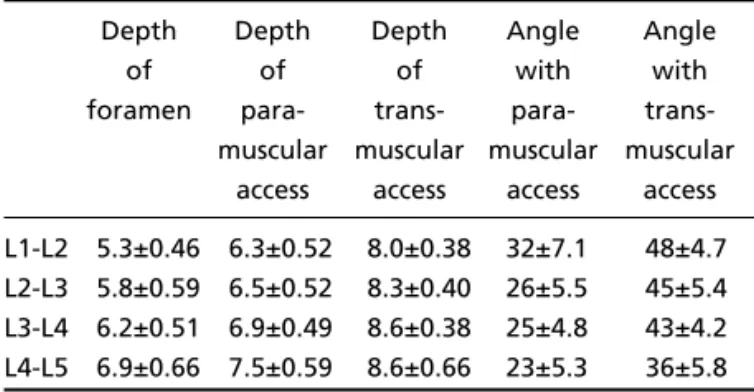
Detailed values for each level are exposed in the Table 2.
At all studied levels the angle of vision was lar-ger in the trans-muscular approach (trans-muscu-lar - L1-L2: 48˚, L2-L3: 45˚, L3-L4: 43˚, L4-L5: 36˚,
Table 2. Average depth of foramen, of each approach, and of angles, with the respective confidence interval (95%).
Depth Depth Depth Angle Angle
of of of with with
foramen para- trans- para- trans-muscular muscular muscular muscular
access access access access L1-L2 5.3±0.46 6.3±0.52 8.0±0.38 32±7.1 48±4.7 L2-L3 5.8±0.59 6.5±0.52 8.3±0.40 26±5.5 45±5.4 L3-L4 6.2±0.51 6.9±0.49 8.6±0.38 25±4.8 43±4.2 L4-L5 6.9±0.66 7.5±0.59 8.6±0.66 23±5.3 36±5.8
Arq Neuropsiquiatr 2005;63(1) 49
tebral innervation and concluded that the corre-spondent branches may be stretched and dam-aged if the muscles are retracted laterally to the articular processes, a required procedure in the para-muscular technique. Thus, the trans-muscu-lar approach, besides providing a better angle of vision, is also less likely to endanger the para-ver-tebral muscles and their innervation. A prospective study comparing patients submitted to surgery with each of these approaches should be conducted in order to establish if there is any impact on outcome.
REFERENCES
1. Abdulah AF, Dito EW III, Byrd EB, Williams R. Extreme lateral lum-bar disc herniation: clinical syndrome and special problems of diagno-sis. J Neurosurg 1974;41:229-234.
2. Porchet F, Cholet-Bornand A, de Tribolet N. Long-term follow up of patients surgically treated by the far-lateral approach for foraminal and extraforam-inal lumbar disc herniations. J Neurosurg 1999;(Spine 1);90:59-66. 3. Basile R Junior, Mendonça ABF Netto, Napoli MMM, Bonetti CL, Barros
TEP Filho. Hérnia de disco lombar lateral: foraminal e extraforaminal. Rev Hosp Clin. Fac Med S Paulo 1986;41:177-180.
4. Epstein NE. Evaluation of varied surgical approaches used in the man-agement of 170 far-lateral disc herniations: indications and results. J Neurosurg 1995;83:648-653.
.5 Mixter WJ, Barr JS. Rupture of the intervertebral disc with involvement of the spinal canal. N Engl J Med 1934;211:210-215.
6. Donaldson WF, Star MJ, Thorne RP. Surgical treatment for the far late-ral herniated lumbar disc. Spine 1993;18:1263-1267.
7. Maroon JC, Kopitnik TA, Schulhof LA, Abla A, Wilberger JE. Diagnosis and microsurgical approach to far lateral disc herniation in the lum-bar spine. J Neurosurg 1990;72:378-382.
8. Reulen HJ, Pfaunder S, Ebeling U. The lateral microsurgical approach to the “extracanalicular” lumbar disc herniation. I: a technical note. Acta Neurochir (Wien) 1987;84:64-67.
9. Reulen HJ, Muller A, Ebeling U. Microsurgical anatomy of the lateral approach to extraforaminal lumbar disc herniations. Neurosurgery 1996;39:345-350.
10. Schlesinger SM, Fankhauser H, de Tribolet N. Microsurgical anatomy and operative technique for extreme lateral lumbar disc herniations. Acta Neurochir (Wien) 1992;118:117-129.
11. Haher TR, O’Brien M, Dryer JW, Nucci R, Zipnick R, Leone DJ. The role of the lumbar facet joints in spinal stability. Spine 1994;19:2667-2671. 12. Watkins MB. Posterolateral fusion of the lumbar and lumbosacral spine.
J Bone Joint Surg. Am 1953;35-A:1014-1018.
13. Wiltse LL, Bateman JG, Hutchinson RH, Nelson WE. The paraspinal sacrospinalis-splitting approach to the lumbar spine. J Bone Joint Surg. Am 1968;50A:919-926.
14. Recoules-Arche D. La chirurgie de la hernie discale du canal de cunju-gaison lombaire. Neurochirurgie 1985;31:61-64.
15. Fankhauser H, de Tribolet N. Extreme lateral disc herniation. Br J Neurosurg 1987;1:111-129.
16. Muller A, Reulen HJ. A paramedian tangential approach to lumbosacral extraforaminal disc herniations. Neurosurgery 1998;43:854-862. 17. Airaksinen O, Herno A, Kaukanen E, Saari T, Sihvonen T, Suomalainen
O. Density of lumbar muscles 4 years after descompressive spinal sur-gery. Eur Spine J 1996;5:193-197.
18. Sihvonen T, Herno A, Paljarvi L, Airaksinen O, Partanen J, Tapaninaho A. Local denervation atrophy of paraspinal muscles in postoperative failed back syndrome. Spine 1993;18:575-581.