Periodontics
Leonardo Silveira Damasceno Fernanda da Silva Gonçalves Edson Costa e Silva
Elton Gonçalves Zenóbio Paulo Eduardo Alencar Souza Martinho Campolina Rebello Horta
Department of Dentistry, Pontifícia Universidade Católica de Minas Gerais, Belo Horizonte, MG, Brazil.
Corresponding author: Martinho Campolina Rebello Horta E-mail: martinhohorta@pucminas.br
Received for publication on Feb 27, 2012 Accepted for publication on Apr 25, 2012
Stromal myofibroblasts in focal reactive
overgrowths of the gingiva
Abstract: Focal reactive overgrowths are among the most common oral mucosal lesions. The gingiva is a signiicant site affected by these lesions, when triggered by chronic inlammation in response to microorganisms in dental plaque. Myoibroblasts are differentiated ibroblasts that active-ly participate in diseases characterized by tissue ibrosis. The objective of this study was to evaluate the presence of stromal myoibroblasts in the main focal reactive overgrowths of the gingiva: focal ibrous hyperpla-sia (FFH), peripheral ossifying ibroma (POF), pyogenic granuloma (PG), and peripheral giant cell granuloma (PGCG). A total of 10 FFHs, 10 POFs, 10 PGs, and 10 PGCGs from archival specimens were evaluated. Samples of gingival mucosa were used as negative controls for stromal myoibroblasts. Oral squamous cell carcinoma samples, in which stromal myoibroblasts have been previously detected, were used as positive con-trols. Myoibroblasts were identiied by immunohistochemical detection of alpha smooth muscle actin (α-sma). Myoibroblast immunostaining was qualitatively classiied as negative, scanty, or dense. Differences in the presence of myoibroblasts among FFH, POF, PG, and PGCG were analyzed using the Kruskal-Wallis test. Stromal myoibroblasts were not detected in FFH, POF, PG, or PGCG. Consequently, no differences were observed in the presence of myoibroblasts among FFH, POF, PG, or PGCG (p > 0.05). In conclusion, stromal myoibroblasts were not de-tected in the focal reactive overgrowths of the gingiva that were evalu-ated, suggesting that these cells do not play a signiicant role in their pathogenesis.
Descriptors: Gingival Diseases; Gingival Overgrowth; Myoibroblasts; Immunohistochemistry.
Introduction
Focal reactive overgrowths are among the most common lesions of the oral mucosa.1 The gingiva is an important area affected by these
le-sions,1,2 primarily triggered by chronic inlammation in response to
mi-croorganisms in dental plaque.2,3-7 These lesions are composed of one or
more of the following connective tissue components:
• collagen,
• bone,
• endothelial cells, and
• multinucleated giant cells.1,2,7
The most common focal reactive overgrowths of the gingival connective tissue are:
• focal ibrous hyperplasia (FFH),
• peripheral ossifying ibroma (POF),
• pyogenic granuloma (PG), and
• peripheral giant cell granuloma (PGCG).4,7,8
FFH, also known as irritation ibroma, is a fo-cal reactive hyperplasia of ibroblasts with over-production of collagen.2,5,7 POF is a focal reactive
hyperplasia of ibrous connective tissue presenting bone formation.7,9 PG is a focal reactive growth of
granulation tissue with marked proliferation of en-dothelial cells and blood vessel formation.5,7 PGCG
is a focal overgrowth composed of mononuclear and multinucleated giant cells.3,6,7
Myoibroblasts are differentiated ibroblasts that express alpha smooth muscle actin and that have in-termediate characteristics of both classic ibroblasts and smooth muscle cells.10,11 Transdifferentiation by
TGF-β1 stimulation is its most common origin.11,12
Myoibroblasts synthesize and degrade extracel-lular matrix components during inlammation and during the process of tissue repair and remodel-ing.10,11,13-15 Therefore, these cells actively participate
in diseases characterized by the ibrosis of organs and tissues.14,15 Although the presence of
myoblasts has been reported in hereditary gingival ibro-matosis16 and drug-induced gingival hyperplasia,17,18
few studies have evaluated its presence in focal reac-tive overgrowths of the gingiva.19-22
Therefore, the aim of this study was to evaluate the presence of stromal myoibroblasts in the main focal reactive overgrowths of the gingiva (FFH, PG, POF, and PGCG). Differences in the presence of myoibroblasts among FFH, POF, PG, and PGCG were also analyzed.
Methodology
Tissues and samplesA total of 10 FFHs, 10 PGs, 10 POFs and 10 PGCGs, taken from archival formalin-ixed, paraf-in-embedded specimens, were evaluated. This study was approved by the local ethics committee (CAAE - 0161.0.213.000-07).
Immunohistochemistry
Myoibroblasts were identiied by the immuno-histochemical detection of alpha smooth muscle actin (α-sma), a marker for myoibroblasts. Four-micrometer sections from the parafin-embedded samples were used. Tissue sections were dewaxed with xylene, hydrated using graded alcohols, and treated with 0.6% H2O2 to eliminate endogenous peroxidase activity. Antigen retrieval was conducted by heating in a 0.01 M citrate buffer (pH 6.0) for 30 minutes. Subsequently, an anti-α-sma monoclo-nal antibody was used (clone 1A4, diluted 1:100; Dako Corporation, Carpinteria, USA). The LSAB+ kit (Dako Corporation, Carpinteria, USA) was used for the application of the biotinylated link antibody and of peroxidase-labeled streptavidin, according to the manufacturer’s instructions. The reactive prod-ucts were visualized by immersing the sections for 3 min in 0.03% diaminobenzidine solution, con-taining 2 mM H2O2. The sections were then coun-terstained with Mayer’s hematoxylin, dehydrated, and mounted.
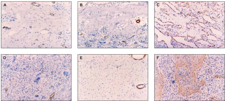
Normal vessels’ smooth muscle immunoreac-tivity for α-sma was used as an internal positive control. Samples of oral squamous cell carcinoma, showing numerous and densely arranged stromal myoibroblasts, were used as a positive control (Fig-ure 1F). Samples of gingival mucosa were used as a negative control for myoibroblasts (Figure 1E). The negative control for α-sma immunoreactivity was performed by omission of the primary antibody.
Scoring of immunostaining results
Alpha smooth muscle actin-positive stromal cells, showing cytoplasmic immunostaining, were considered to be myoibroblasts. Light microscopy was used to evaluate the immunohistochemical re-actions. The myoibroblast immunostaining was qualitatively classiied as negative (samples in which no stromal myoibroblasts were detected), scanty (samples showing sporadic stromal myoibroblasts), or dense (samples showing numerous and densely arranged stromal myoibroblasts).
Statistical analysis
version 5.0 (Optical Digital Technology, Belém, Bra-zil). Differences in the presence of myoibroblasts among FFH, POF, PG, and PGCG were analyzed using the Kruskal-Wallis test. The level of signii-cance was established at 5%.
Results
The results are illustrated in Table 1 and Figure 1.
No stromal myoibroblasts were observed in FFH (Figure 1A), POF (Figure 1B), PG (Figure 1C), or PGCG (Figure 1D). Consequently, no differenc-es were observed in the prdifferenc-esence of myoibroblasts among FFH, POF, PG, or PGCG (p > 0.05).
Discussion
Myoibroblasts are differentiated ibroblasts that have morphologic and immunophenotypic features similar to those of smooth muscle cells.10,11 In
addi-tion to alpha smooth muscle actin, myoibroblasts show immunopositivity for vimentin, non-muscle myosin, and ibronectin.23 These cells show a
spin-dle-cell or stellate-cell morphology, an eosinophilic cytoplasm and an abundant pericellular matrix.23
Moreover, these cells display the typical
ultrastruc-tural features of secreting cells (a prominent rough endoplasmic reticulum and Golgi apparati produc-ing secretion granules), as well as peripheral myo-ilaments, ibronexus junctions, and gap junctions.23
Myoibroblasts synthesize and secrete cytokines, inlammatory mediators, extracellular matrix pro-teins, matrix metalloproteinases and tissue inhibi-tors of matrix metalloproteinases (TIMPs).11-15
Due to their ability to secrete and degrade extra-cellular matrix components, myoibroblasts actively participate in the morphogenesis of tissues or or-gans,11 wound healing,10,11,13 ibrosis,14,15 and tumor Table 1 - Presence of stromal myofibroblasts in focal fibrous hyperplasia (FFH), peripheral ossifying fibroma (POF), pyo-genic granuloma (PG), and peripheral giant cell granuloma (PGCG).
Samples Presence of myofibroblasts P-value 1 Negative Scanty Abundant
FFH (n = 10) 10 (100%) 0 0
> 0.05 POF (n = 10) 10 (100%) 0 0
PG (n = 10) 10 (100%) 0 0
PGCG (n = 10) 10 (100%) 0 0 1P-value was obtained using the Kruskal-Wallis test.
invasion.24,25
Despite the relevance of myoibroblasts in dis-eases characterized by ibrosis,14,15 few studies have
evaluated these cells in gingival overgrowths.16-20 In
granulation tissue, myoibroblasts undergo apop-tosis after wound healing.11,13 However, during
i-brosis, the continuous presence of TGF-β1 should inhibit myoibroblast apoptosis,26 resulting in their
accumulation, mainly in tissues presenting unremit-ting inlammation.11,13 As TGF-β1 levels are
100-fold greater in gingival inlammatory processes, such as periodontitis,27 and as gingival ibroblast
transdifferentiation into myoibroblasts, through TGF-β1 stimulation, has been reported,28 the
evalu-ation of myoibroblasts in gingival inlammatory lesions is required. Therefore, this study aimed to evaluate the presence of myoibroblasts in the main focal reactive overgrowths of the gingiva.
FFH is characterized by the hyperplasia of ibro-blasts with overproduction of collagen.2,5 POF is
characterized by hyperplasia of ibrous connective tissue, nevertheless presenting bone formation.9 In
this study, no myoibroblasts were detected in FFH or POF. These results are in agreement with those of previous reports20,21 and suggest that
myoibro-blasts are not signiicant in the pathogenesis of FFH or POF, despite their high ibroblast activity. This inding can be explained by the low TGF-β1 levels in these lesions or by the presence of myoibroblast inhibitors, such as INF-γ, which can inhibit gingival myoibroblast transdifferentiation.28
This is the irst study evaluating myoibroblasts in PG, a focal reactive overgrowth with marked pro-liferation of endothelial cells and blood vessel for-mation.5 Despite its similarity to granulation tissue,
an important site of myoibroblasts,11,13 these cells
were not detected in any of the 10 PG samples that were evaluated.
Although previous reports have detected myo-ibroblasts in PGCG,19,21,22 no myoibroblasts were
observed in the 10 PGCG samples evaluated in this
study. This divergence is likely a consequence of methodological differences because one study used a histochemical marker for myosin, as well as elec-tron microscopy, to detect myoibroblasts,19 and the
other used immunohistochemical detection of HHF-35, a muscle-actin-speciic antibody.21 Nevertheless,
another one of the studies22 detected myoibroblasts
in PGCG using electron microscopy and immuno-histochemical detection of alpha smooth muscle ac-tin. An additional hypothesis is that myoibroblasts have been occasionally identiied in PGCG because the former report evaluated only 5 samples,19 and
the second study detected myoibroblasts in just 2 of 10 samples.21 In fact, myoibroblasts have also
been sporadically detected in central giant cell gran-uloma,29 a lesion histologically similar to PGCG.
Moreover, it is possible that myoibroblasts arise as cells in healing processes due to the ulceration of the primary lesions and not as a major player in the pathogenesis of these gingival overgrowths. Finally, it is important to emphasize that PGCG is composed of mononuclear and multinucleated giant cells3,4,6 that show immunohistochemical markers of
macrophages and osteoclasts.30
Conclusion
Stromal myoibroblasts were not detected in the focal reactive overgrowths of the gingiva that were evaluated, suggesting that these cells do not play a signiicant role in their pathogenesis.
Acknowledgements
This study was supported by grants from Con-selho Nacional de Desenvolvimento Cientíico e Tec-nológico - CNPq, Fundação de Amparo à Pesquisa do Estado de Minas Gerais - FAPEMIG, and Pro-grama Institucional de Bolsas de Iniciação Cientíica (PROBIC) - PUC Minas, Brazil.
References
1. Stablein MJ, Silverglade LB. Comparative analysis of biopsy specimens from gingival and alveolar mucosa. J Periodontol. 1985 Nov;56(11):671-6.
2. Zhang W, Chen Y, An Z, Geng N, Bao D. Reactive gingival lesions: a retrospective study of 2439 cases. Quintessence Int. 2007 Feb;38(2):103-10.
3. Giansanti JS, Waldron CA. Peripheral giant cell granuloma: review of 720 cases. J Oral Surg. 1969 Oct;27(10):787-91. 4. Bhaskar SN, Cutright DE, Beasley JD, Perez B. Giant cell
reparative granuloma (peripheral): report of 50 cases. J Oral Surg. 1971 Feb;29(2):110-5.
5. Kfir Y, Buchner A, Hansen LS. Reactive lesions of the gingival: a clinicopathological study of 741 cases. J Periodontol. 1980 Nov;51(11):655-61.
6. Bodner L, Peist M, Gatot A, Fliss DM. Growth potential of peripheral giant cell granuloma. Oral Surg Oral Med Oral Pathol Oral Radiol Endod. 1997 May;83(5):548-51. 7. Damasceno LS, Gonçalves FS, Costa e Silva E, Zenóbio EG,
de Souza PEA, Horta MC. [Focal reactive overgrowths of the gingiva – a review of the literature]. Perionews. 2011 Mar-Apr;5(2):169-76. Portuguese.
8. Buchner A, Calderon S, Ramon Y. Localized hyperplastic le-sions of the gingiva: a clinicopathological study of 302 lele-sions. J Periodontol. 1977 Feb;48(2):101-4.
9. Buchner A, Hansen L. The histomorphologic spectrum of peripheral ossifying fibroma. Oral Surg Oral Med Oral Pathol. 1987 Apr;63(4):452-61.
10. Gabbiani G,Ryan GB, Majno G. Presence of modified fibro-blasts in granulation tissue and their possible role in wound contraction. Experientia. 1971 May;27(5):549-50.
11. Tomasek JJ, Gabbiani G, Hinz B, Chaponnier C, Brown RA. Myofibroblasts and mechano-regulation of connective tissue remodelling. Nat Rev Mol Cell Biol. 2002 May;3(5):349-63. 12. Desmoulière A, Geinoz A, Gabbiani F, Gabbiani G. Trans-forming growth factor-beta 1 induces alpha-smooth muscle actin expression in granulation tissue myofibroblasts and in quiescent and growing cultured fibroblasts. J Cell Biol. 1993 Jul;122(1):103-11.
13. Hinz B. Formation and function of the myofibroblast during tissue repair. J Invest Dermatol. 2007 Mar;127(3):526-37. 14. Wight TN, Potter-Perigo S. The extracellular matrix: an active
or passive player in fibrosis? Am J Physiol Gastrointest Liver Physiol. 2011 Dec;301(6):G950-5.
15. Sarrazy V, Billet F, Micallef L, Coulomb B, Desmoulière A. Mechanisms of pathological scarring: role of myofibro-blasts and current developments. Wound Repair Regen. 2011 Sep;19(Suppl 1):10-5.
16. Bitu CC, Sobral LM, Kellermann MG, Martelli-Junior H, Zec-chin KG, Graner E, et al. Heterogeneous presence of myofibro-blasts in hereditary gingival fibromatosis. J Clin Periodontol. 2006 Jun;33(6):393-400.
17. Dill RE, Iacopino AM. Myofibroblasts in phenytoin-induced hyperplastic connective tissue in the rat and in human gingival overgrowth. J Periodontol. 1997 Apr;68(4):375-80.
18. Bullon P, Pugnaloni A, Gallardo I, Machuca G, Hevia A, Battino M. Ultrastructure of the gingiva in cardiac patients treated with or without calcium channel blockers. J Clin Peri-odontol. 2003 Aug;30(8):682-90.
19. Dayan D, Buchner A, David R. Myofibroblasts in peripheral giant cell granuloma. Light and electron microscopic study. Int J Oral Maxillofac Surg. 1989 Oct;18(5):258-61. 20. Lombardi T, Morgan PR. Immunohistochemical
characteriza-tion of odontogenic cysts with mesenchymal and myofilament markers. J Oral Pathol Med. 1995 Apr;24(4):170-6. 21. Miguel MCC, Andrade ESS, Rocha DAP, Freitas RA, Souza
LB. [Immunohistochemical expression of vimentin and HHF-35 in giant cell fibroma, fibrous hyperplasia and fibroma of the oral mucosa.] J Appl Oral Sci. 2003 Mar;11(1):77-82. Portuguese.
22. Filioreanu AM, Popescu E, Cotrutz C, Cotrutz CE. Immu-nohistochemical and transmission electron microscopy study regarding myofibroblasts in fibroinflammatory epulis and giant cell peripheral granuloma. Rom J Morphol Embryol. 2009 Jul;50(3):363-8.
23. Eyden B. The myofibroblast: phenotypic characterization as a prerequisite to understanding its functions in translational medicine. J Cell Mol Med. 2008 Jan-Feb;12(1):22-37. 24. Kalluri R, Zeisberg M. Fibroblasts in cancer. Nat Rev Cancer.
2006 May; 6(5):392-401.
25. De Assis EM, Pimenta LG, Costa e Silva E, Souza PE, Horta MC. Stromal myofibroblasts in oral leukoplakia and oral squamous cell carcinoma. Med Oral Patol Oral Cir Bucal. Forthcoming 2012.
26. Zhang HY, Phan SH. Inhibition of myofibroblast apoptosis by transforming growth factor beta(1). Am J Respir Cell Mol Biol. 1999 Dec;21(6):658-65.
27. Steinsvoll S, Halstensen TS, Schenck K. Extensive expression of TGF-β1 in chronically-inflamed periodontal tissue. J Clin
Periodontol. 1999 Jun;26(6):366-73.
28. Sobral LM, Montan PF, Martelli-Junior H, Graner E, Col-leta RD. Opposite effects of TGF-beta1 and IFN-gamma on transdifferentiation of myofibroblast in human gingival cell cultures. J Clin Periodontol. 2007 May;34(5):397-406. 29. Vered M, Nasrallah W, Buchner A, Dayan D. Stromal
myo-fibroblasts in central giant cell granuloma of the jaws cannot distinguish between non-aggressive and aggressive lesions. J Oral Pathol Med. 2007 Sep;36(8):495-500.
