A macroscopic classification of the embryonic development of the
one-sided livebearer
Jenynsia multidentata
(Teleostei: Anablepidae)
Nathalia C. López-Rodríguez
1, Cíntia M. de Barros
2and Ana Cristina Petry
2This study proposes eight stages according to the main discernible changes recorded throughout the embryonic development of Jenynsia multidentata. The development of morphological embryo structures, pigmentation, and changes in tissues connecting mother and embryo were included in the stage characterization. From the fertilized egg (Stage 1), an embryo reaches the intermediary stages when presenting yolk syncytial layer (Stage 2), initial pigmentation of the outer layers of the retina and dorsal region of the head (Stage 3), and the sprouting of the caudal (Stage 4), dorsal and anal fins (Stage 5). During the later stages, the ovarian folds enter the gills, and the body pigmentation becomes more intense (Stage 6), the body becomes elongated (Stage 7), and there is a greater intensity in body pigmentation and increased muscle mass (Stage 8). The dry weight of the batches varied between 0.6 ± 0.3 mg (Stage 3) to 54.6 ± 19.7 mg (Stage 8), but the dry weight of the maternal-embryonic connecting tissues remained almost constant. After controlling the effect of those reproductive tissues, the gain in dry weight of the batches throughout development increased exponentially from Stage 6, reflecting the increase in size and weight of the embryos due to matrotrophy.
Keywords: Developmental stages, Dry weight, Matrotrophy, Morphology, Viviparity.
Este estudio propone una clasificación constituida por ocho etapas, establecidas con base en los principales cambios macroscópicos registrados a lo largo del desarrollo embrionario de Jenynsia multidentata. El desarrollo de estructuras embrionarias, los patrones de pigmentación y los cambios en los tejidos de conexión entre la madre y el embrión fueron incluidos en la caracterización de cada etapa. Inicia con el huevo fertilizado (Etapa 1), siguiendo con etapas intermedias del embrión en las cuales es visible la capa sincitial del vitelo (Etapa 2), la pigmentación inicial de las capas externas de la retina y el dorso de la cabeza (Etapa 3), y la aparición de las aletas caudal (Etapa 4), dorsal y anal (Etapa 5). En las etapas posteriores, los pliegues del tejido ovárico se introducen por las branquias y se intensifica la pigmentación corporal (Etapa 6), el cuerpo se extiende (Etapa 7) y hay un marcado aumento en la pigmentación corporal y masa muscular (Etapa 8). El peso seco de los lotes varió entre 0,6 ± 0,3 mg (Etapa 3) y 54,6 ± 19,7 mg (Etapa 8); sin embargo, el peso seco del tejido que conecta a la madre con el embrión se mantuvo prácticamente constante. Después de controlar el efecto del peso seco de estos tejidos, la ganancia de peso seco de los lotes de embriones a lo largo del desarrollo aumentó de forma exponencial a partir de la Etapa 6, evidenciándose en el aumento en tamaño y peso de los embriones debido a la matrotrofia.
Palabras clave: Etapas del desarrollo, Matrotrofia, Morfología, Peso seco, Viviparidad.
1Programa de Pós-Graduação em Ciências Ambientais e Conservação, Universidade Federal do Rio de Janeiro - UFRJ, Campus Macaé, Avenida São José do Barreto, 764, 27965-045 Macaé, RJ, Brazil. [email protected]
2Núcleo em Ecologia e Desenvolvimento Socioambiental de Macaé - NUPEM, Universidade Federal do Rio de Janeiro - UFRJ, Avenida São José do Barreto, 764, 27965-045 Macaé, RJ, Brazil. (CMB) [email protected], (ACP) [email protected] (corresponding author)
Introduction
Viviparity defines a reproductive pattern characterized by the development of embryos inside a female’s body under a varied level of post-fertilization maternal provisioning (Wourms, 1981). Viviparity represents one of the most extreme and complex forms of non-behavioral parental care, which has evolved independently in several vertebrate and invertebrate taxa (Blackburn, 2005; Lodé, 2012). Among
Maternal provisioning begins before fertilization when oocytes are enlarged with yolk during vitellogenesis and may last until birth (Wourms, 1981; Lodé, 2012). Species whose embryos depend exclusively on yolk storage are known as
lecithotrophic, whereas species whose embryos gain maternal nutrients beyond their yolk reserves are known as matrotrophic
(Kunz, 2004). Depending on the proportion of maternal resources devoted to the brood after fertilization, species fall along an increasing gradient of matrotrophy, which is usually inferred from the ratio of the dry weight between late- and early-term embryos (Thibault, Schultz, 1978; Meisner, Burns, 1997; Lodé, 2012; but see Riesch et al., 2010). Weight losses of 30-40% are recorded during the embryonic development of lecithotrophics, whereas varying degrees of weight gain occur in matrotrophics (Wourms, 1981; Reznick et al., 2007). In the latter, the yolk content is significantly reduced and embryo development depends almost entirely on alternative sources of nutrition throughout the maternal tissues (Siccardi, 1940; Amoroso, 1960; DeMarais, Oldis, 2005). However, the degree of matrotrophy depends largely on the maternal and embryonic adaptations to provide and absorb nutrients, respectively (Schindler, de Vries, 1988; Meisner, Burns, 1997; Reznick et al., 2007).
In viviparous teleosts, a cystovarian and a placenta-like structure are morphophysiological adaptations involved in bearing and nourishing live young. The former represents a fused pair of ovaries, whose lumen is covered by germinal epithelium where oocytes are released and fertilized, whereas the latter is an apposition of the maternal (the follicle) and embryonic tissues that sustain embryos throughout development (e.g.,a modified yolk sac, externalized pericardial membrane) (Turner, 1940; Schindler, de Vries, 1988). The intense vascularization and secretory capacities of such structures are involved in the maintenance of oocytes, sperm and embryos, not necessarily concomitantly (Wourms, 1981; Schindler, de Vries, 1988). Morphological characterization of the maternal related structures and staging of embryonic development has been extensively employed in studying both evolution of a complex of reproductive traits involving nutritional provisioning and synthesis of life-history traits in fish (Meisner, Burns, 1997; Reznick et al., 2007; Lodé, 2012).
Although viviparity is found in 13 families of Teleostei, most of these studies were focused on the subfamily Poecilinae, in which almost all of the 273 species (except the facultative viviparous Tomeurus gracilis Eigenmann, 1909)
occurring in the Americas are viviparous (Thibault, Schulz, 1978; Haynes, 1995; Lucinda, 2003a; Nelson et al., 2016). Haynes (1995) proposed a standardized classification of embryonic development of poeciliids for life-history studies using the least killifish Heterandria formosa Girard, 1859 and
the western mosquitofish Gambusia affinis (Baird, Girard, 1853) as model organisms. The major contribution of his study was to gather previous rating criteria developed by several authors for more than a dozen species and encompassing them in 11 stages of embryonic development, which started with the immature egg and ended with the mature embryo.
For the less specious Neotropical subfamily Anablepinae, which includes two genera of livebearers (Jenynsia - 14 species and Anableps - three species) (Nelson et al., 2016), a classification of the embryonic development remains mostly unexplored, at least in the light of morphological studies (Turner, 1940; Schindler, de Vries, 1988). Within its genus, the one-sided livebearer Jenynsia multidentata (Jenyns, 1842) has the widest range of geographical distribution, inhabiting mostly shallow and brackish but also highly saline environments along the eastern South American coast (21° S - 35° S) from the northern plains of Rio de Janeiro in Brazil to the lowland areas around the la Plata River in Argentina, and on the northwest and central Argentinian territories (Ghedotti, Weitzman, 1996; Lucinda, 2003b; Di Dario et al., 2013). Ghedotti, Weitzman (1996) removed J. multidentata
from synonymy with J. lineata (Jenyns, 1842) (limited to the Río Cebollati watershed, Uruguay); therefore, most of the few studies with J. lineata up to the last 20 years refer currently to J. multidentata (e.g.,Hylton Scott, 1918;Siccardi, 1940; Schindler, de Wries, 1988; Fontoura et al., 1994).
In J. multidentata, the number of offspring vary geographically (Goyenola et al., 2011) and possibly maternal provisioning as well; therefore, the existence of a methodological framework of embryonic developmental staging would encourage studies on the reproductive life-history traits of J. multidentata of both intra and interpopulational levels, such as those carried out for poeciliids (e.g., Zúñiga-Vega et al., 2011). This study provides a methodological framework developed for the essentially matrotrophic anablepids, using the widely spread one-sided livebearer J. multidentata as a model. Females from a northernmost wild population of J. multidentata
inhabiting a typical saline coastal lagoon of Rio de Janeiro State (Caliman et al., 2010) were maintained up to six months under controlled mating to follow the timing of fecundation and the further chronology of the embryonic development. We provide a morphological characterization of the early development of J. multidentata and a classification tool
for embryonic development in the visually detected stages. The maternal provisioning was inferred by variations in dry weight of batches and maternal reproductive tissues of females from three populations.
Material and Methods
similar of that recorded on sampling location (35 ppt), and photoperiod (10D:14L), replicated in six units. Considering that anablepids may store sperm for a much shorter time than most poeciliids and that the gestation of J. multidentata
should be shorter than the 60 days of some poeciliids (Bisazza
et al., 2000; Betito, 2006; Goyenola et al., 2011; Bassar et al., 2014), this procedure aimed to ensure that a pregnant female would give birth and begin the reproductive cycle before the experimentally induced pregnancy.
Afterwards, females from each aquarium were exposed to five adult males seined in October 2013 in Catingosa lagoon (i.e., from the same population), which were maintained in a perforated cage for five days and subsequently released in the aquaria for additional five days. This procedure aimed to control the timing of mating to chronologically follow the embryonic development in these females. To verify the occurrence of synchrony in ovarian development, all females from one of the six aquaria (n= 19) were euthanized with an overdose of benzocaine (250 mg L-1 previously diluted in 5 ml ethanol) at
the end of the time allowed to mating. Females from the other five aquaria were employed to determine the chronology of the embryonic development. Every 48 h during 58 days, a female of each aquarium was randomly selected and euthanized (alternating in two aquaria one day, in three aquaria on another), and 2-3 potentially pregnant females were obtained daily.
To standardize the conditions inside the aquaria, some procedures were performed daily, i.e.,the ad libitum feeding with commercial fish food and Artemia spp. nauplii at 9 h
and 18 h, respectively, adjustments of temperature (< 0.5°C) and dissolved oxygen (>6 mg L -1), and others weekly, i.e.,
feces removal and renewal of 30% of the water volume. After euthanasia with an overdose of benzocaine (250 mg L-1 previously diluted in 5 ml ethanol), the total length of the
females was recorded with a digital caliper (TL mm).
Histological processing and image recording of the embryos. An ovary from each female was dissected, dehydrated in increasing concentrations of ethanol from 70% to 100% for ten min, clarified in xylene for five min infiltrated and embedded in Paraplast®. Longitudinal sections
of 5 mm were obtained in a Microtome (Leica RM 2245) and stained with Harris Hematoxylin and alcoholic Eosin. The images were obtained using Olympus DP71 camera of 12.5 mega pixels coupled to an Olympus BX51 microscope. For morphological characterization, entire embryos were photographed on a Leica DFC550 camera of 12.5 mega pixels coupled to the stereomicroscope Leica M205 FA.
Chronology of embrionary development. The following aspects were considered for the characterization of the embryonic stages: the occurrence of yolk syncytial layer (herein named YSL), retina pigmentation, the degree of development of structures (i.e.,eyes and fins), body length, shape and pigmentation patterns. Whenever necessary, additional ovaries and batches (herein defined as a group of embryos within a female) of females sampled in the same
location were included in the analyses. Batches under initial development (i.e.,blastula and gastrula stages) were excluded from the analyses due to their low occurrence (<2% of the dissected ovaries) and difficulties in correctly discriminating early stage individuals, which might incur an underestimation of embryo number.
Relationships between the total dry weight of a batch and the dry weight of other reproductive tissues, which include ovary, germinal cells and maternal-embryonic connections (total reproductive mass, sensu Reznick et al., 2007), were explored on 93 batches covering the range of embryonic stages of J. multidentata from the beginning of eye pigmentation onwards. These batches were obtained from females with 54.18 ± 3.94 mm in TL, which were seined in three coastal lagoons located in northern of Rio de Janeiro State (Catingosa lagoon 22°11’13.308” S - 41°23’49.920” W; Garças lagoon 22°12’49.680” S - 41°29’34.116” W; Pitanga lagoon 22°9’40.716” S - 41°17’54.204” W; Geocentric Reference System of the Americas - SIRGAS 2000). After the determination of the embryonic development stages, based on the framework developed herein, the embryos and ovaries were dehydrated at 65°C during 12 h to obtain their dry weight (mg).
Voucher specimens were deposited at the Fish Collection of the Núcleo em Ecologia e Desenvolvimento Socioambiental de Macaé (NPM2938-2940).
Data analysis. An Analysis of Covariance (ANCOVA) was performed to examine whether the relationship between the dry weight of other reproductive tissues (covariate) and the total dry weight of developing batch (response variable) differed among the stages of embryonic development (predictor categorical variable). A post hoc Tukey test was employed as significant differences were detected in ANCOVA. The significant differences in the interaction between the dry weight of other reproductive tissues and the stages of embryonic development were assumed to indicate a differing degree of maternal provisioning to the batch. The assumptions of normality and homocedasticity of residuals (respectively Shapiro-Wilk and Levene tests) were reached and a P <0.05 was considered the significance level.
Results
Despite recording the mating and harassments of females by males, it was not possible to determine whether females were fertilized if they presented ovaries in folliculogenesis, or even if they would be able to become pregnant under captive conditions. Additionally, spermatozoa were not detected in any of the 80 histological slides analyzed.
Characterization of the embryonic development stages.
According to the morphological analysis of the embryos, it was possible to discriminate eight stages of development, beginning from Stage 1, the fertilized egg (ovum) through Stage 8, in which the embryos are ready to birth (Tab. 1; Fig. 1).
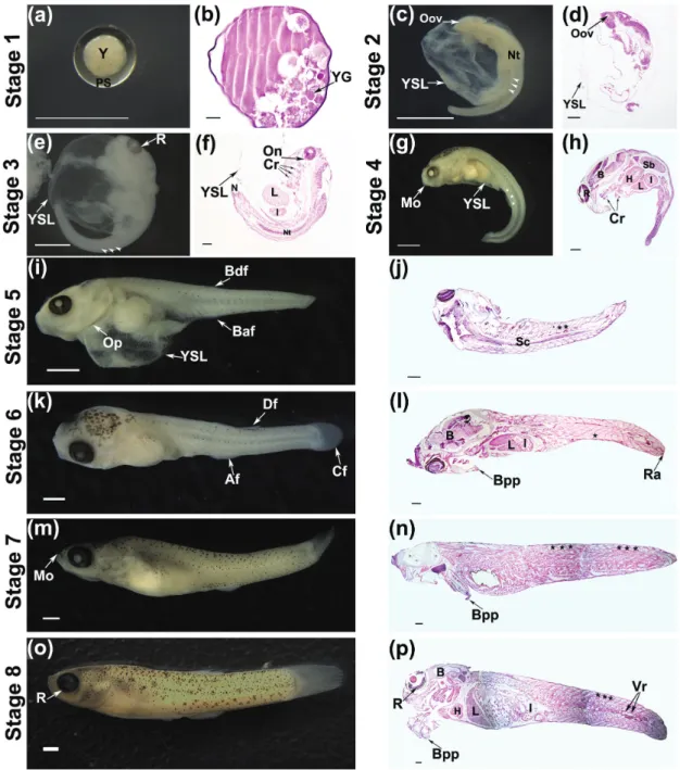
Stage 1: Fertilized egg. Fertilized eggs measured approximately 0.3 mm in diameter, with yolk granules and a well-defined perivitelinic zone. Although the eggs were found free inside the ovary, they were also seen grouped, forming sets on the cephalic region, from where they seemed to migrate along the ovary wall until the central cavity to continue the development (Figs. 1 a-b).
Stage 2: Outline of the optic vesicle. The embryo was unpigmented and the body curved with a diameter of approximately 0.9 mm. The YSL was abundant. It was possible to distinguish the following three regions of the body: cephalic, corporal, and terminal. The outline of the optic vesicle was evident, and the somites had a pointed look, indicating its future division in dorsal and ventral
muscle mass (epiaxial and hypoaxial, sensu Kimmel et al., 1995) (Figs. 1c-d).
Stage 3: Beginning of eye pigmentation. The embryo was still curved with a diameter of approximately 1.5 mm. The YSL was present in most embryos. The greater development of the cephalic region, which represented approximately 30% of body size, was evident. The eye pigmentation begins in the layers of the retina. Pigmentation on the back of the head was first detected in few embryos. The optic nerve, three to five crests of branchial arches with hyaline cartilaginous tissue and neural tube were identified. Moreover, the liver occupying a large part of the abdominal cavity, and the portions of the intestine were observed. From this stage, it was possible to count the embryos, which could be differentiated based on the pigmentation of the retina and/ or iris (Figs. 1 e-f).
Stage 4: Sprout of the caudal fin.The body of the embryo began to distend, was thin, translucent and had little muscle mass. The low frequency of embryos at this stage and the greater variability in their size made it difficult to estimate their total body length. The head accounted for 25% of the body size. The mouth opening and the early development of the caudal fin were observed. The retinas were pigmented and the first punctate melanophores were identified in the dorsal region. Remnants of the YSL were still detected (Figs. 1 g-h).
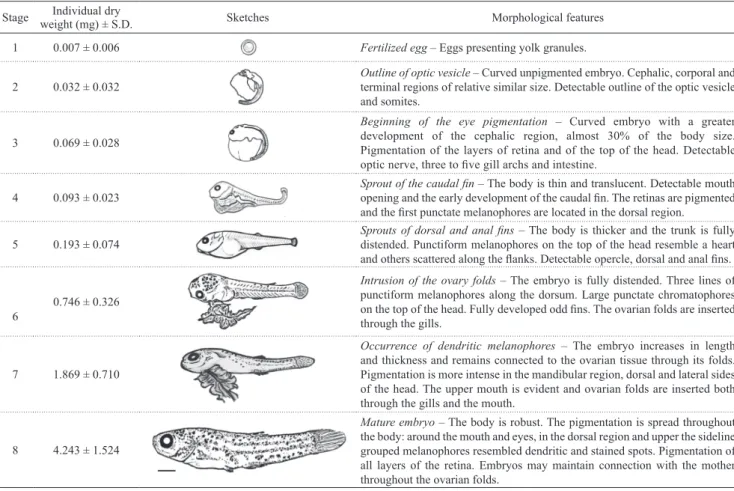
Table 1. Characterization of the embryos of the one-sided livebearer Jenynsia multidentata at the proposed eight stages of development. Scale bar of 0.5 mm.
Stage Individual dry
weight (mg) ± S.D. Sketches Morphological features
1 0.007 ± 0.006 Fertilized egg – Eggs presenting yolk granules.
2 0.032 ± 0.032
Outline of optic vesicle – Curved unpigmented embryo. Cephalic, corporal and terminal regions of relative similar size. Detectable outline of the optic vesicle and somites.
3 0.069 ± 0.028
Beginning of the eye pigmentation – Curved embryo with a greater development of the cephalic region, almost 30% of the body size. Pigmentation of the layers of retina and of the top of the head. Detectable
optic nerve, three to five gill archs and intestine.
4 0.093 ± 0.023
Sprout of the caudal fin – The body is thin and translucent. Detectable mouth
opening and the early development of the caudal fin. The retinas are pigmented and the first punctate melanophores are located in the dorsal region.
5 0.193 ± 0.074 Sprouts of dorsal and anal finsdistended. Punctiform melanophores on the top of the head resemble a heart – The body is thicker and the trunk is fully
and others scattered along the flanks. Detectable opercle, dorsal and anal fins.
6
0.746 ± 0.326
Intrusion of the ovary folds – The embryo is fully distended. Three lines of punctiform melanophores along the dorsum. Large punctate chromatophores
on the top of the head. Fully developed odd fins. The ovarian folds are inserted
through the gills.
7 1.869 ± 0.710
Occurrence of dendritic melanophores – The embryo increases in length and thickness and remains connected to the ovarian tissue through its folds. Pigmentation is more intense in the mandibular region, dorsal and lateral sides of the head. The upper mouth is evident and ovarian folds are inserted both through the gills and the mouth.
8 4.243 ± 1.524
Fig. 1. Stereoscopic (a, c, e, g, i, k, m, o) and light microscopy (b, d, f, h, j, l, n, p) images stained with Hematoxylin and Eosin of the stages of embryonic development of the one-sided livebearer Jenynsia multidentata. Y: yolk; PS: perivitelinic space;
YG: yolk granule; Oov: outline optic vesicle; YSL: syncytial layer of the yolk; R: retine; Arrow head: somites; Nt: neural tube; L: liver; I: intestine; On: optic nerve; Cr: crest of branchial arch; Mo: mouth opening; *: miomers; B: brain; H: heart; Sb: swimbladder; Op: operculum; Baf: bud of the anal fin; Bdf: bud of the dorsal fin; Sc: spinal cord; Df: dorsal fin; Af: anal fin; Cf: caudal fin; Ra: rays of caudal fin; Bpp: branchial Pseudoplacenta; Vr: vertebra. Scale bars of 0.5 mm (a, c, e, g, i, k, m, o) and of 0.2 mm (b, d, f, h, j, l, n, p).
Stage 5: Sprouts of the dorsal and anal fins. The head was more developed. The arrangement of the dorsally punctiform melanophores resembled a heart. The body gets thicker, and in most embryos, the trunk is fully distended. The total length was approximately of 5 mm. Punctiform melanophores were scattered along the flanks. The remnants of YSL were still visible in some embryos. The operculum and the dorsal and anal fins were visible (Figs. 1 i-j).
The odd fins were much more developed and were observed as rays on the rounded caudal fin. The remnants of the YSL were not more evident and ovarian folds were inserted through the gills of all embryos to constitute the branchial pseudoplacenta (Figs. 1 k-l).
Stage 7: Occurrence of dendritic melanophores. The embryo increased in thickness and total length (which ranged between 6.9 and 9.0 mm) and remained connected to the ovarian tissue through its folds that resembled a cord. The muscles were more developed. Dendritic melanophores were detectable, and the pigmentation was more intense in the mandibular region and the dorsal and lateral sides of the head, advancing to the caudal direction. The upper mouth was evident, and in some embryos, the ovarian folds were inserted through the mouth (Figs. 1 m-n).
Stage 8: Mature embryo. The body was robust; the caudal
peduncle was thicker compared to the previous phase, and TL varied between 11.6 and 14.3 mm. The pigmentation was spread throughout the body – around the mouth and eyes. In the dorsal region of the head, there was a cluster of punctiform melanophores resembling a heart. In the dorsal region and the upper sideline, the grouped melanophores
(with greater intensity) resembling spots showed the following two patterns: dendrite (or branched/ramified/star-shaped) and staining (punctate melanophores together). The pigmentation of all retinal layers was completed. Through the thin ovarian membrane, it was possible to identify embryos that maintain their cord connection with the mother (Figs. 1o-p).
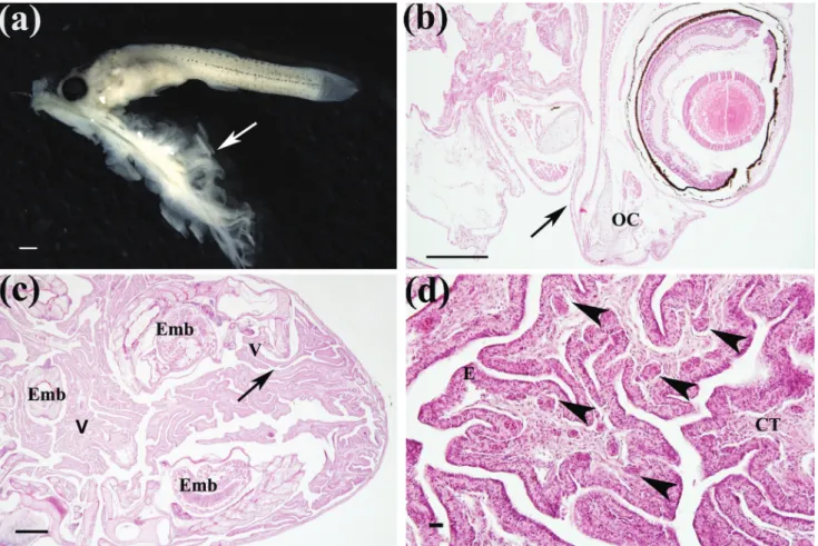
Structure connecting female-embryo. The first detectable maternal-embryonic connection was a cord like structure, which was first recorded in almost elongated and highly pigmented embryos from Stage 6 onwards. This connection lay on the ovarian tissue folds (villus) and extended throughout the gills either via the oral (less frequently recorded) or pharyngeal cavity (most frequently recorded) of the embryo (Figs. 2a-b). In a single batch, all embryos were connected to the ovarian tissue (villus) through a cord-like structure, which was maintained until the final stages of development, when embryos were mature and ready to birth (Fig. 2c). Histological analyses of villus revealed the occurrence of blood vessels located adjacent to epithelium in the connective tissue (Fig. 2d).
Variation in the weight of batches and reproductive tissues through embryonic development. According to the dry weight recorded in 15-18 batches of each developmental stage, significant increases occurred in the total developing embryos despite minor changes in maternal reproductive tissues. On an average, the batches of embryos in the initial stages of development showed substantially smaller weight than those that are close to
maturity or mature (Tab. 2). While the total dry weight of these mature embryos (Stage 8) increased steeply with the developmental stage (an increment of 66-folds, Tab. 2), it increased only slightly in the other reproductive tissues. The significant interaction term between predictor variables (Tab. 3) indicated an effect on the stage of the development of the relation between the dry weight of the batch and the other reproductive tissues.
After controlling the effect of reproductive tissues, almost 90% of the variation in the total dry weight of the batch was explained by the developmental stages (ANCOVA,
F = 62.424, d.f. = 11;81, r2 = 0.880, P <0.001) (Tab. 2).
According to the comparison of the adjusted means, total dry weight of developing embryos from the Stage 6 onwards was significantly heavier than those in Stages 3-4 (Fig. 3). Macroscopic records of the ovaries and embryos revealed that a thin layer (ovarian membrane) covers the batches of mature embryos. However, other maternal-embryonic structures (i.e., the cord-like structure) may override the losses represented by the thinning of the ovarian wall in the later developmental stages on the weight in maternal reproductive tissues.
Table 3. Results of the ANCOVA that evaluated the relationship between the weight of the batches (n = 93) and their respective ovarian tissue across stages of embryonic development of the one-sided livebearer Jenynsia multidentata.
SS d.f. MS F P-value
Stage 358.83 5 71.77 1.07 0.3833
Ovarian tissue 3390.15 1 3390.15 50.54 <0.0001
Stage x Ovarian tissue 5349.12 5 1069.82 15.95 <0.0001
Error 5433.52 81 67.080
Fig. 3. Variation in dry weight of batches of developing embryos (adjusted mean ± 95% confidence interval) of the one-sided livebearer Jenynsia multidentata through the progressive stage of development, after controlling the effect of the dry weight of reproductive tissues. The values of the x-axis range from the beginning of eye pigmentation (Stage 3) to the mature embryo (Stage 8), according to the stages proposed by this study. Different letters (A-D) identify averages that differed significantly between stages of development according to Tukey’s test.
Discussion
This study presents a classification of embryonic development of the one-sided livebearer J. multidentata
based on the morphological characteristics recorded by macroscopic analyses, which were supported by microscopic documentation of eggs and embryos of reared
wild-caught females. To the best of our knowledge, this is the first study describing the embryonic staging of J. multidentata supported by morphological characterization, although there are important studies on the early development of this species (Hylton Scott, 1918; Siccardi, 1940). On the classification of embryonic development for poeciliids, Haynes (1995) put less emphasis on
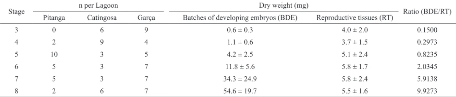
Table 2. Variation in dry weight of the batch of developing embryos (n) and reproductive tissues (mean ± S.D.) and their ratio across the developmental Stages 3 and 8 of the one-sided livebearer Jenynsia multidentata inhabiting coastal lagoons of the northern of Rio de Janeiro State, Brazil.
Stage n per Lagoon Dry weight (mg) Ratio (BDE/RT)
Pitanga Catingosa Garça Batches of developing embryos (BDE) Reproductive tissues (RT)
3 0 6 9 0.6 ± 0.3 4.0 ± 2.0 0.1500
4 2 9 4 1.1 ± 0.6 3.7 ± 1.5 0.2973
5 10 3 5 4.2 ± 2.5 5.1 ± 2.4 0.8235
6 5 3 7 11.8 ± 5.6 5.8 ± 1.7 2.0345
7 5 3 7 34.3 ± 24.9 5.8 ± 2.4 5.9138
the cleavage, morphogenesis and organogenesis; the development of the eye is an important feature for stage identification of poeciliids. The three processes above were neither common nor easy to detect even in the present study. Therefore, the feasibility of macroscopically detecting the main morphological structures throughout embryonic development of J. multidentata was pursued on the present staging framework.
We recorded the development of structures (e.g., fins, eyes) and changes in body size and weight made in poeciliids (Haynes, 1995; Turcotte et al., 2008), which contribute to our understanding of the embryo progress supported by the post-fertilization maternal provisioning of J. multidentata. Since the first scientific records of fish viviparity in the XVII century (see Kunz, 2004 for details), the retention of the batch until term in the maternal reproductive system draws attention to the structures and mechanisms involved in the embryonic development. Extending such approaches to close relatives with similar but independently evolved fertilization mode might provide new insights in the related maternal and environmental processes.
In addition to the major changes in the morphology and pattern of pigmentation of the embryos, the development of other reproductive tissues, which putatively play a role in embryo maintenance were considered for staging in the present study. As viviparity is directly related to the nutritional maternal-embryonic connections, it seemed reasonable to study the development of such structures and relate them to the increase in weight throughout the embryonic development (Schindler, de Vries, 1988). The weight of the batch should reflect the number of embryos and both individual decrease in weight due to the metabolism of yolk and gain in weight due to matrotrophy. Therefore, the exponential increase in dry weight of the batch suggests the continuous matrotrophic nourishment throughout the embryonic development of J. multidentata, as first mentioned by Hylton Scott (1918).
During the initial stages of J. multidentata development, until the sprouting of the unpaired fins, the increases in embryonic dry weight were moderately possibly due to the energetic investment in organogenesis; embryos presented remnants of the syncytial layer and the yolk is probably their first source of nourishment. As the yolk reserves deplete with the progression of development, other maternal sources for nutrition and metabolic exchanges are required (Hylton Scott, 1918; Siccardi, 1940). From Stage 6 onwards, the intraovarian space becomes reduced, and the modification of ovarian folds to a follicular pseudoplacenta intruding embryos by their gills (and mouth, as recorded by Turner, 1940) in the form of strands was first recorded. Although Hylton Scott (1918) associated these maternal-embryonic structures with the oxygen demands, the highest gain in weight was recorded when organogenesis was completed and the dry weight of the embryos increased five-folds (from Stages 6 to 8). This weight gain may reflect both extra nutrition and better feeding, which is converted
in muscle mass, and consequently, size. Pires et al. (2007) reported a higher contribution of maternal resources on later embryonic development for the blackstripe livebearer
Poeciliopsis prolifica Miller, 1960. However, compared with most poeciliids, there was a greater intensity of transferences occurring at the later developmental stages for J. multidentata even with those differing in maternal provisioning.
By injecting suspensions of fluorescent microspheres in the caudal peduncle of gravid females of poeciliids, DeMarais, Oldis (2005) qualitatively evaluated the location and intensity of these substances on embryo tissues across developmental stages. Due to the high levels of transferences restricted to the initial stage, to the high levels across all stages and the variable transfer within broods from Stage 1 onwards, obligate matrotrophy was attributed to H. formosa, facultative matrotrophy to the Gambusia species
G. affinis and the lagerspring gambusia G. geiseri Hubbs,
Hubbs, 1957, and lecithrotrophy to the guppy Poecilia reticulata Peters, 1859.
Despite major studies evaluating post-fertilization maternal provisioning focused on variations in embryonic dry weight, morpho-anatomical studies reveal that matrotrophy relies on embryonic-maternal structures specialized for the absorption and transfer of resources during development. These include both the tissue-tissue transfer of nutrients, which might be the case in J. multidentata, and the secretion of nutrients into the lumen, which are then actively ingested by the embryos (Reznick
et al., 2007). For embryos of matrotrophic species, a failure to catch a villus ovary and/or in establishing a connection to the maternal structures related to nourishment might incur death. Therefore, the low frequency of embryos in different developmental stages within a batch (<2% of all ovaries analyzed), suggests that a failure in establishing the cord (pseudoplacenta follicular), although rare, might occur in
J. multidentata.
Water temperature and salinity seem to play a major role in the reproductive dynamics and growth rates of
J. multidentata. The breeding season of subtropical populations in South Brazil and Uruguay is closely associated to the warmer months, and specimens reach larger sizes in estuaries (Fontoura et al., 1994; Garcia et al., 2004; Goyenola et al., 2011). Mai et al. (2005)
experimentally tested the effect of salinity on the rates of survival and growth in length and mass of newborns. Significantly higher rates were recorded under increases of salinity (6 to 16) when compared to decreases (6 to 0), suggesting a high affinity of J. multidentata to brackish waters. However, the effect of temperature and salinity on the embryonic development of the species is still lacking.
between the aquaria units. Therefore, the characterization of the embryonic development of J. multidentata was restricted to the appearance of morphological features and associated dry weight variation. The time ratio relative to each of the eight stages and the potential effect of environmental factors within the complete embryonic development should be properly addressed in future studies, which could benefit from the inclusion of temperature and salinity in the experimental design (e.g.,Trexler, 1997).
Other not mutually exclusive factors might be involved in the failure in achieving controlled pregnancy. Fish used in experimental trials generally descend from wild caught individuals, which may slightly reduce the negative consequences of stress due to captivity, such as the agonistic behavior recorded among the wild caught females in this study compared with others (Bisazza et al., 2000). Contrary to some Cyprinodontiformes from other studies elsewhere, whose abdominal distension and/or a visible anal spot are indicators of advanced pregnancy (Bisazza et al., 2000;
DeMarais, Oldis, 2005; Marsh-Matthews, Deaton, 2006), the thin females of J. multidentata in this study had embryos that were either ready to birth or were in intermediate stages of development as were depleted of embryos or eggs. This may have hampered the visual monitoring of females next to parturition. Another reason could be related to the ability of females to store sperm from the recesses of the ovaries and, thus, be able to modulate fertilization batches of oocytes as reported for some species of poeciliids, which can gestate multiple broods at different stages of development (Pires et al., 2007; Turcotte et al., 2008; Bassar et al., 2014). However, this alternative seems more remote for
J. multidentata, as there are no reports of the occurrence of a spermatheca or a special intraovarian compartment to house the sperm and there was no occurrence of sperm in the histological slides analyzed. Further studies with
J. multidentata should consider these above-mentioned characteristics when delineating the experimental design.
There are several examples in the literature supporting the view that strictly viviparous embryos show markedly morphophysiological modifications during early development, including dramatic increases in weight (Reznick et al., 2007). Hylton Scott (1918) first recorded
embryos immersed in ovarian fluid through which occurs the gas exchange in early stages. However, in the later stages of development, the embryos, due to their larger volume, are positioned head-tail fin and are attached to the ovarian membrane. Thus, the fluid renewal is negligible, and the maternal structures in close contact with the embryo provide the necessary oxygen for their breathing. The records from almost a century ago (Hylton Scott, 1918) and from this study support the hypothesis that J. multidentata is primarily a matrotrophic species, with vascularized specialized structures for the nutritional maternal transferences and maintenance of the embryos. The vascularized, specialized structures are developed and maintained until the end of pregnancy.
The eight stages of embryonic development of J. multidentata proposed herein might be conveniently divided into two intervals of gain in weight. The first one, until Stage 5, when embryos relied basically on yolk content stored in the egg prior to fertilization and the dry weight was almost invariable until it had developed the sprouts of the dorsal and anal fins; and the second one, from Stage 6 to Stage 8, when maternal structures related to embryo nourishment, such as the intruding ovarian folds, develop, and the dry weight increases manifold. The maintenance of matrotrophy in widely distributed species but of limited dispersal ways such as the continental fish might involve some degree of variation in the maternal nourishment of the embryos (Garcia
et al., 2004; Goyenola et al., 2011). Therefore, this aspect continues to be an interesting topic to investigate throughout the embryonic development of the one-sided livebearer J. multidentata.
Acknowledgments
This study is a part of a master thesis by Nathalia López-Rodríguez in the Programa de Pós-Graduação em Ciências Ambientais e Conservação (PPG-CiAC), Universidade Federal do Rio de Janeiro (UFRJ). Lais Correia and Julia Iorio assisted in the field and keeping females in animal facilities, and Juliana Silva in sample staining protocols. Grant for NCLR was provided by the Coordenação de Aperfeiçoamento de Pessoal de Nível Superior (CAPES). All experimental procedures were approved by the Comissão de Ética em Experimentação Animal at the Universidade Federal do Rio de Janeiro - Campus Macaé (MAC017). Funding was partially provided by the Conselho Nacional de Desenvolvimento Científico e Tecnológico (CNPq) (PELD Sítio RLaC – Restingas e Lagoas Costeiras do Norte Fluminense, 403841/2012-7).
References
Amoroso EC. Viviparity in fishes. In: Jones IC, editor. Hormones in Fish. London: Symposium Zoological Society; 1960. p.153-181.
Bassar RD, Auer SK, Reznick DN. Why do placentas evolve? A test of the life-history facilitation hypothesis in two clades in the genus Poeciliopsis representing two independent origins of placentas. Funct Ecol. 2014; 28(4):999-1010.
Betito R. Comparação da complexidade das adaptações bioecológicas de dois peixes (Jenynsia multidentata e Poecilia vivipara) (Cyprinodontiformes) no estuário da Lagoa dos Patos (RS - Brasil). Revista Didática Sistêmica. 2006; 3:71-100. Bisazza A, Manfredi S, Pilastro A. Sexual competition, coercive
mating and mate assessment in the one-sided livebearer, Jenynsia multidentata: are they predictive of sexual dimorphism? Ethology. 2000; 106(11):961-78.
Caliman A, Carneiro LS, Santangelo JM, Guariento RD, Pires APF, Suhett AL, Quesado LB, Scofield V, Fonte ES, Lopes PM, Sanches LF, Azevedo FD, Marinho CC, Bozelli RL, Esteves FA, Farjalla VF. Temporal coherence among tropical coastal lagoons: a search for patterns and mechanisms. Braz J Biol. 2010; 70(3-Suppl.):803-14.
DeMarais A, Oldis D. Matrotrophic transfer of fluorescent microspheres in poeciliid fishes. Copeia. 2005; 3:632-36. Di Dario F, Petry AC, Pereira MMS, Mincarone MM, Agostinho
LS, Camara EM, Caramaschi EP, Britto MR. An update on the fish composition (Teleostei) of the coastal lagoons of the Restinga de Jurubatiba National Park and the Imboassica Lagoon, northern Rio de Janeiro State. Acta Limnol Bras. 2013; 25(3):257-78.
Fontoura NF, Braun AS, Lewis DS, Souto GDB. Dinâmica populacional da ictiofauna da lagoa Fortaleza, Cidreira, Rio Grande do Sul. II. Jenynsia lineata (Jenyns, 1842) (Teleostei, Anablepidae). Biociências. 1994; 2(1):79-93.
Garcia AM, Vieira JP, Winemiller KO, Raseira MB. Reproductive cycle and spatiotemporal variation in abundance of the one-sided livebearer Jenynsia multidentata in Patos Lagoon, Brazil. Hydrobiologia. 2004; 515(1-3):39-48.
Goyenola G, Iglesias C, Mazzeo N, Jeppesen E. Analysis of the reproductive strategy of Jenynsia multidentata (Cyprinodontiformes, Anablepidae) with focus on sexual differences in growth, size, and abundance. Hydrobiologia. 2011; 673(1):245-57.
Ghedotti MJ, Weitzman SH. A new species of Jenynsia (Cyprinodontiformes: Anablepidae) from Brazil with comments on the composition and taxonomy of the genus. Sci Pap Univ Kansas Nat Hist Mus. 1996; 179:1-25.
Haynes JL. Standardized classification of Poeciliid development for life-history studies. Copeia. 1995; 1:147-54.
Hylton Scott MI. 1918. Sobre el desarrollo intraovarial de “Jenynsia lineata” (Nota preliminar). Anales Soc Ci Argent. 1918; 86:349-54.
Kimmel CB, Ballard WW, Kimmel SR Ullmann B, Schilling TF. Stages of embryonic development of the zebrafish. Dev Dyn. 1995; 203:253-310.
Kunz YW. Developmental biology of teleost fishes. Dordrecht: Springer; 2004. (Fish and Fisheries Series; 28).
Lodé T. Oviparity or viviparity? That is the question… Reprod Biol. 2012; 12(3):259-64.
Lucinda PH. Family Poeciliidae. In: Reis RE, Kullander SO, Ferraris CJ, Jr., organizers. Check list of the freshwater fishes of South and Central America. Porto Alegre: Edipucrs; 2003a. p.555-581.
Lucinda PH. Family Anablepidae. In: Reis RE, Kullander SO, Ferraris CJ, Jr., organizers. Check list of the freshwater fishes of South and Central America. Porto Alegre: Edipucrs; 2003b. p.582-585.
Mai ACG, Garcia AM, Vieira JP. Influência da salinidade no crescimento de juvenis de Jenynsia multidentata Jenyns (Pisces). Rev Bras Zool. 2005; 22(3):780-83.
Marsh-Matthews E, Deaton R. Resources and offspring provisioning: a test of the Trexler-DeAngelis model for matrotrophy evolution. Ecology, 2006; 87(12):3014-20. Meisner AD, Burns JR. Viviparity in the halfbeak genera
Dermogenys and Nomorhamphus (Teleostei: Hemiramphidae).
J Morphol. 1997; 234(3):295-317.
Nelson JS, Grande TC, Wilson MVH. Fishes of the World. 5th ed. Hoboken (NJ): J. Wiley & Sons; 2016.
Pires MN, McBride KE, Reznick DN. Interpopulation variation in life-history traits of Poeciliopsis prolifica: implications for the study of placental evolution. J Exp Zool. 2007; 307(A):113-25. Reznick D, Meredith R, Collette BB. Independent evolution of
complex life history adaptations in two families of fishes, live-bearing halfbeaks (Zenarchopteridae, Beloniformes) and Poeciliidae (Cyprinodontiformes). Evolution. 2007; 61(11):2570-83.
Riesch R, Plath M, Schlupp I, Marsh-Matthews E. Matrotrophy in the cave molly: an unexpected provisioning strategy in an extreme environment. Evol Ecol. 2010; 24(4):789-801. Schindler JF, de Vries U. Ovarian structural specializations
facilitate aplacental matrotrophy in Jenynsia lineata
(Cyprinodontiformes, Osteichthyes). J Morphol. 1988; 198(3):331-39.
Siccardi EM. Algunos hechos relativos a las primeras fases del desarrollo de Jenynsia lineata (Jenyns) Berg. Rev Soc Argent Biol. 1940; 15(2):3-7.
Thibault RE, Schultz RJ. Reproductive adaptations among viviparous fishes (Cyprinodontiformes: Poeciliidae). Evolution. 1978; 32(2):320-33.
Trexler JC. Resource availability and plasticity in offspring provisioning: embryo nourishment in sailfin mollies. Ecology. 1997; 78(5):1370-81.
Turcotte MM, Pires MN, Vrijenhoek RC, Reznick DN. Pre- and post-fertilization maternal provisioning in livebearing fish species and their hybrids. Funct Ecol. 2008; 22(6):1118-24. Turner CL. Adaptations for viviparity in Jenynsiid fishes. J
Morphol. 1940; 67(2):291-97.
Wourms JP. Viviparity: the maternal-fetal relationship in fishes. Am Zool. 1981; 21(2):473-515.
Zúñiga-Vega JJ, Suárez-Rodríguez M, Espinosa-Pérez H. Johnson JB. Morphological and reproductive variation among populations of the Pacific molly Poecilia butleri. J Fish Biol. 2011; 79(4):1029-46.