Endophytic
Actinobacteria from
Laminaria ochroleuca:
a new source of
bioactive compounds
Mariana Girão Silva Martins
Dissertação de Mestrado apresentada à
Faculdade de Ciências da Universidade do Porto em
Biologia Celular e Molecular
2018
E nd op hy tic A cti no ba cteri a from La mi na ri a oc hroleuc a : a ne w so urce of bioac tiv e co mpou nd s M ar iana G ir ão S ilv a M ar tins FCUP 2018 2.º CICLOEndophytic
Actinobacteria
from Laminaria
ochroleuca: a new
source of bioactive
compounds
Mariana Girão Silva Martins
Mestrado em Biologia Celular e Molecular
Departamento de Biologia da Faculdade de Ciências da Universidade do Porto (FCUP)2017/1018
Orientador
Maria de Fátima Carvalho, Investigadora auxiliar, Centro Interdisciplinar de Investigação Marinha e Ambiental (CIIMAR) e Professora auxiliar convidada FCUP
Coorientador
Pedro Leão, Investigador auxiliar, Centro Interdisciplinar de Investigação Marinha e Ambiental (CIIMAR) e Professor Auxiliar Convidado FCUP
Todas as correções determinadas pelo júri, e só essas, foram efetuadas. O Presidente do Júri,
Acknowledgements
The accomplishment of this master thesis would not have been possible without the guidance and support of several people, to whom I am truthfully grateful.
First of all, thank you to my supervisor Dr. Fátima Carvalho, for having welcomed me so kindly and guided me along this journey. Thank you for the sharing of knowledge, trust and dedication that were undoubtedly important to the success of this work. I would also like to thank my co-supervisor Dr. Pedro Leão, for all the teachings, enthusiasm and trust in my work.
I am also grateful to Dr. Filipe Pereira for being always available to help, and by all the teachings in molecular biology, and to Dr. Ralph Urbatzka for the help in the anticancer assays. Thank you also to Dr. Ana Paula Mucha, for the opportunity to join the EcoBioTec Laboratory, and CIIMAR for providing the equipment and installations.
I cannot forget to recognise my friends at EcoBioTec laboratory for their contagious joy, motivation, support and sharing of knowledge. A special thanks to Inês Ribeiro for her fellowship and always available help. To my colleagues from BBE and CNP laboratories, I also appreciate all your help, especially from Tiago Ribeiro and Nelly Brugerolle.
Last but not least, thank you to my parents for always being present with a word of motivation. Thank you also to Diogo, for all his support, and to Beatriz, Teresa and Rita, who were part of this journey with me.
Abstract
Nature is the major reservoir of biologically active molecules. The urgent need of finding novel molecules with pharmaceutical interest is prompting the research of underexplored environments, such as marine ecosystems. In this regard, marine actinobacteria represent a remarkable source of biologically active compounds. Marine actinobacteria often live in association with other organisms, representing unique ecological niches for the discovery of new bioactive substances. Despite the proven potential of actinobacteria as source of secondary metabolites with pharmacologically-relevant activities, only few studies have focused on actinobacteria associated with macroalgae. This study aimed to investigate the cultivable community of endophytic actinobacteria associated with the macroalgae Laminaria ochroleuca and assess its potential to produce compounds with antimicrobial and anticancer activities. Fragments of tissues from different parts of L.
ochroleuca (holdfast, stipe and blade), collected in a rocky shore in northern Portugal,
were surface sterilized and plated in three culture media selective for actinobacteria. A total of 90 actinobacterial strains were isolated, with the majority being affiliated with the genus Streptomyces. Isolates associated with the genera Isoptericola, Rhodococcus,
Nonomuraeae, Nocardiopsis, Microbispora and Microbacterium were also obtained.
Extracts from all actinobacterial isolates were tested for their antimicrobial activity using the agar-based disk diffusion method, followed by determination of MIC values for extracts showing relevant activities. Forty-five isolates inhibited, to some degree, the growth of Candida albicans and/or Staphylococcus aureus, with MIC values ranging from < 0.487 to 1000 µg/mL. The actinobacterial isolates were also tested for their anticancer potential in two human carcinogenic cell lines (breast carcinoma T-47D and neuroblastoma SH-SY5Y) and compared to their cytotoxicity on a non-carcinogenic endothelial cell line (hCMEC/D3). Thirty isolates exhibited anticancer activity in at least one of the tested cell lines, decreasing their viability to less than 50%. Four isolates were identified with strong cytotoxic activity on SH-SY5Y cells, but with low activity on hCMEC/D3 cells. According to LC-MS/MS dereplication data, the activity of most extracts may be associated with the presence of secondary metabolites antimycins. The active strain Streptomyces sp. KENR25 was selected for a bioassay-guided fractionation, following part of a traditional natural product discovery pipeline, with several fractions showing mainly antimicrobial activity. This study reveals that L. ochroleuca is a rich source of actinobacteria with promising antimicrobial and anticancer activities and further supports that macroalgae may be a valuable resource of actinobacteria and, consequently, of new molecules with biotechnological importance.
Keywords
marine actinobacteria, endophytic actinobacteria, bioactivity, antimicrobial, anticancer, macroalgae, kelp, Laminaria ochroleuca
Resumo
A natureza constitui o maior reservatório de moléculas biologicamente ativas. A urgente necessidade de encontrar novas moléculas com interesse farmacêutico tem incentivado a investigação de ambientes pouco explorados, como os ecossistemas marinhos. Neste sentido, as actinobactérias marinhas são uma fonte notável de compostos biologicamente ativos. As actinobactérias marinhas vivem muitas vezes em associação com variados organismos, representando nichos ecológicos únicos para a descoberta de novas substâncias bioativas. Apesar do enorme potencial das actinobactérias como fonte de metabolitos secundários com atividades farmacológicas relevantes, poucos estudos se têm focado no potencial de actinobactérias associadas a macroalgas. Este estudo teve como objetivo investigar a comunidade cultivável de actinobactérias endófitas associadas à macroalga Laminaria ochroleuca e avaliar o seu potencial para produzir compostos com atividades antimicrobiana e anticancerígena. Fragmentos de tecidos de diferentes partes da alga L. ochroleuca (rizoide, estipe e lâmina), recolhida na costa rochosa norte de Portugal, foram esterilizados na sua superfície e plaqueados em três meios de cultura seletivos para actinobactérias. Um total de 90 estirpes de actinobactérias foram isoladas, estando a a maioria afiliada com o género Streptomyces. Isolados associados com os géneros Isoptericola, Rhodococcus, Nonomuraeae,
Nocardiopsis, Microbispora e Microbacterium também foram obtidos. Todos os extratos
actinobacterianos foram testados quanto à sua atividade antimicrobiana usando o método de difusão em agar, seguindo-se a determinação do MIC para extratos com atividade relevante. Quarenta e cinco isolados inibiram o crescimento de Candida
albicans e/ou Staphylococcus aureus, com valores de MIC entre < 0.487 e 1000 µg/mL.
O potencial anticancerigeno dos isolados foi também testado em duas linhas celulares carcinogénicas humanas (carcinoma da mama T-47D e neuroblastoma SH-SY5Y) e a sua citotoxicidade comparada numa linha celular não-carcinogénica (hCMEC/D3). Trinta isolados exibiram atividade anticancerígena em pelo menos uma das linhas celulares testadas, diminuindo a viabilidade das células para menos de 50%. Quatro isolados foram identificados com uma forte atividade citotóxica nas células SH-SY5Y, mas com pouca atividade nas células hCMEC/D3. De acordo com os dados de desreplicação, a atividade da maioria dos extratos está associada com a presença dos metabolitos secundários antimicinas. A estirpe ativa KENR25 foi selecionada para um fracionamento guiado por ensaios de bioatividade, seguindo parte do procedimento tradicional para a descoberta de produtos naturais, onde várias frações mostraram principalmente atividade antimicrobiana. Este estudo revela que a alga L. ochroleuca é uma fonte rica
de actinobactérias com promissoras atividades antimicrobianas e anticancerígenas, suportando a ideia de que as macroalgas podem ser um recurso valioso de actinobactérias e, consequentemente, de novas moléculas com importância biotecnológica.
Palavras-chave
actinobactérias marinhas, actinobactérias endófitas, bioatividade, atividade antimicrobiana, atividade anticancerígena, macroalga, kelp, Laminaria ochroleuca
Table of Contents
Acknowledgements ... iii
Abstract ... iv
Resumo ... vi
List of Figures ... x
List of Tables ... xiii
List of Abreviations ... xiv
I. INTRODUCTION
... 21. Phylum Actinobacteria ... 3
2. Marine Actinobacteria ... 4
3. Marine Actinobacteria as a Source of Bioactive Compounds ... 5
3.1. Antimicrobial Activity ... 8
3.2. Anticancer Activity ... 8
4. Actinobacteria in Kelps ... 9
5. Aim and outline of this thesis ... 10
II. MATERIALS AND METHODS
... 1111. Sampling and Bacterial Isolation ... 11
2. Taxonomic Identification of the Isolates ... 12
3. Bioactivity Assays ... 13
3.1. Preparation of Crude Extracts ... 13
3.2. Screening of Antimicrobial Activity ... 14
3.3. Screening of Anticancer Activity ... 16
4. Dereplication of Active Crude Extracts ... 17 5. Bioactivity-Guided Study of Bioactive Molecules - Streptomyces sp. KENR25 . 18
5.1. Large-Scale Cultivation ... 19
5.2. Organic Extraction ... 19
5.3. Bioassay-guided fractionation ... 20
5.3.1. Vacuum Liquid Chromatography (VLC) ... 20
5.3.2. Flash Chromatography (FC) ... 22
5.3.3. High-Performance Liquid Chromatography (HPLC) ... 23
5.4. Molecular Networking ... 24
III.
RESULTS
... 251. Phylogenetic Identification of Actinobacteria Isolated from L. ochroleuca ... 25
2. Bioactive Potential of the Actinobacterial Isolates ... 29
3. Bioactive Compounds Produced by Actinobacterial Strains ... 33
4. Bioassay-guided Study of Bioactive Molecules Produced by Streptomyces sp. KENR25 ... 35
IV. DISCUSSION
... 43V. CONCLUSION
... 49VI. REFERENCES
... 50List of Figures
Figure 1. Distribution of actinobacteria in the marine environment, according to a total of
10 400 16S rRNA gene sequences retrieved from marine actinobacteria. Adapted from [22]. ………..………....….... 5
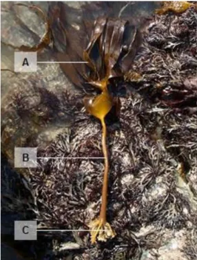
Figure 2. Specimen of L. ochroleuca with the indication of (A) blade, (B) stipe and (C)
holdfast. ……… 12
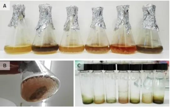
Figure 3. Liquid cultures of some actinobacterial isolates (A) bacterial growth in the
Erlenmeyer flasks, (B) Amberlite XAD16N resin added to the cultures and (C) crude extracts obtained from some liquid cultures. ………..……… 14
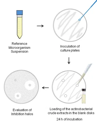
Figure 4. Illustrative diagram of the agar-based disk diffusion assay. ………..……… 15
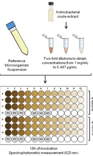
Figure 5. Illustrative diagram of the minimal inhibitory concentration (MIC) assay. PC
and NC indicate positive and negative controls, respectively. ……….……… 16
Figure 6. Illustrative diagram of the MTT assay. ………... 17
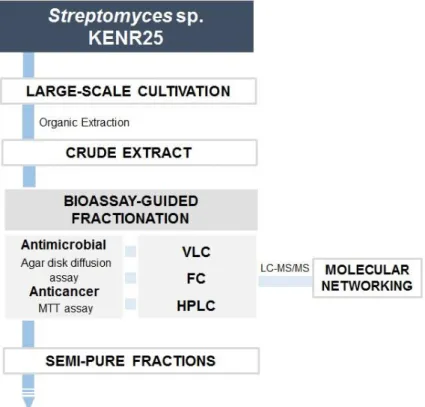
Figure 7. Diagram of the bioassay-guided workflow to study bioactive molecules. .... 19
Figure 8. Vacuum Liquid Chromatography fractionation of Streptomyces sp. KENR25
crude extract. ………...……… 21
Figure 9. Sub-fractioning of the DEFG fraction. (A) Pre-chromatography Thin-Layer
Chromatography and (B) Flash Chromatography column. …..……… 23
Figure 10. Actinobacterial isolates recovered from L. ochroleuca. (A) Percentage of
actinobacterial strains isolated from holdfasts, stipes and blades of L. ochroleuca and (B) Distribution of the isolates by the selective culture media used in the study (SCN: Starch-Casein-Nitrate agar; RH: Raffinose-Histidine; NPS: Nutrient-Poor Sediment).
Figure 11. Morphological diversity of some actinobacterial strains isolated from L.
ochroleuca. Strain (A) KENS1, (B) KENR91, (C) KENR92, (D) KENR94, (E) KENRB10,
(F) KENR56, (G) KENR81 and (H) KENB8. ………--……… 26
Figure 12. Actinobacterial genera recovered from L. ochroleuca. (A) Percentage of
actinobacterial genera isolated from L. ochroleuca, (B) genera distribution in the holdfast, stipe and blade of the macroalgae and (C) genera distribution according to the selective culture media used for the isolation. ……… 27
Figure 13.Phylogenetic relationship of the 87 actinobacterial isolates recovered from L.
ochroleuca, based on 16S rRNA gene homology with their GenBank nearest neighbours.
Numbers at nodes represent bootstrap values when higher than 60%. Numbers in parenthesis correspond to GenBank accession numbers. Bacillus subtilis was used as outgroup. ………..… 28
Figure 14. Antimicrobial activity of actinobacterial strains isolated from L. ochroleuca.
(A-E) Examples of inhibition halos against C. albicans and (F-G) against S. aureus. (A) Strain KENR13A, (B) strain KENR25, (C) strain KENR6, (D) strain KENR16B, (E) strain KENR21, (F) strain KENR64 and (G) strain KENR60. …….……… 29
Figure 15. Actinobacterial strains isolated from L. ochroleuca with anticancer activity.
The graphs show the extracts able to decrease cancer cells viability to less than 50% of the control, after 24 and/or 48 h of exposure, for (A) SH5Y-SH, (B) T-47D, and (C) hCMEC/D3 cell lines. PC and SC indicate positive and solvent controls, respectively. Error bars represent standard deviation of the mean of at least duplicate assays performed in triplicates each. ……… 32
Figure 16. Inhibition halos of the antimicrobial active VLC fractions: (A) fraction D, (B)
fraction E, (C) fraction F, (D) fraction G, and (E) fraction I2. ……… 36
Figure 17. MTT results of VLC fractions. The graphs show the cellular viability after 24
and/or 48 h of exposure for (A) SH5Y-SH, (B) T-47D cell lines. PC and NC indicate positive and negative controls, respectively. Error bars represent standard deviation of the mean of at least duplicate assays. ……….………...……… 36
Figure 19. Inhibition halos caused by of the antimicrobial active FC fractions: (A) fraction
DEFG-A, (B) fraction DEFG-B, (C) fraction DEFG-C, (D) fraction DEFG-D and (E) fraction DEFG-E. ……….…….… 38
Figure 20. 1H NMR spectra of Streptomyces sp. KENR25 FC fractions DEFG-A –
DEFG-E. ………...……… 39
Figure 21. Molecular networking developed with the MS/MS data obtained for the
fractions DEFG-A – DEFG-E. (A) Complete molecular networking and (B) cluster corresponding to the antimycins. The numbers within the nodes correspond to the mass of each fragment. The size of the nodes indicates the abundance of the fragment. The similarity between two nodes was computed based on the cosine score defining the connecting edges. The colours match the fragments found in each of the DEFG sub-fractions. ………--……… 40
Figure 22. HPLC spectra with the peaks corresponding to the fractions DEFG-A-A to
List of Tables
Table 1. Bioactive metabolites produced by marine actinobacteria, until 2017 (Adapted
from [13, 14, 25]) ………..………...…...… 6
Table 2. Solvent mixtures used for elution of Streptomyces sp. KENR25 crude extract
on Vacuum Liquid Chromatography. Due to the high polarity of the extract, the last solvent mixture was repeated (fraction I2) ………..……… 21
Table 3. Solvent mixtures used for elution of the DEFG fraction on FC ……...……… 23
Table 4. Actinobacteria isolated from L. ochroleuca with antimicrobial activity. The
diameter of inhibition halos obtained by the agar disk diffusion method and MIC values are shown ………..…..………… 30
Table 5. Dereplication results for the 35 actinobacterial crude extracts selected,
indicating the compounds recorded for each one and the correspondent cosine value. ………..…..…………...34
Table 6. VLC fractions with the indication of the mass amount yield for each one ... 35
Table 7. FC fractions with the indication of the mass amount yield for each one …… 38
Table 8. HPLC fractions with the indication of the mass amount yield for each one (BL -
List of Abreviations
16S rRNA 16S ribosomal RNA
BLAST Basic Local Alignment Search Tool
BP Base pair
CTAB Cetyl trimethylammonium bromide
DMEM Dubelco’s Modified Eagle Medium
DMSO Dimethyl sulfoxide
EtOAc Ethyl acetate
FC Flash Chromatography
GNPS Global Natural Product Social Molecular Networking
HPLC High performance liquid chromatography
LC-MS/MS Liquid chromatography-tandem mass spectrometry MIC Minimal Inhibitory Concentration
MH Mueller-Hinton
MTT (3-(4,5-Dimethylthiazol-2-yl)-2,5-Diphenyltetrazolium Bromide)
NCBI National Center for Biotechnology Information
NPS Nutrient-Poor Sediment
OD Optical Density
PCA Plate Count Agar
PCR Polymerase Chain Reaction
PMA Phosphomolybdic Acid
RH Raffinose-Histidine
SCN Starch-Casein-Nitrate
SD Sabouraud Dextrose
TLC Thin-Layer Chromatography
I.
Introduction
Human health is constantly and increasingly under the threat of several disorders that demand urgent solutions, such as cancer pathologies or the increasing incidence of antibiotic multi-resistant microbial species. The search for novel bioactive compounds is an effective approach to tackle this problematic, with the natural environment still being the major source to find such molecules [1, 2]. Taking cancer as an example, from 1940 to the end of 2014, about half of the approved drugs for combating this disease were either natural products (NPs) or compounds directly derived from them [3]. Among the different natural sources from which bioactive molecules may derive, microorganisms play a prominent role: from bacteria to fungi, microorganisms have proved their ability to produce a wide range of NPs exhibiting numerous biological activities [4]. Within the domain Bacteria, the phylum Actinobacteria plays a major role in the production of bioactive compounds, especially the species belonging to the order Actinomycetales, commonly known as actinomycetes. So far, more than 10.000 different bioactive compounds produced by actinomycetes have been identified, representing about 50% of all bioactive metabolites obtained from microorganisms, with most of them being derived from the genus Streptomyces and a smaller fraction from the so-called rare actinomycetes [5, 6]. From the pharmaceutical, biotechnological, industrial and economic point of view, actinomycetes are amongst the most valuable microorganisms, being capable of producing antibiotics, antimicrobials, anti-inflammatory, anticancer and antitumor agents, immunosuppressive compounds, etc. [3, 7]. Despite the wide range of known bioactive compounds and their significant applications, the discovery of new natural molecules is coming to a stagnation point. However, this reality is not in agreement with the data obtained from genomic analysis which suggest that only about 10% of microbial secondary metabolites are known [8]. The decrease in the discovery of new bioactive compounds and the increase in the re-isolation of known ones, may be explained by the fact that in the last decades, most of the bioactivity screening programmes have focused on highly explored environments, mainly terrestrial ones [7, 9]. Considering the need of finding novel secondary metabolites with bioactive action, it is highly promising to investigate actinobacteria from unexplored or underexplored habitats, such as the marine environment. The conditions found in marine environments are very different from those occurring in terrestrial ones, which translates in different microbial evolutionary mechanisms and, consequently, in different metabolisms that may lead to the production of novel differentiated chemical molecules [10].
1. Phylum Actinobacteria
Actinobacteria are filamentous Gram-positive bacteria, usually with a high guanine/ cytosine content in their DNA that can vary between 50% and 70% according to the species. The phylum Actinobacteria represents one of the largest taxonomic units within the domain Bacteria, both in number and diversity, including 6 classes, 25 orders, 52 families, and 232 genera [7, 11]. Microorganisms belonging to this phylum exhibit a wide variety of morphologies, from coccoid to fragmenting hyphal forms or highly differentiated branched mycelium, with several species being able to reproduce by sporulation. Actinobacteria species also exhibit diverse metabolic, physiological and ecological properties that result in the production of several secondary metabolites, many with biological activity [11]. These microorganisms are widely disseminated in both terrestrial and aquatic habitats, including marine ecosystems. They are usually more common in the soil where they play an important role in the recycling of organic matter but can also be found as plant symbionts (e.g., Frankia sp.), animal or plant pathogens (e.g.,
Corynebacterium sp., Mycobacterium sp., Propionibacterium sp. and Nocardia sp.) or
even gastrointestinal commensals (e.g., Bifidobacterium sp.) [11, 12]. Since the 1950s, Actinobacteria from different sources, fundamentally from terrestrial ecosystems, have been studied for their high potential to produce bioactive compounds, resulting in the discovery of a wide range of clinically-relevant compounds. Anticancer, antitumor or antibacterial compounds are representative examples of the wide range of biological active molecules produced by these microorganisms [13, 14]. Actinobacteria also have a role as plant growth-promoting agents [15, 16] and play an important part in bioremediation processes, since they are able to metabolize complex organic compounds and to remove xenobiotics, such as pesticides and heavy metals, from the environment [17, 18]. One other characteristic of these microorganisms is their ability to produce and accumulate polyhydroxyalkanoates (PHAs), biodegradable and renewable polymers that may be used to produce disposable and biodegradable plastics or as scaffold material for tissue engineering or controlled drug delivering [19]. Given the immense biotechnological, industrial, ecological and biopharmaceutical applications of Actinobacteria, there is high interest in studying these microorganisms as potential sources of new compounds. Within the phylum Actinobacteria, the order Actinomycetales comprehends most of the taxonomic units responsible for the synthesis of bioactive secondary metabolites, and the terms actinobacteria or actinomycetes will be hereafter used as synonymous of microorganisms affiliated with this order. Among these taxonomic units, the genus
Streptomyces is particularly relevant as these microorganisms are considered the
with almost all the natural antibiotics currently in clinical use being derived from secondary metabolites of these bacteria [6, 20]. Recognizing the unmatched potential of actinomycetes to produce compounds with relevant bioactivities, these organisms continue to be primary targets to discover novel useful natural products [21].
2. Marine Actinobacteria
Historically, actinobacteria were thought to inhabit exclusively terrestrial environments and it was believed that their occurrence in the oceans was due to the transport of terrestrial spores to those regions. Today, it is known that this is false and that actinobacteria are widely distributed in the marine environment, with several indigenous marine actinobacteria being described. Among several others, representative genera of marine actinobacteria include Actinomadura, Aeromicrobium, Dietzia, Gordonia,
Marinophilus, Micromonospora, Nonomuraea, Rhodococcus, Saccharomonospora,
Salinispora, Streptomyces, Solwaraspora, Williamsia, and Verrucosispora [7]. Although
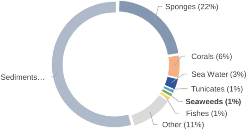
their prevalence in sediments, they also exist in association with fishes, sponges, seaweeds or corals (Fig. 1) [7, 22]. Nevertheless, the ecological role, diversity, distribution and bioactive potential of marine actinobacteria are still largely unknown and require further study [13, 23]. The environmental conditions found in marine ecosystems are very different from terrestrial ones, driving unparalleled evolutionary mechanisms. For marine actinobacteria, this is reflected in novel capacities to produce secondary metabolites [7]. Given the fact that the discovery of novel bioactive compounds is reaching a stagnation point, marine actinobacteria pose as a promising source of novel natural products [13].
Figure 1. Distribution of actinobacteria in the marine environment, according to a total of 10 400
16S rRNA gene sequences retrieved from marine actinobacteria. Adapted from [22].
Sediments… Sponges (22%) Corals (6%) Sea Water (3%) Tunicates (1%) Seaweeds (1%) Fishes (1%) Other (11%)
3. Marine Actinobacteria as a Source of Bioactive
Compounds
The discovery of new drugs to help treating serious human diseases is a constant demanding and, in this respect, marine actinobacteria constitute a precious source for the search of these so needed compounds [1]. It is known that the biosynthesis of these compounds is usually triggered by a wide variety of environmental and physiological signals [24] and, since the last decade, marine actinobacteria have been attracting the attention of the scientists for their ability to produce novel compounds with antibacterial, antifungal, antiviral, antitumor, anticancer, cytotoxic, anti-inflammatory, anti-parasitic, anti-malarial and immunosuppressive activities [13, 14, 25, 26]. Table 1 presents a summary of bioactive compounds reported from marine actinobacteria, until 2017.
Table 1. Bioactive metabolites produced by marine actinobacteria, until 2017 (Adapted from [13, 14, 25])
Compound Source Biological Activity Ref
Abyssomycins Verrucosispora sp. Antibacterial [27]
Actinofuranones Streptomyces sp. Cytotoxic [28]
Ammosamides Streptomyces sp. Cytotoxic [29]
Antimycin A Streptomyces sp. Antifungal; Anticancer; Anti-inflammatory;
[30]
Arenamides Salinipora arenicola Cytotoxic [31]
Arenicolides Salinispora arenicola Antitumor [32]
Aureolic acid Streptomyces sp. Antitumor; Antibacterial [33]
Aureoverticillactam Streptomyces aureoverticillatus
Anticancer [34]
Avermectins Streptomyces avermitilis Anti-parasitic [35]
Benzastatin C Streptomyces nitrosporeus Antiviral [36]
Bisanthraquinone Streptomyces sp. Antibacterial [37]
Bonactin Streptomyces sp. Antibacterial; Antifungal [38]
Butenolides Streptomyces sp. Antiviral; Cytotoxic [39]
Caboxamycin Streptomyces sp. Anticancer [40]
Caprolactones Streptomyces sp. Anticancer [41]
Chalcomycin A Streptomyces sp. Antibacterial [42]
Chandrananimycins Actinomadura sp. Antibacterial; Antifungal; Anticancer
[43]
Chartreusin Streptomyces chartreusis Antitumor [44]
Chinikomycins Streptomyces sp. Anticancer [45]
Chloro-dihydroquinones Novel actinomycete Antibacterial; Anticancer [46]
Chromomycins Streptomyces griseus Antitumor [33]
Curvularin-7-O-dglucopyranoside
Pseudonocardia sp. Antibacterial; Anticancer [47]
Cyclomarin A Streptomyces sp. Anti-inflammatory; Anti-Malarial; Anti-Tuberculosis
[48]
Daryamides Streptomyces sp. Antitumor; Antifungal [49]
Diazepinomicin Micromonospora sp. Antibacterial; Anticancer; Anti-inflammatory
[50]
Essramycin Streptomyces sp. Antibacterial [51]
Frigocyclinone Streptomyces griseus Antibacterial [52]
Glaciapyrroles Streptomyces sp. Antibacterial [53]
Gutingimycin Streptomyces sp. Antibacterial [54]
Helquinoline Janibacter limosus Antibacterial [55]
Himalomycins A, B Streptomyces sp. Antibacterial [56]
IB-00208 Actinomadura sp. Anticancer [57]
Komodoquinone A Streptomyces sp. Neuritogenic activity [58]
Lajollamycin Streptomyces nodosus Antibacterial [59]
Lagumycin B Micromonospora sp. Antibacterial; Cytotoxic [60]
Lipoxazolidinones A, B Marinispora sp. Antibacterial [62]
Lucentamycins Nocardiopsis lucentensis Cytotoxic [63]
Lynamicins Marinispora sp. Antibacterial [64]
Manumycins Streptomyces sp. Antibacterial; Antitumor [45]
Mansouramycin C Streptomyces sp. Cytotoxic [65]
Marinomycins Marinispora sp. Antibacterial; Cytotoxic; [66]
Marinopyrroles Streptomyces sp. Antibacterial; Anticancer [67]
Mechercharmycins Thermoactinomyces sp. Anticancer [68]
Monacyclinones Streptomyces sp. Antibacterial; Cytotoxic [69]
N-acetyl-N-demethylmayamycin
Streptomyces sp. Antibacterial; Antitumor [70]
Neomarinone Strain CNH-099 C Cytotoxic [71]
N-(2-hydroxyphenyl)-2- phenazinamine (NHP)
Nocardia dassonvillei Antifungal; Anticancer [72]
Pacificanones Salinispora pacifica Cytotoxic [73]
Phenazines 1,2 Streptomyces sp. Anti-inflammatory; Anticancer [74]
Piericidins Streptomyces sp. Anticancer [75]
Piperazimycins Streptomyces sp. Cytotoxic [76]
Proximicins Verrucosispora sp. Cytostatic [77]
Pyridinium Amycolatopsis alba Antibacterial; Cytotoxic [78]
Resistoflavine Streptomyces chibaensis Cytotoxic [79]
Saadamycin Streptomyces sp. Antifungal [80]
Salinamides Streptomyces sp. Antibacterial; Anti-inflammatory [81]
Saliniketals Salinispora arenicola Anticancer [32]
Salinisporamycin Salinispora arenicora Antibacterial; Cytotoxic [82]
Salinipyrones Salinispora pacifica Cytotoxic [73]
Salinosporamide A* Salinispora tropica Anticancer [83]
Selina-4(14),7(11)-diene-8,9-dio
Streptomyces sp. Anticancer [84]
Sisomicin Streptomyces sp. Antibacterial [85]
Staurosporine Streptomyces sp. Anti-parasitic [86]
Strepsesquitriol Streptomyces sp. Anticancer [87]
Streptoanthraquinone A Streptomyces sp. Antibacterial; Anticancer [70]
Streptochlorin Streptomyces sp. Anticancer [88]
Streptokordin Streptomyces sp. Anticancer [89]
Streptomyceamide C Streptomyces sp. Cytotoxic [90]
Thalassospiramides A, B Thalassospira sp. Immune-suppressive [91]
Thiocoraline Micromonospora sp. Antitumor; Antimicrobial [92]
Tirandamycin C Streptomyces sp. Antibacterial [93]
Trioxacarcins Streptomyces sp. Antibacterial; Anticancer; Anti-Malarial
[94]
Valinomycin Streptomyces sp. Anti-Parasitic [86]
Violapyrones Streptomyces sp. Antibacterial; Cytotoxic [95]
3.1. Antimicrobial Activity
The increasing of antibiotic-resistant bacteria is a worldwide problem and a worrying phenomenon. The microbial resistance to antibiotics, is mainly attributed to their overuse, misuse and to the lack of development of new effective drugs by the pharmaceutical industry [96]. This growing incidence of multi-drug resistant pathogenic microorganisms leads to the urgent need of searching for new antimicrobial compounds [97]. Marine actinobacteria have proved to produce numerous secondary metabolites with antimicrobial activity (Table 1). Some relevant examples illustrated in the literature include compounds with activity against Gram-positive pathogenic bacterial strains, such as abyssomycin C, a polycyclic polyethylene produced by an actinobacteria belonging to the genus Verrucosispora exhibiting antibacterial activity against methicillin-resistant
Staphylococcus aureus (MRSA) and Mycobacterium tuberculosis [98, 99] or tirandamycin
C, a dienoyl tetramic acid produced by a marine Streptomyces with activity against vancomycin-resistant Enterococcus faecalis (VRE) [93]. Other examples include compounds with a broader spectrum of action, being effective against both Gram-positive and Gram-negative strains, such as essramycin, a pyrimidine produced by a marine strain of Streptomyces effective against Escherichia coli, Pseudomonas
aeruginosa, Bacillus subtilis, Staphylococcus aureus and Micrococcus luteus [51] or
bonactin, an ester also produced by a marine strain of Streptomyces with activity against
Bacillus megaterium, Micrococcus luteus, Kleibsiella pneumoniea, Staphylococcus
aureus, Alicagenes faecalis and Escherichia coli [38]. There are also some compounds
described with antifungal activity, as saadamycin, a polyketide produced by an endophyte marine Streptomyces [80] or chandrananimycin, a phenoxazin-3-one produced by a marine strain of Actinomadura with activity against Candida albicans [43]. Considering the numerous antimicrobial compounds produced by marine actinobacteria, these bacteria constitute a hotspot for the screening of novel compounds capable of responding to the urgent problem of antibiotic-resistant strains.
3.2. Anticancer Activity
Cancer is one of the most significant public health problems in the world, being one of the main causes of human death [100]. The pursuit of compounds exhibiting novel anticancer activities and/or that may be more effective while causing less negative effects on healthy cells is one of the major focuses of current scientific research [101]. In this field, marine actinobacteria are also rising to prominence and numerous compounds
synthesized by these organisms have been described to hold activity against cancer cells (Table 1). One of the best examples of success is salinosporamide A. This compound is a rare bicyclic γ-lactam-β-lactone isolated from the marine actinobacteria
Salinispora tropica. Salinosporamide A revealed an inhibitory effect in several malignant
cell types and has entered in clinical trials for the treatment of lymphoma, solid tumors, and multiple myeloma [14]. Another example of a relevant anticancer compound produced by a marine actinobacteria is streptoanthraquinone A, a polycyclic quinone obtained from a marine Streptomyces which induces apoptosis in glioma cells and suppresses the proliferation of four different glioma cell lines [70]. Considering the high potential of marine actinobacteria to produce anticancer compounds, bioprospection of these microorganisms in unexplored marine habitats offers very promising results.
4. Actinobacteria in Kelps
Kelps are large brown marine algae that belong to the class Phaeophyceae and to the order Laminariales. These algae usually form complex structures, known as kelp forests, which dominate shallow rocky coasts of cold-water marine habitats distributed worldwide, including the north coast of Portugal. Countless organisms live in association with these kelp forests such as marine mammals, fishes, crabs, sea urchins, molluscs and other algae, making this one of the most diverse and productive ecosystems of the world [102]. Kelps are a rich source of bioactive compounds that include polysaccharides, peptides, omega‐3 fatty acids, carotenoids, phenolics and vitamins [103]. Many of these molecules have a relevant role in the most varied sectors, such as in the pharmaceutical, agricultural, nutritional and cosmetic industries [104-106].
Macroalgae, including kelps, are known to host diverse species of actinobacteria, both epiphytic and endophytic, but very few studies have been carried out to assess their biotechnological potential, with the majority of the bioactivity screenings targeting epiphytic microorganisms, mainly fungi [107]. Nevertheless, some studies demonstrated that actinobacteria isolated from macroalgae are capable of producing diverse bioactive compounds, including antibiotics and antitumor and anti-inflammatory compounds [108, 109]. Focusing in the endophytic microorganisms, these are characterized by living in the inner tissues of a plant/algae without causing negative damages to the host [110]. During this symbiotic association, endophytes produce secondary metabolites that improve the fitness of the host plant/algae and its resistance against environmental stressors, obtaining in return nutrients and shelter from their host [111]. The interaction between endophytic microorganisms and their host may result in the production of secondary
metabolites with relevant bioactivities [112]. Considering the many bioactive properties of kelp extracts, known and well established for years, one possibility that remains open is the fact that the production of these metabolites can be driven by the community of actinobacteria present in the macroalgae.
Thus, the bioprospection of endophytic actinobacteria in the kelp L. ochroleuca is an innovative study that may contribute to the discovery of novel compounds with pharmacological, industrial or ecological interest.
5. Aim and outline of this thesis
Novel drugs are urgently needed to tackle serious illnesses that affect millions of people worldwide, such as cancer pathologies or infections caused by antibiotic-resistant microorganisms. Secondary metabolites produced by marine actinobacteria constitute a promising solution for these alarming problems, due to their clinical relevant properties.
In this context, the present master’s thesis aimed to explore the diversity and potential of actinobacterial endophytes of the macroalgae L. ochroleuca to produce secondary metabolites with antimicrobial and anticancer activities. The study had as starting point a collection of 112 microbial strains isolated by the student during a work conducted in the last year of her BSc degree.
This thesis is organised in five sections. It starts with an Introduction where the main characteristics of the phylum Actinobacteria, with special emphasis on marine actinobacteria, and their recognized value as producers of antimicrobial and anticancer compounds are addressed. This section is followed by the Materials and Methods where the whole methodology associated to the work performed is detailed, namely the methodology employed for the taxonomic identification of the isolated microorganisms, the bioactivity assays and chemical elucidation of bioactive compounds. The subsequent two sections are relative to the Results and to the Discussion, where the results obtained under the scope of this thesis are presented and discussed. Finally, the last section consists on the main conclusions of the performed work.
II. Materials and Methods
As mentioned in the previous section, this work had as starting point a collection of 112 microbial strains isolated from L. ochroleuca in a study conducted by the student during the last year of her BSc degree. Though no actinobacterial strains were isolated in the scope of present thesis, the methodology employed in the previous BSc study to achieve this goal will be here referred in order to allow a better understanding of the whole work performed.
1. Sampling and Bacterial Isolation
Samples of L. ochroleuca were collected in the intertidal zone of Mindelo rocky shore, located in northern Portugal (41.309298°; -8.742228°), transported to the laboratory under refrigeration and processed on the same day. The collected specimens were washed with sterile sea water and segmented into three distinct parts: holdfast, stipe and blade (Fig. 2). Each part was cut into small pieces (ca. 2 cm in length) that were subjected to a chemical and enzymatic disinfection treatment for the elimination of the epiphytic microbial community. In this process, samples were incubated in a CTAB buffer solution (diluted 1:100) containing proteinase K (20 mg/mL) for 30 min at 60 °C, and washed for three consecutive times with sterile sea water for 1 min [113]. The effectiveness of the disinfection treatment was evaluated by plating the final wash water and the disinfected tissues of each part of the algae on Plate Count Agar (PCA). To increase the isolation of endophytic actinobacteria, grinded tissues from the different parts of L. ochroleuca were inoculated in duplicate in three selective culture media: Starch-Casein-Nitrate agar (SCN: 10 g of soluble starch, 0.3 g of casein, 2 g of K2HPO4, 2 g of KNO3, 2 g of NaCl, 0.05 g of MgSO4.7H2O, 0.02 g of CaCO3, 0.01 g of FeSO4.7H20 and 17 g of agar, per litre of distilled water), Raffinose-Histidine agar (RH: 10 g of raffinose, 1 g of L-Histidine, 1 g of K2HPO4, 0.5 g of MgSO4.7H2O, 0.01 g of FeSO4.7H20 and 17 g of agar, per litre of distilled water) and Nutrient-Poor Sediment Extract agar (NPS: 100 mL of marine sediment extract obtained by washing 900 mL of sediments with 500 mL of seawater and 17 g of agar, per litre of seawater). All culture media were supplemented with cycloheximide (50 mg/L) and nalidixic acid (50 mg/L) to inhibit the growth of fungi and Gram-negative bacteria. The plates were incubated at 28 °C for a period of up to 6
weeks. Morphologically distinct colonies were isolated and cryopreserved at -80 ºC in 30% (v/v) of glycerol.
Figure 2. Specimen of L. ochroleuca with the indication of (A) blade, (B) stipe and (C) holdfast.
2. Taxonomic Identification of the Isolates
All isolates were taxonomically identified through 16S rRNA gene sequencing. For each isolate, genomic DNA was extracted using the E.Z.N.A. Bacterial DNA Kit (Omega Biotek, Norcross, GA) according to the manufacturer's recommendation. The 16S rRNA gene was amplified by PCR using the universal primers 27F (5'-GAGTTTGATCCTGGCTCAG-3') and 1492R (5'-TACGGYTACCTTGTTACGACTT-3') [114, 115]. The PCR mixture (final volume of 10 μL) consisted in: 5 μL of Qiagen Multiplex PCR Master Mix (Qiagen, Valencia, CA), 1 μL of each primer (2 µM), 2 μL of DNA template and 1 μL of nuclease-free water. The reaction was started with an initial denaturation at 95 °C for 15 min followed by 30 cycles of denaturation at 94 °C for 30 s, annealing at 48 °C for 90 s and extension at 72 °C for 90 s, and a final extension at 72 °C for 10 min. PCR products were separated on a 1.4% agarose gel containing SYBR Safe (ThermoFisher Scientific, USA). Purification and sequencing of the samples was performed by GenCore, I3S (Instituto de Investigação e Inovação em Saúde, Portugal) as follows: 10 µL reactions were prepared by combining 0.8 µL of Big Dye Terminator v3.1 Cycle Sequencing Kit (Applied Biosystems, Life Technologies, United Kingdom) with 0.8 µL of 10 µM primers (the same primers used in the PCR), 1 mL of BigDye
Terminator v1.1 & v3.15X Sequencing Buffer and water DNase, RNase-free (Gibco, USA). Thermal cycler conditions were: 96 °C for 2 min, 35 cycles at 96 °C for 30 s, 50 °C for 15 s and 60 °C for 4 min and one final hold at 60 °C for 10 min. Sequencing reaction products were purified using Sephadex™G-50 Fine DNA Grade columns (GE Healthcare, United Kingdom) according to the manufacturer’s recommendations. Purified samples were added to 12 µL Hi-Di™ formamide (Life Technologies, USA). Sequencing was performed in a Genetic Analyzer 3130xl sequencer (Applied Biosystems), according to the manufacturer’s recommendations. The obtained 16S rDNA sequences were analysed using the Geneious software, version 11.1.4. The taxonomic affiliation of the isolates was established using the 16S ribosomal RNA (Bacteria and Archaea) database from NCBI BLAST tool (https://blast.ncbi.nlm.nih.gov/Blast.cgi) and confirmed using the Identify tool from EzTaxon (http://www.ezbiocloud.net/) and the Sequence Match tool from the Ribosomal Database Project (https://rdp.cme.msu.edu/index.jsp). To complement the taxonomic evaluation of the isolates, a phylogenetic tree was elaborated. According to BLAST results, the 3 closest neighbour sequences for each isolate were selected, choosing only organisms belonging to different species. Once the sequences were obtained for all isolates (128 sequences), a Geneious alignment was performed resulting in an alignment with 1281 bp. The phylogenic tree was made using the Maximum Likelihood method with 1000 bootstraps based on the Tamura-Nei model. Strains KENR90, KENR91 and KENR92 were not included in the tree since their nucleotide sequences were too short (590 bp, 617 bp and 833 bp, respectively). The tree was constructed using the Molecular Evolutionary Genetics Analysis program Version 7.0 (MEGA7) [116].
3. Bioactivity Assays
3.1. Preparation of Crude Extracts
Each actinobacterial isolate was grown in a 100 mL Erlenmeyer flask containing 30 mL of liquid culture medium with a composition identical to the solid medium from which the isolate was obtained (without the addition of cycloheximide and nalidixic acid) (Fig. 3A). The flasks were incubated at 28 ºC, 100 rpm, in the dark. After four days of incubation, 0.5 g of Amberlite XAD16N resin (Sigma-Aldrich, St. Louis, Mo.) were added to the culture medium and the flasks were incubated for another three days (Fig. 3B). The obtained biomass and resin were then harvested by centrifugation (4.600 rpm for 10 min), freeze-dried and preserved at -20 ºC. Each culture was extracted using 30 mL of a
solution of acetone/methanol 1:1 (v/v) and the organic layer was dried in a rotary evaporator. The resulting crude extract (Fig. 3C) was dissolved in dimethyl sulfoxide (≥ 99.9%, DMSO, Sigma-Aldrich, USA) to obtain stock solutions at 10 mg/mL, 3 mg/mL and 1 mg/mL for the bioactivity assays.
Figure 3. Liquid cultures of some actinobacterial isolates (A) bacterial growth in the Erlenmeyer
flasks, (B) Amberlite XAD16N resin added to the cultures and (C) crude extracts obtained from some liquid cultures.
3.2. Screening of Antimicrobial Activity
The antimicrobial activity of the crude extracts obtained from the actinobacterial isolates was tested against five reference microorganisms - Escherichia coli (ATCC 25922), Bacillus subtilis (ATCC 6633), Staphylococcus aureus (ATCC 29213),
Salmonella typhimurium (ATCC 25241) and Candida albicans (ATCC 10231) - using the
agar-based disk diffusion method (Fig. 4). The bacterial strains were grown in Mueller-Hinton agar (MH) and C. albicans in Sabouraud Dextrose agar (SD). For the bioassay, reference microorganisms were suspended in the corresponding liquid medium and their turbidity was adjusted to 0.5 McFarland standard (OD625 = 0.08−0.13). The suspensions were then used to inoculate agar plates (MH or SD, according to the reference microorganism) by evenly streaking the plates with a swab dipped in the suspension. Blank paper disks (6mm in diameter; Oxoid) were placed on the surface of the inoculated medium and 15 μL of each actinobacterial crude extract (10 mg/mL) were loaded into the disks. Positive control consisted in 15 μL of enrofloxacin (10 mg/mL) for the bacterial
strains, and 15 μL of nystatin (10 mg/mL) for C. Albicans, and the negative control consisted in 15 μL of DMSO. Antimicrobial activity was determined by measuring the diameter of the inhibition halo formed around each disk after 24 h of incubation at 37 °C. For each extract, assays were conducted in duplicate against each reference microorganism. For extracts exhibiting antimicrobial activity, the minimal inhibitory concentration (MIC) (Fig. 5) was also determined. Inoculum suspensions of the reference strains were prepared as mentioned above. For each extract, stock solutions at 1 mg/mL were prepared in the appropriate culture medium. Two-fold dilutions in the same medium were sequentially obtained from these stocks, resulting in extracts solutions with concentrations ranging from 1 mg/mL to 0.487 µg/mL. The assay was conducted in 96 well plates by adding 50 μL of the microbial inoculum (diluted 1:100) and 50 μL of each extract dilution to each well, in triplicate for each extract dilution. The MIC was determined by spectrophotometric analysis (625 nm), after 18 h of incubation at 37 °C. The growth control consisted in 50 μL of microbial inoculum and 50 μL of medium broth and the sterility control in 100 μL of medium broth. For each extract, MIC was determined from two independent experiments.
Figure 5. Illustrative diagram of the minimal inhibitory concentration (MIC) assay.
PC and NC indicate positive and negative controls, respectively.
3.3. Screening of Anticancer Activity
The anticancer activity of each actinobacterial crude extract was tested in two human cancer cell lines - breast ductal carcinoma (T-47D) and neuroblastoma (SH-SY5Y) (Sigma-Aldrich, St. Louis, Missouri, USA) - using the MTT assay (Fig. 6). T-47D cells were grown in Dulbeco's Modified Eagle Medium (DMEM) (Gibco, Thermo Fischer Scientific, Waltham, Massachusetts, USA) and SH-SY5Y in a 1:1 mixture of MEM/F12 medium, both supplemented with 10% (v/v) fetal bovine serum (Biochrom, Berlin, Germany), 1% (v/v) antibiotics (100 mg/L streptomycin), 100 IU/mL penicillin (Biochrom, Berlin, Germany) and 0.1% (v/v) amphotericin (GE Healthcare, Little Chafont, United Kingdom), at 37 °C in an incubator with 5% carbon dioxide. The cells were seeded in 96-well plates at 6.6×10⁴ cells/mL, let to adhere overnight and exposed to the extracts (15 µg/mL) for 24 and 48 h. Positive control consisted in 20% DMSO and solvent control in
0.5% DMSO. After 24 and 48 h of exposure, 20 μL of MTT (final concentration: 0.2 mg/mL) were added per well and the plates were incubated for an additional period of 4 h at 37 °C. After this step, the culture medium was removed from each well and 100 μL of DMSO were added to the wells. The results were obtained by reading the OD of the plates at 570 nm in a plate reader (Biotek, Synergy HT). The assays were performed in triplicate and two independent experiments were performed for each extract. Cellular viability was expressed as a percentage relative to the solvent control. Extracts exhibiting anticancer activity were additionally tested on a non-tumour cell line, human brain capillary endothelial cells (hCMEC/D3) (kindly donated by Dr. P. O. Courad (INSERM, France) following the same procedure described above.
Figure 6. Illustrative diagram of the MTT assay.
4. Dereplication of Active Crude Extracts
A set of 35 actinobacterial crude extracts was selected for dereplication analysis. The selection of these extracts was made according to their performance in the bioactivity screenings: inhibition halos > 1 cm and capability to decrease at least one cancer cell line viability to less than 30% (Annex 1). The selection also took into account the phylogenetic relationship of the actinobacterial isolates producing the bioactive extracts, which was evaluated through the construction of a phylogenetic tree (Annex 2). From this analysis, seven strains (KENR16B, KENR19, KENR21, KENR34, KENR69, KENR79 and KENR93) were discarded once they proved to be highly similar to others,
both in terms of bioactive profile and taxonomic classification. The crude extract of each selected isolate was analysed by Liquid chromatography-tandem mass spectrometry (LC-MS/MS). The samples were prepared in 1.5 mL vials at 1 mg/mL. The eluents used were water/methanol/formic acid 95:5:0.1 (v/v) and acetone/methanol/formic acid. 95:5:0.1 (v/v). The separation was carried out on an ACE UltraCore 2.5 Super C18 (75 × 2.1 mm) column. The obtained data were processed using the dereplication workflow from Global Natural Product Social Molecular Networking (GNPS) (https://gnps.ucsd.edu/) [117] using the default parameters, except for the ion mass tolerance precursor which was set at 0.01 Da and fragment ion mass tolerance which was set at 0.04 Da. All compounds that fulfilled the following conditions were considered as likely identities: natural compounds with relevant biological activity (i.e., antimicrobial, anticancer, cytotoxic) and cosine value > 0.85. The dereplication data was matched with the strain’s bioactivity profiles, allowing ultimately to select which strains would be more promising to proceed to a bioactivity-guided isolation of novel bioactive compounds.
5. Bioactivity-Guided Study of Bioactive Molecules -
Streptomyces sp. KENR25
The strategy employed within the present topic corresponds to part of a traditional natural product discovery pipeline relying on the combination of fractionation techniques and submission of the resulting fractions to bioassays (Fig. 7). The fractionation techniques are based on chromatographic procedures which allow the separation, based on selected physical parameters, of different components present in a sample. Strain KENR25, identified as Streptomyces sp., was selected due to its performance in the bioactivity screenings: both antimicrobial and cytotoxic activity against the two cancer cell lines tested. This selection was made before the dereplication procedure, therefore the composition of the crude extract and presence of known bioactive compounds was not yet known.
Figure 7. Diagram of the bioassay-guided workflow to study bioactive molecules.
5.1. Large-Scale Cultivation
The cultivation was performed following the previously described procedure for growing actinobacterial isolates in liquid culture by scaling-up the culture volume. The strain was grown at 28 ºC, 100 rpm and in the dark, in a total of 15 L, using 1 L Erlenmeyer flasks, each containing 500 mL of SCN liquid medium. After four days of incubation, 8.3 g of resin were added to each flask and the flasks incubated for three additional days. The obtained biomass and resin were then harvested by centrifugation (4.600 rpm for 10 min), freeze-dried and preserved at -20 ºC until organic extraction and further fractionation.
5.2. Organic Extraction
The extraction apparatus was prepared by assembling a Büchner funnel containing two Whatman grade 1 filter paper (Whatman, Sigma-Aldrich, USA) and a cloth, a vacuum adapter and a round bottom (RB) flask. The freeze-dried biomass and the resin obtained from large-scale cultivation were firstly placed in a stainless-steel goblet and macerated with a spatula. Then, the biomass and resin were entirely immersed in a solution of 500 mL of acetone/methanol 1:1 (v/v). This mixture was left under constant stirring for 2 h, at
30 °C. The ensuing steps consisted in decanting the solvent content into the Büchner funnel, collect the extract in the RB flask and recover into the goblet the biomass that was retained in the cloth. This process was repeated, adapting the solvent mixture and stir time, until the solvent content run out of colour. After the extraction was complete, the RB flask was removed from the assembly and the solvents dried in a rotary evaporator. The content of the RB flask was dissolved using acetone and methanol and filtered through cotton to retain any remaining cells. The filtered extract was finally dried under vacuum to evaluate the obtained mass of the crude extract.
5.3. Bioassay-guided fractionation
5.3.1. Vacuum Liquid Chromatography (VLC)
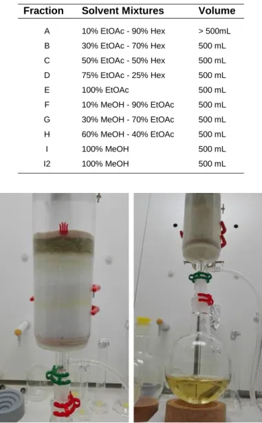
The crude extract was fractionated by normal phase Vacuum Liquid Chromatography (VLC) (Fig. 8) using a solvent polarity gradient (Table 2) on a glass fritted chromatography column. A thin layer of sand (ca. 1 cm in height) and 300 g of silica gel 60 (0.015-0.040 mm) (Merck, Sigma-Aldrich, USA) were placed in the column and then a mixture of hexane/ethyl acetate 9:1 (v/v) was used to pack the column. The crude extract was dissolved using a mixture of hexane/ethyl acetate 9:1 (v/v) and gently loaded onto the top of the silica column. A second layer of sand was positioned over the extract. The different eluents were sequentially added to the column without letting the silica surface become exposed to air. A total of ten fractions (A-I2, in order of increasing polarity) were collected in RB flasks and dried in a rotary evaporator. The chromatographic fractions were resuspended using dichloromethane and ethyl acetate 1:1 (v/v) (A-E) or methanol (F-I2), filtered through cotton, transferred to pre-weighted 16 mL vials and dried under vacuum. The mass of each fraction was determined, and the vials stored at -80 ºC until further use. The antimicrobial and anticancer activity of the ten VLC fractions was tested following the same protocols previously described for these assays. To gather information on the structure of the potentially active compounds, each active fraction, as well as its adjacent ones, was analysed by proton nuclear magnetic resonance (1H NMR) spectroscopy. Each sample was prepared by dissolving the mass
in a deuterated solvent methanol or chloroform, according to their polarity, and transferred into 400MHz NMR tubes (Norell Standard SeriesTM 5 mm, Sigma-Aldrich, USA). The samples were then sent to the Laboratory for Structural Elucidation (LAE) of the Materials Centre of the University of Porto (CMUP) where a Resonance Spectrometer Bruker Avance III 400 MHz, 9.4 Tesla was used for the analysis. The data
was analysed using the Mnova software allowing to verify the fractions spectra similarity. At this point it was decided to focus attention on the antimicrobial activity of four fractions - D, E, F and G - that proved to be active against C. albicans. Considering the antibiogram results, together with the 1H NMR spectra similarity, these four fractions were dissolved
using dichloromethane, pooled together in the same vial and processed as a single fraction (DEFG).
Table 2. Solvent mixtures used for elution of Streptomyces sp. KENR25 crude extract on
Vacuum Liquid Chromatography. Due to the high polarity of the extract, the last solvent mixture was repeated (fraction I2)
Fraction Solvent Mixtures Volume
A 10% EtOAc - 90% Hex > 500mL B 30% EtOAc - 70% Hex 500 mL C 50% EtOAc - 50% Hex 500 mL D 75% EtOAc - 25% Hex 500 mL E 100% EtOAc 500 mL F 10% MeOH - 90% EtOAc 500 mL G 30% MeOH - 70% EtOAc 500 mL H 60% MeOH - 40% EtOAc 500 mL I 100% MeOH 500 mL I2 100% MeOH 500 mL
Figure 8. Vacuum Liquid Chromatography fractionation of Streptomyces
5.3.2. Flash Chromatography (FC)
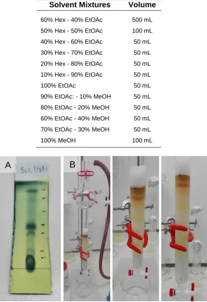
The antimicrobial-active DEFG fraction was fractionated using normal phase Flash Chromatography (FC). A Thin-Layer Chromatography (TLC) (Fig. 9A) was initially performed as a polarity indicator for the compounds present in the DEFG fraction, allowing determining the appropriate first solvent mixture to initiate the FC (Fig. 9B). For the TLC procedure, the fraction was applied onto a silica gel plate using a glass capillary and a mixture of ethyl acetate/hexane 1:1 (v/v) was used as mobile phase. To allow the detection of the compounds, the plate was stained using phosphomolybdic acid (PMA) and heated to activate the stain for visualization. The active DEFG fraction was further fractionated by normal phase FC. According to the TLC results it was decided that the first elution solvent mixture for FC would be ethyl acetate/hexane 2:3 (v/v). This mixture was used to solvate the silica gel (0.040-0.063 mm) in the column. The sample was added to the top of the silica layer, followed by the addition of cotton to protect the silica surface from the impact of solvents addition. Elution was performed using a gradient of increasing polarity (Table 3) and the sample was recovered in 10 mL tubes. Tubes with a similar sample coloration and profile on the TLC plates were combined in separated RB flasks, leading to a total of nine fractions (DEFG-A - DEFG-I). These sub-fractions were transferred to 8 mL pre-weighted vials and dried under vacuum. The mass of each sub-fraction was determined, and the vials stored at -20 ºC. To identify the fraction containing the active compounds responsible for the activity against C. albicans, the nine FC fractions were tested following the same protocol previously described for the antimicrobial assay, and the active fractions were also analyzed by 1H NMR.
Table 3. Solvent mixtures used for elution of the DEFG fraction on FC
Solvent Mixtures Volume
60% Hex - 40% EtOAc 500 mL 50% Hex - 50% EtOAc 100 mL 40% Hex - 60% EtOAc 50 mL 30% Hex - 70% EtOAc 50 mL 20% Hex - 80% EtOAc 50 mL 10% Hex - 90% EtOAc 50 mL 100% EtOAc 50 mL 90% EtOAc: - 10% MeOH 50 mL 80% EtOAc - 20% MeOH 50 mL 60% EtOAc - 40% MeOH 50 mL 70% EtOAc - 30% MeOH 50 mL 100% MeOH 100 mL
Figure 9. Sub-fractioning of the DEFG fraction. (A) pre-chromatography
Thin-Layer Chromatography and (B) Flash Chromatography column.
5.3.3. High-Performance Liquid Chromatography (HPLC)
The data obtained with all the procedures described above for the active fractions, the higher amount of available extract and the chromatogram simplicity of the sample, led to the selection of the fraction DEFG-A for semi-preparative High Performance Liquid Chromatography (HPLC) fractionation in a Waters Alliance HPLC combined with a Photodiode Array (PDA) detector. The sample was first dissolved in approximately 5 mL, using dichloromethane and methanol, and filtered through cotton into a vial. In each run, a sample volume of 250 μL was injected and the separation was carried out on a Luna 10u C18 column (100A, 4 μm, 250 × 10 mm, Phenomenex), with a flow rate of 3 mL/min. The chromatogram was monitored with fixed wavelengths at 225 nm and 254 nm. The elution was performed using HPLC grade acetonitrile and ultra-pure water according to
the following program: acetonitrile/water 3:7 (v/v) for 40 min followed by a linear gradient to 95% acetonitrile, held for 15 min before returning to the initial conditions. The 10 HPLC fractions were transferred to 8 mL pre-weighted vials and dried under vacuum. The mass of each fraction was determined, and the vials stored at -20 ºC.
5.4. Molecular Networking
A molecular networking was performed as an additional approach to study the metabolomic profile of DEFG sub-fractions that showed positive result in the antimicrobial assay. The first step was to analyse the selected fractions by LC-MS/MS, following the same protocol previously described for this technique. The obtained data followed then the “Data analysis” workflow from GNPS using the default parameters, except for the ion mass tolerance precursor which was set at 0.01 Da and fragment ion mass tolerance which was set at 0.04 Da. The GNPS tools were used to get an overview of identified compounds/clusters and then the MS/MS data was downloaded to Cytoscape software, version 3.6.1., where the molecular networking was generated (http://www.cytoscape.org/). A colour layout was applied to the network to allow an easier visualization and interpretation of the results.
III. Results
1. Phylogenetic Identification of Actinobacteria Isolated from
L. ochroleuca
At the beginning of the present MSc project, a total of 112 bacterial strains were available for the study, being 90 identified as actinobacteria (and the remaining identified as Firmicutes, Proteobacteria and Fungi). These actinobacteria were previously isolated from disinfected tissue fragments obtained from holdfasts, stipes and blades of L.
ochroleuca. Most of the strains (89%) were isolated from the holdfasts (Fig. 10A). The
highest percentage of the isolates was recovered from the medium SCN (63%), followed by RH (27%) and NPS (10%) (Fig. 10B). The isolates exhibited diverse morphological features, with several strains presenting characteristics typical of actinobacteria, such as slow growth, formation of hyphae and production of spores and pigments, the latter sometimes leading to the change of the colour of the culture medium (Fig. 11).
Figure 10. Actinobacterial isolates recovered from L. ochroleuca. (A) Percentage of actinobacterial strains isolated
from holdfasts, stipes and blades of L. ochroleuca and (B) Distribution of the isolates by the selective culture media used in the study (SCN: Starch-Casein-Nitrate agar; RH: Raffinose-Histidine; NPS: Nutrient-Poor Sediment).
Figure 11. Morphological diversity of some actinobacterial strains isolated from L. ochroleuca: (A) strain KENS1, (B) strain
KENR91, (C) strain KENR92, (D) strain KENR94, (E) strain KENRB10, (F) strain KENR56, (G) strain KENR81 and (H) strain KENB8.
The disinfection treatment was not entirely effective, as five isolates were recovered from the disinfection controls. Two of these isolates were grown in a PCA plate inoculated with a sterilised holdfast fragment being identified as Pseudomonas sp. and
Lysinibacillus sp., two were obtained in a PCA plate inoculated with the final wash water
from the disinfection of a holdfast fragment being identified as Bacillus sp. and
Staphylococcus sp., and one was found in a PCA plate inoculated with the final wash
water from the disinfection of a blade fragment being identified as a Penicillium sp. Most actinobacterial isolates identified by 16S rRNA gene sequencing belonged to the Streptomyces genus. However, strains belonging to other genera (including some rare) were also retrieved: Isoptericola, Rhodococcus, Nonomuraeae, Nocardiopsis,
Microbispora and Microbacterium (Figs. 12A and 13). All of these genera, except for Microbispora, were found in holdfasts of L. ochroleuca, while only two actinobacterial
genera were recovered from stipes (Microbispora and Microbacterium) and blades (Streptomyces and Microbacterium) (Fig. 12B). The selective medium SCN allowed the retrieval of strains belonging to all genera identified in this study, revealing to be the most efficient medium (Fig. 12C). Growth of Streptomyces and Microbacterium strains was observed in all culture media used. Several of the isolated strains, many of them affiliated with the genus Streptomyces, were found to group very closely with high bootstrap values (Fig. 13), indicating a high similarity between them. In addition, the obtained isolates consistently clustered with species already described in the literature, discarding the possibility of some of these isolates constitute a new species (Fig. 13).
![Table 1. Bioactive metabolites produced by marine actinobacteria, until 2017 (Adapted from [13, 14, 25] )](https://thumb-eu.123doks.com/thumbv2/123dok_br/15962489.1099663/21.892.129.761.145.1157/table-bioactive-metabolites-produced-marine-actinobacteria-adapted.webp)