COVID-19 laboratory diagnosis: the whole truth, so far
Edna Ribeiro, Miguel Brito
H&TRC – Health & Technology Research Center, ESTeSL – Escola Superior de Tecnologia da Saúde de Lisboa, Instituto Politécnico de Lisboa. Lisboa, Portugal. [email protected]
ABSTRACT: Coronavirus disease 2019 (COVID-19) is the most recent worldwide biological threat to humans worldwide with a severe impact in all areas of human development particularly health, economy, and mobility, caused by a virus belonging to the Coronaviridae family (SARS-CoV-2). Currently, the definite diagnosis of COVID-19 is based on the viral isolation or positive result of polymerase chain reaction (PCR) performed from sputum, nasal swab, or throat swab, although the virus has also been detected in blood and stool. Biological samples collection is performed based on the existing guidelines and PCR protocols had to be adapted. Both sampling and PCR must be performed by specialized professionals in order to avoid false negatives which have been reported in several published papers. Furthermore, considering the limitations of molecular tools such as highly skilled professionals, infrastructure limitations, and supply shortages, rapid diagnostic tests have also been developed based on the detection of viral components (Direct; antigen detection) and in the host immune response (Indirect; antibody detection). Titers of SARS-CoV-2 antibodies may be used as an indicator of COVID-19 prognosis and to discriminate asymptomatic carriers which allows the establishment of the COVID-19 spectrum; however, the persistence, reduction, and duration of SARS-CoV-2 immunity antibodies require further investigation. In a period of a pandemic without a vaccine or specific medications to stop the virus progression, testing is the most important task to perform in order to identify and isolate infected persons, even if they don’t present symptoms.
Keywords: COVID-19; Laboratory diagnosis; Biological sampling; rRT_PCR; Immunologic rapid tests.
Diagnóstico laboratorial de COVID-19: toda a verdade, até agora
RESUMO: A COVID-19 é a ameaça biológica mais recente para os seres humanos a nível mundial, com um impacto severo em todos os setores do desenvolvimento, particularmente na saúde, economia e mobilidade, causada por um vírus pertencente à família Coronaviridae (SARS-CoV-2). Atualmente, o diagnóstico definitivo de COVID-19 é baseado no isolamento viral ou resultado positivo da deteção de material genético viral através da reação em cadeia da polimerase (polymerase chain reaction – PCR) realizada a partir de amostras de expetoração, esfregaço nasal ou esfregaço na garganta, embora o vírus também tenha sido detetado no sangue e nas fezes. A colheita de amostras biológicas é realizada com base em diretrizes existentes e os protocolos de PCR tiveram de ser adaptados. Estas metodologias devem ser realizadas por profissionais especializados, a fim de evitar falsos negativos, os quais foram relatados em vários artigos publicados. Além disso, considerando as limitações inerentes às metodologias moleculares, como a necessidade de profissionais altamente qualificados, limitações de infraestruturas e escassez de reagentes, também foram desenvolvidos testes rápidos de diagnóstico com base na deteção de componentes virais (direto; deteção de antigénios) e na resposta imune do hospedeiro (indireto; deteção de anticorpos). Os títulos de anticorpos SARS-CoV-2 podem ser usados como um indicador do prognóstico do COVID-19 e para discriminar portadores assintomáticos, o que permite o estabelecimento do espectro do COVID-19; no entanto, a persistência, redução e duração dos anticorpos da imunidade à SARS-CoV-2 requerem investigação adicional. Atualmente, num período de uma pandemia sem vacina ou medicamentos específicos para interromper a progressão do vírus, testar é a tarefa mais importante a ser realizada para identificar e isolar indivíduos infetados, incluindo assintomáticos.Palavras-chave: COVID-19; Diagnóstico laboratorial; Amostras biológicas; rRT_PCR; Testes imunoló-gicos rápidos.
specimen and thus sputum can be induced trough specific procedures4.
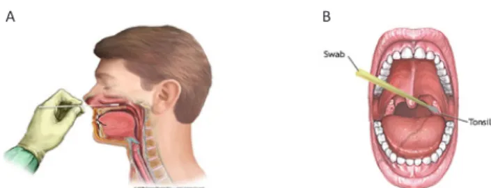
Nasal swab specimen collection: WHO determined that nasopharyngeal and oropharyngeal swab are two of the minimum respiratory samples that should be collected for SARS-CoV-2 testing2. In this procedure the health professional
enter a flexible swab, preferable with 3 different thicknesses, ending in a ‘furry’ or flock tip, several centimetres along the floor of the nose until the posterior nasopharynx has been reached and once resistance is encountered as exemplified in Figure 1-A. The swab should then be rotated several times and after pull out it should be inserted and kept in a transport medium6-7.
Throat swab specimen collection: Both tonsils and the posterior pharynx are swabbed vigorously using a flexible swab as exemplified in Figure 1-B also preferable with 3 diffe-rent thicknesses, ending in a ‘furry’ or flock tip and withdraw the swab into a transport medium6.
Introduction
Coronavirus disease 2019, abbreviated to COVID-19, is the most recent worldwide biological threat, caused by a virus from the Coronaviridae family. This virus, defined/designated as “severe acute respiratory syndrome coronavirus 2” (SARS--CoV-2), presents a genome sequence with extremely high sequence identity (i.e. up to 80%) to those of the homolo-gous virus that caused the SARS outbreak in 2003 (i.e. SARS--CoV)1. This ongoing epidemic respiratory diseases caused by
COVID-19, SARS-CoV2 started in Wuhan, Hubei, in China at the end of December 2019 and at the time of writing this article, SARS-CoV-2 has already infected over 11,425.209 people in 213 different countries, causing 534,062 related deaths. The registered official data in Portugal by General Directorate of Health (DGS) indicate 44,129 infected people, 29,166 reco-vered and 1,620 deaths (https://covif19.min-saude.pt/).
Efficient COVID-19 laboratory testing is crucial for the pandemic mitigation and for supporting decisions on infec-tion control strategies and patient management at health-care facilities, including asymptomatic cases.
The World Health Organization (WHO) have determined that specimens to be collected for SARS-CoV-2 laboratory diagnosis are nasopharyngeal and oropharyngeal swab or wash in ambulatory patients (upper respiratory specimens) and/or sputum (if produced) and/or endotracheal aspirate or bronchoalveolar lavage (lower respiratory specimens)2.
Nevertheless, it is important to notice that SARS-CoV-2 has also been detected in patients’ blood and stool2.
Currently, the definite diagnosis of COVID-19 is based on the viral isolation or positive result of polymerase chain reaction (PCR) with detection of the SARS-CoV-2 virus RNA, performed from sputum, or nasal swab, or throat swab. However, since viral pneumonia usually does not result in the proper produc-tion of purulent sputum, the nasopharyngeal swab has been the preferred collect method to obtain quality specimens for testing.
Biological specimen collection for SARS-CoV-2 definite diagnosis
Sputum specimen collection: Sputum is mucous that humans cough up from deep inside the lungs, usually thick, cloudy, and sticky. Sputum specimens are commonly collected to identify the etiology of lower respiratory tract infections caused by virus and bacteria3. Briefly, patients
are instructed to gargle and rinse the mouth with water immediately before the specimen collection (preferably in the morning). This procedure facilitated the elimination of gathered cells and commensal bacteria that could inter-fere with test results. Then patients are instructed to inhale repeatedly to the full lungs capacity and then exhale the air in a forceful cough which should induce the mucus to be expectorated into the previously provided sterile container. Although the specimens are collected by the patients, health professionals must give strict indications in order to ensure the sample quality3. Moreover, in some cases, particularly in
children, patients are unable to produce an adequate sputum
Figure 1 (A-B). Biological specimen collection for SARS-CoV-2 laboratory diagnosis nasal (A) and throat (B) swab.
Molecular tools for SARS-CoV-2 virus RNA detection SARS-CoV-2 is an enveloped virus containing a single strand of positive-sense RNA. Therefore, the molecular diag-nosis must include isolation of the virus RNA, which is then used as a template to synthesize a double-strand DNA mole-cule (cDNA) by a mechanism of reverse transcription using a reverse transcriptase enzyme in a process called RT-PCR (reverse transcription polymerase chain reaction). After these two phases, it is possible to detect the presence of the virus using the polymerase chain reaction (PCR) technique with a specific pair of primers (complementary to a conserved region of the virus nucleic acid sequence). There is a diversity of PCR methods, however, WHO suggest the use of real-time quantitative PCR (rRT_PCR)7, due to their unique
characteris-tics, namely the amplification and analysis are done simul-taneously in a closed system minimizing the false-positive results associated with amplification product contamination. Moreover, in addition to the diagnosis, the real-time PCR can be a quantitative PCR, giving the possibility of determining the viral load.
RNA extraction from de virus
Currently, there are many specialized methods of extrac-ting RNA. Generally, these methods are divided into
-based or column-based protocols8. Most of these protocols
have been developed into commercial kits allowing more rapid and easier extraction processes. The isolation of the virus RNA is the most crucial step in the diagnosis process, In fact, it requires high-quality nucleic acids to avoid the presence of downstream steps inhibitors (RT-PCR or PCR reaction), and cross-contamination of samples (false posi-tives). Moreover, despite the SARS-CoV-2 virus being extre-mely resistant to the environment, in the laboratory, the RNA is an unstable molecule and has a very short half-life once extracted from the virus capsule. Consequently, extreme care should be taken in order to avoid false-negative results due to RNA degradation.
In the particular situation of a pandemic, that needs rapid results from a huge quantity of samples, it is recommended, for molecular diagnosis, the use of commercial kits to extract RNA from the SARS-CoV-2 virus. Moreover, it is essential, due to security reasons, to ensure that the reagents used for RNA extraction are effective in the inactivation of SARS-CoV-2. For instance, the Center for Disease Control of the United States of America have tested several kits and confirmed that the external lysis buffer is effective for inactivation of the virus9
(e.g. QIAamp Viral RNA Mini Kit, Roche MagNA Pure -DNA and Viral NA Kit, BioMérieux NucliSENS® easyMAG, among others). The selection of the commercial kit takes also in consider-ation its availability in the country and the price. For instance, in Portugal, several laboratories are using the NZY Viral RNA Isolation Kit supplied by one national enterprise NZYTeck genes and enzymes (https://www.nzytech.com/).
For laboratory technician safety, due to the type of virus, the isolation of RNA should be performed in a biosafety cabinet in a BSL-2 or equivalent.
Reverse transcription and real-time quantification of extracted RNA
Once RNA is isolated, it should be converted into cDNA, and then it can be used in a real-time PCR reaction using specific
primers. For the study of RNA by real-time PCR, there are two common methods that can be applied: one-step RT-PCR and two-step RT PCR. In the first one, the reverse transcriptase step is performed in the same tube as the primer specific PCR reaction (one-step). In the second method cDNA must be firstly obtained by reverse transcription reaction in one tube and then the specific primers reaction will be performed in another PCR reaction (two-step). There are advantages and disadvantages to both methods, however, in the case of the present COVID-19 pandemic, it should be used the easier and the lowest cost reaction method. One-step RT-PCR is certainly easier to set up with less overall hands-on. For that reason, all protocols that WHO has on the webpage are one-step RT-PCR10. In this method, the PCR kit includes both enzymes,
namely Reverse Transcriptase enzyme that performs the first step of the reaction to convert the RNA single strand in a DNA single strand, and then, the Taq DNA polymerase, an heat resistant enzyme, responsible for the amplification steps with the specific primers and probes.
One important decision for the application of this mole-cular methodology is the assay selection for molemole-cular detection of SARS-CoV-2. It means that the exact nucleo-tide sequence of the virus must be known in order to design primers and probes that can anneal and rigorously amplify the virus cDNA. Deep sequencing methods (also called Next--Generation Sequencing methods) played a major role in the initial identification of SARS-CoV-211, since research teams
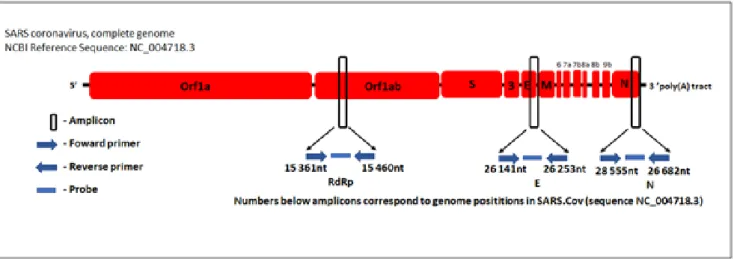
sequenced all the virus genome, and published in public databases. In each country, next-generation sequencing will continue to be needed to determine future mutations of SARS-CoV-2. Based on the sequences of SARS-CoV-2 available on databases primers and probes were designed to target specific regions of the virus genome. The regions generally used for primers and probes include glycoproteins spike gene (S), envelope gene (E), transmembrane gene (M), helicase gene (Hel), and nucleocapsid gene (N), RNA-dependent RNA polymerase gene (RdRp), hemagglutinin-esterase gene (HE), and open reading frames ORF1a and ORF1b10-12 (Figure 2). The
Figure 2. Relative positions of primers and probes on the virus genome (E = Envelope protein gene; M = Membrane protein gene; N = Nucleocapsid protein gene; ORF = Open reading frame; RdRp = RNA dependent RNA polymerase gene; S = Spike protein gene, the numbers 3, 6, 7, 7a, 8a, 8b and
selection of primers and probes should have in consideration the most prevalent variant of the virus in the country, taking into account possible mutations. Moreover, cross-reactivity with other coronaviruses should be taken into account and should be tested by in silico analysis and by laboratory expe-rimental analysis.
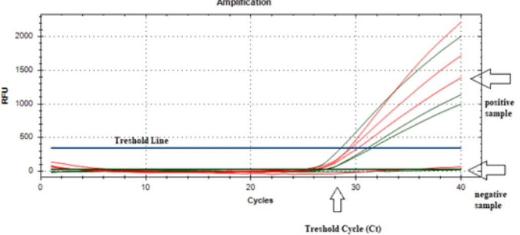
In the real-time PCR procedure, the probe (a small oligonu-cleotide with a fluorophore and a quencher in the 3’ and 5’ ends) anneals to a specific target sequence located between the forward and reverse primers. Due to the 5’ nuclease acti-vity of Taq polymerase, the probe is degraded during the extension phase of the PCR cycle, causing the reporter dye to separate from the quencher dye, generating a fluores-cent signal. This signal is detected in each cycle by the PCR apparatus and registered in a plot. A positive sample creates an amplification curve that crosses the threshold line in a specific threshold cycle (Ct), whereas a negative sample does not have amplification and doesn’t cross the threshold line (Figure 3).
What is a positive result?
According to WHO, routine confirmation of positive cases should be based on the detection of unique sequences of virus RNA by rRT-PCR but with confirmation by nucleic acid sequencing when necessary. These rules were created at the very beginning of the epidemy in China. In that situation, if the diagnosis was to be performed in a country with unknown cases a positive result (that situation just occurred in January and February 2020, at the beginning of the outbreak) should include a positive PCR result for at least two different targets on the virus genome7. However, after pandemic outbreak
identification, the rule screening by rRT-PCR of a single discri-minatory target is considered enough to consider a positive case.
A negative result in rRT-PCR using a single region, or even in a multiplex set, doesn’t rule out the possibility of virus infection. As mentioned earlier, several factors could lead to a negative result in an individual with COVID-19. The factors include: i) poor quality of the specimen, containing little patient material (in this case the inclusion of a human target test as control can overcome this problem); ii) the specimen was collected late or very early in the infection and the PCR reac-tion doesn’t have enough sensi-bility to detect virus RNA; iii) the specimen was not handled and shipped appropriately (this includes heat destruction of the RNA or inhibitors in the sample); iv) technical reasons inherent to the test, that includes a muta-tion in the virus, or quality of the reagents and protocols.
Relevantly, false-negative rRT--PCR tests have been reported worldwide13 which indicates
diagnosis limitations. Evidence has demonstrated that some patients with clinically distinct SARS-CoV-2 disease have initial negative rRT-PCR test results, which turned to positive results in later stages of the disease13.
These results clearly demonstrate limitations of rRT-PCR sen-sitivity to detect COVID-19, which is described as lower than that of other diagnostic tools as for example chest computed tomography. In a case of a 34-year-old man with a negative diagnosis for COVID-19 based on four sequential rRT-PCR tes-ts from pharyngeal swab samples with severe pneumonia symptoms after 3 days of admission, resulted positive only at the fifth day after admission and at fifth rRT-PCR test14.
Rapid direct/indirect SARS-CoV-2 virus tests
Molecular tests such as rRT-PCR, which isolate and amplify the SARS-CoV-2 virus RNA, are the gold standard for labo-ratory diagnostics. However, these molecular tools require In some protocols, an additional set of primers and probes
can be included, designed to amplify a human gene (e.g. human RNase P gene) that works as a positive control for nucleic acid extraction and amplification. Since the mole-cular diagnosis is directed to human samples, it is expected that in all samples there is plenty of human RNA that has been extracted and may be also amplified. Therefore, if there is no positive signal for the human gene there should be a problem in sampling, RNA isolation, or rRT-PCR.
Most used protocols, during the pandemic, also include two SARSCoV-2 regions in the reaction, in a multiplex reac-tion. This multiplex reaction gives more confidence in case of a mutation in one of the other regions, complementary to the used pair of primers, giving a false negative result, that will be overcome by a positive result due to the amplification of a different region of the virus.
Figure 3. Real-time PCR amplification plot. This plot corresponds to a multiplex reaction that
rapid SARS-CoV-2 antigen detection tests, which are in conformity with the European Union legislation, Directive 98/79/EC on IVDs15.
Indirect antibody detection tests: Indirect detection tests are based on the detection of antibodies associated with the host immune response against the virus. Currently, several CE-marked rapid SARS-CoV-2 antibody tests have been avai-lable, and numerous research groups have also developed and are validating in-house antibody detection tests15.
Gender divergencies in SARS-CoV-2 infection/immune response
Data have demonstrated gender differences regarding women’s and men’s susceptibility to SARS-CoV-2 infection. This difference has been associated with differences in innate immunity, steroid hormones, and sex chromosomes factors. In fact, in women in comparison with men, immune regula-tory genes encoded by the X chromosome are associated with lower viral load levels, decreased inflammation, and higher levels of antibodies which remain longer in circula-tion. The increased immunological activation in women has been correlated with the trigger of TLR7 which is a protein expressed in innate immune cells that is able to recognize single-strand RNA virus with associated production of antibo-dies against the virus and the expression of pro-inflammatory cytokines such as IL-1 and IL-6 that control CoV-19 replication via the production of IFN21. The immune system is affected
by the X chromosome also due to the distinct expression of proteins, including FOXP3, TLR8, CD40L, and CXCR3 which may be over-expressed in women, due to the biallelic expres-sion of the X-linked genes influencing the response to viral infections and vaccinations21.
Laboratory abnormalities in COVID-19 diagnosed patients Hematological parameters such as leukocytosis, neutro-philia, lymphopenia, increased MDW (monocyte volume distribution) have been identified as potential predictors of pathology progression toward severe or critical COVID-19 outcomes. Additionally, increased values of C reactive protein (CRP), lactate dehydrogenase (LDH), erythrocyte sedimenta-tion rate (ESR), and D-dimer, along with a diminished concen-tration of serum albumin has also been reported in COVID-19 patients22. Moreover, regarding prognostication purposes,
increased values of LDH, aspartate aminotransferase (AST), alanine aminotransferase (ALT), total bilirubin, creatinine, cardiac troponins, D-dimer, prothrombin time (PT), procal-citonin and CRP, and decreased serum albumin, have been valuable23.
Concluding remarks
Presently the definite diagnosis of COVID-19 is based on a positive result from rRT-PCR performed in a biological respi-ratory tract sample. However, other diagnostic methods are necessary, namely direct antigen detection, to develop a rapid diagnostic test (or point of care tests) and indirect anti-body detection, to analyze the person immunity so impor-tant in a post quarantine period.
well-equipped laboratory facilities, highly skilled profes-sionals, multiple reagents, and high false-negative rates. Currently, infrastructure limitations and supply shortages are constraining the capacity to perform all necessary tests due to the rising demand for COVID-19 diagnostics.
In this context, rapid diagnostic tests for COVID-19 have been developed based on the detection of viral components and in the host immune response to the virus15. These rapid
tests are limited to qualitative or semi-quantitative in vitro diagnostics, and currently, two types of COVID-19 rapid tests are being used or being developed, namely direct SARS--CoV-2 antigen detection and indirect antibody detection tests.
It is acknowledged by the scientific community that IgM ensures the first line of defense throughout viral infections, which is followed by the generation of adaptive, high-affinity IgG responses crucial for long term immunity and immunolo-gical memory16.
COVID-19 infection induces IgG antibodies against N protein that can be detected by serum as early as day 4 after the onset of disease and with most patients seroconverting by day 14. Laboratory evidence of clinical patients showed that a specific T-cell response against SARS-CoV-2 is impor-tant for the recognition and killing of infected cells, particu-larly in the lungs of infected individuals.
In the particular case of SARS-CoV-2 infection studies have reported IgG antibodies induction against the viral N protein (nucleocapsid protein) detectable in serum as early as day 4 after the beginning of the disease and with several patients seroconverting by day 14 and specific T-cell response, parti-cularly in the lungs of infected individuals17. Additionally,
IgM and IgG antibodies resultant from SARS-CoV-2 immune response have been detected by immunofluorescence assay in samples from days 9, 10, and 20 after the beginning of illness18.
Despite the fact that serology testing cannot overcome rRT-PCR for diagnosing of acute viral infections, combined IgM-IgG rapid immunoassays have been developed against the SARS-CoV-2 virus in human blood with the capacity to evaluate different infection stages and symptomatic or asymptomatic carriers19. These serologic tests were based in
blood samples collected from 397 PCR confirmed COVID-19 patients and 128 negative patients, with high diagnostic precision (i.e. up to 89% sensitivity and up to 91% specificity)19.
Titers of SARS-CoV-2 antibodies enable the evaluation of the progress of viral infection as they are consistent with clinical manifestations which suggest that antibody detec-tion may be used as an indicator of COVID-19 prognosis and to discriminate asymptomatic carriers. On the other hand, it is important to understand that in the early stages of infection a high percentage of positive SARS-CoV-2 patients (81.6%) may test negative for IgM and IgG in serological assays20
and that after recovery the persistence, decrease, and dura-tion of SARS-CoV-2 immunity antibodies requires further investigation.
Direct SARS-CoV-2 antigen detection: Direct detection tests are based on the detection of specific viral components present during the infection (antigen). Organizations such as FIND (https://www.finddx.org/) have listed ten CE-marked
In a phase of a pandemic without a vaccine or specific medications to stop the virus progression, testing is the most important task to perform in order to identify and isolate infected persons, even if they don’t present symptoms. The watchword in this COVID-19 pandemic has been “test, test,
test, and test”.
Moreover, considering the high levels of mortality and infectivity associated with this new coronavirus, there is an urge to develop safe and simple tests, with fast and accurate detection due to the possibility of future outbreaks from this virus and virus family.
Acknowledgments
Authors acknowledge the support of H&TRC – Health & Technology Research Center, ESTeSL – Escola Superior de Tecnologia da Saúde de Lisboa, Instituto Politécnico de Lisboa.
References
1. Ceraolo C, Giorgi FM. Genomic variance of the 2019: nCoV coronavirus. J Med Virol. 2020;92(5):522-8.
2. World Health Organization. Laboratory testing for coronavirus disease (COVID-19) in suspected human cases: interim guidance [Internet]. Geneva: WHO; 2020. Available from: https://apps.who.int/iris/bitstream/ handle/10665/331501/WHO-COVID-19-laboratory-2020. 5-eng.pdf?sequence=1&isAllowed=y
3. British Columbia Centre for Disease Control. Sputum collec-tion for tuberculosis (TB) testing. HealthLinkBC [Internet]. 2017;(51b). Available from: https://www.healthlinkbc.ca/ healthlinkbc-files/sputum-tuberculosis-testing
4. Grant LR, Hammitt LL, Murdoch DR, O’Brien KL, Scott JA. Procedures for collection of induced sputum specimens from children. 2012;54(Suppl 2):S140-5.
5. Yukon Communicable Disease Control. Nasopharyngeal swab procedure [Internet]. UCDC; 2015 Oct. Available from: http://www.hss.gov.yk.ca/pdf/npswab.pdf
6. World Health Organization. WHO guidelines for the collection of human specimens for laboratory diag-nosis of avian influenza infection [homepage]. Geneva: WHO; 2005. Available from: https://www.who.int/influ-enza/human_animal_interface/virology_laboratories_ and_vaccines/guidelines_collection_h5n1_humans/ en/
7. World Health Organization. Laboratory testing for 2019 novel coronavirus (2019-nCoV) in suspected human cases [homepage]. WHO; 2020 Mar 19. Available from: https://www.who.int/publications-detail/laborato- ry-testing-for-2019-novel-coronavirus-in-suspect-ed-human-cases-20200117
8. Tan SC, Yiap BC. DNA, RNA, and protein extraction: the past and the present. J Biomed Biotechnol. 2009;2009:ID574398.
9. Centers for Disease Control and Prevention. CDC 2019-novel coronavirus (2019-nCoV) real-time RT-PCR diag-nostic panel: for emergency use only [Internet]. CDC; 2020 Mar 30. Available from: https://www.fda.gov/ media/134922/download
10. Corman VM, Landt O, Kaiser M, Molenkamp R, Meijer A, Chu DK, et al. Detection of 2019 novel corona-virus (2019-nCoV) by real-time RT-PCR. Euro Surveil. 2020;25(3):2000045.
11. Chen L, Liu W, Zhang QI, Xu K, Ye G, Wu W, et al. RNA based mNGS approach identifies a novel human coro-navirus from two individual pneumonia cases in 2019 Wuhan outbreak. Emerg Microbes Infect. 2020;9(1):313-9. 12. Tang YW, Schmitz JE, Persing DH, Stratton CW. The labo-ratory diagnosis of COVID-19 infection: current issues and challenges. J Clin Microbiol. 2020;58(6):e00512-20. 13. “Pr es s Pr e,” 2019.
14. Feng H, Liu Y, Lv M, Zhong J. A case report of COVID-19 with false negative RT-PCR test: necessity of chest CT. Jpn J Radiol. 2020;38(5):409-10.
15. European Center for Disease Prevention and Control. An overview of the rapid test situation for COVID-19 diagnosis in the EU/EEA [Internet]. Stockholm: ECDC; 2020. Available from: https://www.ecdc.europa.eu/sites/ default/files/documents/Overview-rapid-test-situa-tion-for-COVID-19-diagnosis-EU-EEA.pdf
16. Racine R, Winslow GM. IgM in microbial infections: taken for granted? Immunol Lett. 2009;125(2):79-85.
17. Rokni M, Ghasemi V, Tavakoli Z. Immune responses and pathogenesis of SARS-CoV-2 during an outbreak in Iran: comparison with SARS and MERS. Rev Med Virol. 2020;30(3):e2107.
18. Haveri A, Smura T, Kuivanen S, Österlund P, Hepo-joki J, Ikonen N, et al. Serological and molecular find-ings during SARS-CoV-2 infection: the first case study in Finland, January to February 2020. Euro Surveil. 2020;25(11):2000266.
19. Li Z, Yi Y, Luo X, Xiong N, Liu Y, Li S, et al. Development and clinical application of a rapid IgM-IgG combined antibody test for SARS-CoV-2 infection diagnosis. J Med Virol. 2020;10.1002/jmv.25727. [Epub ahead of print] 20. Cassaniti I, Novazzi F, Giardina F, Salinaro F, Sachs M,
Perlini S, et al. Performance of VivaDiag COVID‐19 IgM/ IgG rapid test is inadequate for diagnosis of COVID‐19 in acute patients referring to emergency room depart-ment. J Med Virol. 2020;10.1002/jmv.25800. [Epub ahead of print]
21. Conti P, Younes A. Coronavirus COV-19/SARS-CoV-2 affects women less than men: clinical response to viral infection. J Biol Regul Homeost Agents. 2020;34(2). [Epub ahead of print]
22. Lippi G, Plebani M. Laboratory abnormalities in patients with COVID-2019 infection [letter to the editor]. Clin Chem Lab Med. 2020;58(7):1131-4.
23. Lippi G, Plebani M. The critical role of laboratory medi-cine during coronavirus disease 2019 (COVID-19) and other viral outbreaks. Clin Chem Lab Med. 2020 Mar 19. [Epub ahead of print]
Conflict of interests
This is a contributed review article submitted upon invitation of the Editorial Board.