Universidade de Lisboa
Faculdade de Ciˆencias
Departamento de Biologia Animal
narcissus is required for the correct
establishment of left-right
asymmetries in the zebrafish brain
Pedro Miguel Dias Henriques
Dissertation
Mestrado em Biologia Evolutiva e do Desenvolvimento
2013
Universidade de Lisboa
Faculdade de Ciˆencias
Departamento de Biologia Animal
narcissus is required for the correct
establishment of left-right asymmetries in
the zebrafish brain
Pedro Miguel Dias Henriques
Dissertation
Mestrado em Biologia Evolutiva e do Desenvolvimento
External supervisor: Prof. Stephen Wilson
Internal supervisor: Prof. S´
olveig Thorsteinsd´
ottir
2013
Man is all symmetry,
Full of proportions, one limb to another, And all to all the world besides; Each part may call the furthest brother,
For head with foot hath private amity, And both with moons and tides.
Acknowledgements
Um especial obrigado ao Steve Wilson, que, por poucas palavras, acre-ditou em mim, e a todos os (muitos!) elementos do laborat´orio, as-sim´etricos ou n˜ao, que raramente recusaram partilhar uma pint e com quem espero partilhar muitas mais.
Um grande obrigado `a minha n˜ao menor coordenadora Ana Faro, que me ensinou muito mais do que pensa, que estava normalmente presente quando as horas eram longas no laborat´orio e com quem posso partilhar hoje de uma boa amizade.
Um obrigado tamb´em `a minha tia, que tornou esta e outras viagens poss´ıveis, fazendo-me ver um pouquinho do mundo que ela j´a conhece, e `a minha irm˜a, que desde pequeno me tem mostrado, consciente ou inconscientemente, um percurso pelo qual seguir.
Quero agradecer adicionalmente a todos os que me acompanharam e apoiaram at´e aqui, n˜ao s´o a n´ıvel profissional e acad´emico, mas tamb´em a n´ıvel emocional. Aos familiares, aos amigos, aos amigos de circunstˆancia, aos que se perderam, aos que nunca foram, aos que ser˜ao e a todos aqueles que me ensinaram, a bem ou a mal, a viver. Acima de tudo quero agradecer aos meus pais, que por fruto do acaso se lembraram de me trazer ao mundo e me tentaram ensinar tudo o que sabiam, apesar das ocasionais resistˆencias, e `a minha av´o e avˆo, que me educaram e me fizeram a pessoa que sou hoje. Amo-vos, apesar de n˜ao o dizer alto...
Abstract
The vertebrate brain is functionally and anatomically left-right (L-R) asym-metric, yet how these asymmetries arise and are maintained during develop-ment is still poorly understood. In zebrafish, the epithalamus comprises some of the most conspicuous asymmetries found in vertebrates, making it a valu-able model to study their development. In it, left and right dorsal habenulae (dHb) develop differently in size, cytoarquitecture and axonal connectivity. Additionally, the parapineal organ migrates to the left side and exclusively projects to the left habenula (lHb), being required for the development of its molecular and subsequent cytoarchitectural left-sided identity. Several studies have shown that Nodal, Wnt and Notch signalling pathways have an impor-tant role in dHb asymmetric specification. However, how they interact with each other to achieve this is still largely unknown. Through a forward genetic screen, our lab has identified the narcissus mutation, which induces defects in the asymmetric specification of dHb neurons. Here, we show that in narcis-sus mutants, both habenulae display symmetric expression of some, but not all, lHb markers, and that the asymmetric afferent projections to the interpe-duncular nucleus (IPN) and efferent innervation from the olfactory bulb and parapineal are disrupted. Additionally, both habenulae display significantly less BrdU incorporation in two tested timepoints, but asymmetric early neu-rogenesis still occurs.
Together, our findings demonstrate that narcissus is required for the cor-rect specification of L-R asymmetries in the zebrafish brain and provide a valuable background for future studies to decipher its role in the establish-ment and maintenance of these asymmetries during embryonic developestablish-ment.
Keywords: Brain asymmetry, Epithalamus, Habenula, Lateralization, Nar-cissus, Zebrafish
Sum´
ario
O c´erebro ´e essencialmente assim´etrico em termos anat´omicos e funcionais, estando descritas assimetrias entre a esquerda e a direita no sistema nervoso de esp´ecies representativas de praticamente todas as classes de vertebrados. De facto, h´a muito se sabe que v´arias funcionalidades se encontram extrema-mente lateralizadas no c´erebro humano, como ´e o exemplo do processamento da linguagem, dominante no hemisf´erio esquerdo, e certas capacidades visuo-espaciais no hemisf´erio direito. Mais recentemente, v´arios estudos mostraram haver uma rela¸c˜ao positiva entre c´erebros pouco lateralizados e deficiˆencias cong´enitas de linguagem, como no caso da dislexia, levando `a hip´otese de que estas poder˜ao estar ligadas a problemas no estabelecimento de assimetrias du-rante o processo ontog´enico. Apesar disto, pouco se sabe atualmente sobre como as assimetrias de esquerda-direita (E-D) aparecem e como s˜ao mantidas durante o desenvolvimento embrion´ario do sistema nervoso.
H´a v´arios anos que o peixe-zebra tem provado ser um modelo valioso na ´
area da biologia do desenvolvimento de vertebrados, muito devido ao seu bem caracterizado sistema gen´etico, `a facilidade de manipula¸c˜ao gen´etica e celular e `as suas propriedades ´oticas que facilitam t´ecnicas de microscopia in vivo. No estudo do estabelecimento de assimetrias E-D, o peixe-zebra apresenta-se como particularmente ´util. Neste organismo, a regi˜ao cerebral do epit´alamo, composta pelas hab´enulas e pelo complexo pineal, apresenta v´arias assimetrias entre a esquerda e a direita a n´ıvel citoarquitect´onico, de express˜ao de genes e connectividade neuronal. A hab´enula dorsal do lado esquerdo ´e ligeiramente maior que a do lado direito, possuindo tamb´em uma maior concentra¸c˜ao de neuropilo e express˜ao diferencial de genes. Esta diferen¸ca de express˜ao ´e na verdade t˜ao acentuada, que permite a divis˜ao dos n´ucleos da hab´enula nos sub-n´ucleos lateral, demarcado pela express˜ao de kctd12.1, e medial, que expressa kctd12.2, e cujo tamanho ´e tamb´em proporcionalmente maior na esquerda e direita, respetivamente. Adicionalmente, o ´org˜ao parapineal, que origina de um subconjunto de c´elulas anteriores da glˆandula pineal, migra para o lado esquerdo e estabelece proje¸c˜oes axonais exclusivamente com a hab´enula
esquerda, sendo inclusivamente necess´ario para a correta especifica¸c˜ao dos neur´onios do sub-n´ucleo lateral nesse lado. Cada hab´enula projeta tamb´em diferencialmente para o n´ucleo interpeduncular, situado no mesenc´efalo, onde a regi˜ao dorsal e ventral ´e inervada preferencialmente pelas hab´enulas esquerda e direita, respetivamente.
Como se estabelecem estas assimetrias ´e ainda um t´opico de grande de-bate. Sabe-se que a sinaliza¸c˜ao de Nodal, um dos primeiros eventos a quebrar a assimetria no embri˜ao e necess´aria para o correto estabelecimento das as-simetrias viscerais, possui tamb´em um papel crucial no estabelecimento das assimetrias epitamˆamicas. Nodal ´e normalmente expresso no lado esquerdo do dienc´efalo durante as primeiras fases do desenvolvimento, atuando como um bias no estabelecimento da lateralidade das assimetrias. De facto, v´arios estudos apontam para uma troca de informa¸c˜ao entre a assimetria visceral e epitalˆamica, sendo a primeira necess´aria para o estabelecimento da ´ultima atrav´es de uma intera¸c˜ao mediada pela via de sinaliza¸c˜ao Wnt.
V´arios estudos apontam na verdade para o requerimento de algumas das maiores vias de sinaliza¸c˜ao para o correto estabelecimento e manuten¸c˜ao de assimetrias no epit´alamo. Para al´em de necess´aria para a quebra de assime-tria inicial, a sinaliza¸c˜ao Wnt tem tamb´em um papel crucial na correta especi-fica¸c˜ao dos sub-n´ucleos habenulares, pois a sua inibi¸c˜ao atrav´es de tratamentos farmacol´ogicos ou por muta¸c˜oes gen´eticas induz a diferencia¸c˜ao de um maior r´acio de neur´onios do sub-tipo dorsal lateral em ambas as hab´enulas, assu-mindo estas uma especifica¸c˜ao caracter´ıstica de esquerda. Adicionalmente, a via de sinaliza¸c˜ao Notch parece estar envolvida na manuten¸c˜ao da cronologia do programa de neurog´enese diferente entre esquerda e direita, mostrando-se essencial para a especifica¸c˜ao diferencial dos neur´onios dos dois lados. De facto, a maior parte dos neur´onios do sub-n´ucleo lateral nasce por volta das 32 horas-p´os-fertiliza¸c˜ao (hpf) enquanto que neur´onios do sub-n´ucleo medial nascem mais tarde `as 48 hpf. Apesar de v´arias hip´oteses serem apontadas para esta assimetria, a mais plaus´ıvel assenta na ideia de que na direita, uma ativa¸c˜ao assim´etrica da sinaliza¸c˜ao Notch leva a uma mais longa manuten¸c˜ao da popula¸c˜ao de c´elulas estaminais, inibindo assim a neurog´enese nesse lado.
estas vias de sinaliza¸c˜ao para o estabelecimento de assimetrias no epit´alamo. Para melhor compreender isto, o nosso laborat´orio realizou um screening gen´etico para identificar muta¸c˜oes que resultem em problemas de assimetria epitalˆamica. Um destes mutantes, narcissus (nss), ´e caracterizado pela bilate-ralidade de marcadores caracter´ısticos da hab´enula esquerda, como kctd12.1, e sub-express˜ao de marcadores da hab´enula direita, como kctd8. Neste estudo, procurei caracterizar em maior detalhe o mutante narcissus e tentei identifi-car os mecanismos atrav´es dos quais este gera as perturba¸c˜oes de assimetria observadas.
Nos mutantes narcissus, todas as proje¸c˜oes aferentes e eferentes assim´etricas para e das hab´enulas est˜ao afetadas. Apesar da parapineal migrar correta-mente para a esquerda, as suas proje¸c˜oes para a hab´enula esquerda possuem uma distribui¸c˜ao mais extensa do que em embri˜oes controlo, cujas proje¸c˜oes tendem a acumularem-se em regi˜oes mais pr´oximas da hab´enula. Adicio-nalmente, a proje¸c˜ao assim´etrica para a hab´enula direita, proveniente de c´elulas mitrais no bolbo olfact´orio e marcadas pela express˜ao do transg´enico Tg(lhx2a:gap-YFP) est˜ao completamente ausentes nos mutantes narcissus. Este defeito n˜ao ser´a no entanto a prov´avel causa da express˜ao sim´etrica de kctd12.1, pois a abla¸c˜ao seletiva destas proje¸c˜oes em embri˜oes controlo an-tes da forma¸c˜ao da enerva¸c˜ao no epit´alamo n˜ao revelou qualquer altera¸c˜ao de express˜ao de kctd12.1 e do marcador da habenula direita kctd8. Ines-peradamente, ambas as hab´enulas projetam tamb´em preferencialmente para a regi˜ao ventral do n´ucleo interpeduncular, um fen´otipo provavelmente cau-sado por uma deficiˆencia da sinaliza¸c˜ao de orienta¸c˜ao axonial mediada por Nrp1a/Sema3D, intimamente respons´avel por esta conex˜ao.
Ao contr´ario de outros mutantes onde marcadores da hab´enula esquerda s˜ao bilateralmente expressos, nos mutantes de narcissus, a express˜ao de Pku558b, que marca maioritariamente uma sub-popula¸c˜ao de neur´onios na hab´enula do sub-tipo dorsal lateral, continua assim´etrica aos 4 dias p´os-fertiliza¸c˜ao (dpf). Por outro lado, a express˜ao de kctd12.1 come¸ca por ser assim´etrica at´e 2 dpf, apenas adquirindo bilateralidade aos 3 dpf. Atrav´es da an´alise da data do nas-cimento dos neur´onios dos diferentes sub-n´ucleos habenulares, identific´amos que nos mutantes narcissus ocorre significativamente menos prolifera¸c˜ao nas
hab´enulas `as 32 e `as 50 hpf, dois est´adios do desenvolvimento em que a mai-oria dos neur´onios dos dois sub-n´ucleos nascem. Este resultado sugere um mecanismo em mutantes onde uma diminui¸c˜ao da popula¸c˜ao de progenitores habenulares pode ser respons´avel pelos fen´otipos observados. Apesar disto, a neurog´enese assim´etrica inicial mant´em-se intacta nos mutantes narcissus.
Em conclus˜ao, neste trabalho demonstr´amos que narcissus ´e crucial para o desenvolvimento das assimetrias no epit´alamo do peixe-zebra, providenciando um valioso conhecimento para o decifrar do seu papel no estabelecimento e manuten¸c˜ao destas assimetrias em estudos futuros.
Palavras chave: Assimetria cerebral, Epit´alamo, Habenula, Lateraliza¸c˜ao, Narcissus, Peixe-zebra
Contents
Contents viii
List of Figures x
Nomenclature xii
1 Introduction 1
1.1 Brain laterality: from genes to behaviour . . . 1
1.2 The epithalamus and the dorsal diencephalic conduction system . 6
1.3 Zebrafish as a model to study brain L-R asymmetries or
How to build an asymmetric brain . . . 9
1.4 the narcissus mutant . . . 15
2 Material and Methods 18
2.1 Zebrafish lines and maintenance . . . 18
2.2 Whole-mount in situ hybridization and immunohistochemistry . . 19
2.3 Morpholino antisense oligonucleotide injections . . . 19
2.4 BrdU pulse labelling of habenular precursors . . . 20
2.5 Lipophilic dye retrograde labelling of habenular efferent axons . . 20
2.6 Wnt agonist/antagonist pharmacological treatments . . . 20
2.7 Axotomies . . . 21
2.8 Quantification of parapineal projections and BrdU pulse labelled nuclei . . . 21
2.9 Microscopy and image manipulation . . . 22
CONTENTS
3 Results 23
3.1 dHbL axons fail to correctly innervate the dIPN in nss mutants . 23
3.2 Parapineal and olfactory bulb efferent projections to the habenu-lae are affected in nss mutants, and projections from the pallium remain asymmetric . . . 28
3.3 Axonal projections in Tg(lhx2a:gap-YFP) are not required for dHbM fate induction . . . 31
3.4 Habenular neurogenesis is affected in nss mutants . . . 34
3.5 Wnt signalling acts upstream or independently of nss to determine habenular neuronal fate . . . 38
4 Discussion 41
A Supplementary Material 48
A.1 BrdU cell counting ImageJ macro . . . 48
B Supplementary Figures 55
List of Figures
1.1 Structural L-R asymmetries in the human brain . . . 3
1.2 Diagram of the afferent and efferent connectivity of the mammalian habenula . . . 7
1.3 Asymmetric circuitry in the zebrafish epithalamus . . . 11
1.4 Current model of the development of epithalamic asymmetry dur-ing embryonic development in zebrafish . . . 14
1.5 narcissus mutants have disrupted L-R asymmetry of the habenular nuclei . . . 17
3.1 dHbL axons innervation pattern is affected in nss mutants . . . . 26
3.2 nss mutants have normal midline tissues development but display defects in commissure formation . . . 27
3.3 Asymmetric afferent projections to the dHb are disrupted in nss mutants . . . 30
3.4 Asymmetric afferent projections from the OB in Tg(lhx2a:gap-YFP) are not required to specify dHbM neurons . . . 33
3.5 Habenular neurogenesis is affected in nss mutants . . . 37
3.6 Modulation of canonical Wnt signalling through pharmacological treatments alters habenular development in both wild-types and nss mutants . . . 40
B.1 Timecourse analysis of Tg(lhx2a:gap-YFP) projections to the epi-thalamus . . . 55
B.2 Axotomised lhx2aTg+ axons degenerate back towards the olfactory
LIST OF FIGURES
B.3 Pharmacological treatments disrupting Wnt signalling affect the development of the lateral line . . . 57
B.4 Expression of sema5a and its receptor plexin B3 in the zebrafish brain . . . 58
Nomenclature
DDC Dorsal Diencephalic Conduction system dHb Dorsal Habenula
dHbL Lateral sub-nucleus of the Dorsal Habenula dHbM Medial sub-nucleus of the Dorsal Habenula dIPN Dorsal Interpeduncular Nucleus
dpf Days-post-fertilization hpf Hours-post-fertilization L Left LHb Lateral Habenula lHb Left Habenula MHb Medial Habenula OB Olfactory Bulb R Right rHb Right Habenula vHb Ventral Habenula
Chapter 1
Introduction
1.1
Brain laterality: from genes to behaviour
George Herbert wasn’t wrong to praise man’s symmetry, for it does indeed seem to be the default condition in the biological world. But beneath this superficial view, it is stunning to find that left-right (L-R) asymmetries not only exist, but are extremely widespread across phyla, conferring an advantage even in the presence of a seemingly symmetrical world. This is indeed true for our internal organs, such as the heart, lungs, stomach and liver, which are asymmetrically arranged, most likely as a way for a more efficient packaging. Not so differently, in the nervous system, both structural and functional L-R asymmetries have been described from roundworms to humans where they’ve been shown to influence physiology, cognition and animal behaviour (Hobert et al., 2002; Frasnelli et al.,
2012; Vallortigara and Rogers,2005).
In humans, one of the most obvious functional asymmetries is hand domi-nance, or handedness. Approximately 90% of the world population is dominant for the right-hand, meaning that in these, most tasks requiring hand manipula-tion are best performed with the right hand than with the left one (Corballis,
2009). Although this may suggest hand structure to be different between sides, it is in fact a difference in skill that is responsible for this dominance, reflecting a cerebral asymmetry, rather than a mechanical one, to be its cause. But despite handedness being known for centuries, interest in brain lateralization was only
sparked in the mid- nineteenth century, when Paul Broca, with the observation that patients with left hemispheric damage were usually accompanied by lan-guage impairments, discovered that lanlan-guage processing in humans is dominant to the left hemisphere (Broca, 1965). Several years later, by studying the so-called split-brain patients, in whom the corpus callosum which connects the two brain hemispheres had been surgically severed to control the spread of epileptic seizures, researchers were able to pave the way to the discovery of many other brain functional lateralizations, allowing them to separately stimulate each hemi-sphere and record its functional output (Gazzaniga, 2005;Wolman, 2012).
Functional brain lateralization is thought to arise from differences in the anatomical structure and neuronal circuitry between hemispheres. A century after Broca’s discovery, Geschwind and Levitsky conclusively demonstrated that a significant L-R size asymmetry existed in the planum temporale, a region asso-ciated with language processing and handedness (Geschwind and Levitsky,1968). More recently, structural magnetic resonance imaging (MRI) studies allowed the visualization and quantification of other anatomical brain asymmetries (Figure
1.1). For example, the right frontal lobe petalia and the left occipital petalia, im-pressions left on the inner surface of the skull by protrusions of one hemisphere relative to the other, were shown to be more pronounced in right-handed indi-viduals (Kertesz et al., 1986). Also, Good and colleagues found that males have a greater leftward asymmetry in the planum temporale and Heschl’s gyrus, when compared to females (Good et al.,2001), following the idea that males possess a more asymmetrical brain than females, a feature thought to underlie differences in motor, visuospatial skills and linguistic performance between genders (Hiscock et al., 1994).
Although much is known about human brain lateralization, it is far from true that we are the only specie that exhibits such feature. To date, an increasing amount of studies strongly argue for functional lateralization to be a highly con-served and universal feature of the vertebrate brain and, in some of these, to be associated with anatomical and molecular asymmetries (Bisazza et al., 1998;
Vallortigara and Rogers, 2005) (Table 1.1). Toads, for example, are more likely to flee away from a predator if it appears in their left visual hemifield (Lippolis et al., 2002). Similarly, many species of teleosts preferentially use the right eye
Figure 1.1: Structural L-R asymmetries in the human brain. A, Three-dimensional rendering of a MRI scan of a human brain showing marked and artificially exaggerated L-R asymmetries. Prominent differences in the protru-sions of both hemispheres and widths of the frontal (F) and occipital (O) lobes are observed. Also, the left occipital lobe spreads across the midline and skews the interhemispheric fissure in a rightward direction. B, Map of surface brain asymmetries produced by averaging of several individual MRI scans. Prominent asymmetries can be identified in the Broca’s anterior speech area and in language regions surrounding the Sylvian fissure. Image in A is from dorsal and in B from ventral perspective. Images adapted from (Toga and Thompson, 2003).
Left hemisphere Right Hemisphere Considered responses: Able to inhibit
responding while deciding between alternative responses; Visuo-spatial
analysis centered on local features
Rapid, species-typical responses; Visuo-spatial analysis centered on relational properties of the spatial layout
Prey discrimination and catching (fish, toads) Predator detection (fish, chicks) Foraging with discrimination and/or manipulation
of food items (birds)
Predator escape (frog tadpoles, fish, toads, chicks, dunnarts)
Approach and manipulation of objects (birds, mon-keys, apes)
Neurochemical changes with predator stress (rats, cats)
Inhibition of aggression (chicks, humans) Avoidance/withdrawal (monkeys, apes, humans) Inhibition of intense emotions, especially negative
emotions (humans)
Fear (chicks, rats)
Aggression (toads, lizards, chicks, monkeys) Recognition of categories/attention to large
changes (birds, rats)
Courtship and copulatory behaviour (newts, birds) Contact/monitoring of conspecifics (fish, tadpoles) Recognition of species-typical vocalizations (birds,
mice, some monkeys, humans for speech)
Expression of intense emotions (monkeys, apes, hu-mans)
Attention to landmarks (birds)
Attention to local cues (birds, monkeys, humans)
Recognition /analysis of faces (sheep, monkeys, hu-mans)
Recognition of individual conspecifics (chicks) Spatial cognition (birds, rats, humans)
Attention to global cues (chicks, monkeys, humans)
Table 1.1: Summary of some reported lateralized functions in different species. Left and right columns represent functions dominant to the left or right cerebral hemisphere respectively. Adapted from (Vallortigara and Rogers, 2005) when approaching a possible predator, while prefer to examine conspecifics with the left eye (Mikl´osi et al.,2001, 1997).
Aggressiveness towards conspecifics also seem to be strongly lateralized to the left hemifield in many species, while prey catching and foraging responses have a rightward bias, particularly when there is need for considerate discrimina-tion between stimuli or careful manoeuvring of objects (Vallortigara and Rogers,
2005). As left and right sensory hemifields are controlled by their contralateral cerebral hemispheres, these findings argue for a right hemispheric control of more emotional and rapid responses and a left control of more considered ones. In agreement with this, impairments of the left hemisphere in humans, forcing the right one to take control, leads to the expression of more intense, and sometimes aggressive, emotions (Nestor and Safer, 1990).
Why is it that so many species exhibit functional brain lateralization? In a physical world that is indifferent to left or right, it seems disadvantageous to have, for example, fleeing responses lateralized to one hemifield, as it is as likely for a predator to appear from either the left or the right. One theory argues that an asymmetrical brain is able to increase neuronal capacity, as the specialization of a particular function to one side leaves the other one free to perform other tasks, thereby allowing the brain to perform parallel processing more efficiently (Levy,
1977). Supporting this hypothesis, Rogers and colleagues found that chicks that have strong lateralized brains are more able to simultaneously perform pecking (left hemisphere dominant) and predator detection (right hemisphere dominant) tasks (Rogers et al., 2004). Not contrary to this view, a lateralized brain might also be a way to avoid inter-hemispheric competition for similar tasks, and to avoid the simultaneous appearance of incompatible responses (Vallortigara and Andrew,1991; Vallortigara,2000). This is particularly important in species that exhibit a great independence of left and right hemifields, as failure to suppress one response induced by two similar but contralateral stimuli might have disastrous consequences (Wallman and Pettigrew, 1985).
If a more asymmetrical brain seems to be an advantage, at least at the individ-ual level, is it possible to attribute the cause of disabilities in strongly lateralized functions, such as language (left) and visuo-spatial analysis (right), to defects in brain lateralization? Several studies, starting almost a decade ago, have suggested that language literacy impairments in humans, such as dyslexia and Specific Lan-guage Impairment (SLI) are linked to poorly lateralized brains (Bishop, 2013). More recently, this link was further supported with the improvement of neu-roimaging techniques such functional Transcranial Doppler Ultrasound (fTCD) and functional Magnetic Resonance (fMRI) (Knecht et al.,1998; Badcock et al.,
2012). As both language impairments and brain laterality are heritable (±70% for dyslexia), one can infer that they might be genetically linked. In this view, several models of association can be deduced: (1) genes might influence language impairments by being directly linked with laterality, while the latter is responsible for the impairments (endophenotype model); or (2) genes affect both language and laterality without being a relation between the last two (pleitropy model). Studies in twins and genetic variants in genes linked to language impairments have
uncovered a correlation between laterality and these impairments, but inconsis-tencies between studies cast doubt on their robustness (Bishop, 2013). Where found, correlations were usually of low effect thus arguing against the endophe-notype model. Nonetheless, polymorphisms in several genes, such as FOXP2 and CNTNAP2, show a promising link with brain structural and functional asymme-tries, arguing, at least in part, for a genetic predisposition to language literacy impairments (Kos et al., 2012; Pinel et al.,2012).
1.2
The epithalamus and the dorsal diencephalic
conduction system
Some of the most conspicuous and best described central nervous system asymme-tries of vertebrates can be found in the epithalamus. This structure arises during embryonic development through a major subdivision of the diencephalon and is constituted by two sets of neuronal conglomerates, the habenular and pineal com-plexes (Concha and Wilson,2001). The pineal complex itself is comprised of the medially located pineal organ (or epiphysis) and the associated parapineal organ (fish), frontal organ (amphibians) or parietal eye (reptiles). With the exception of amphibians, the parapineal sends axonal projections exclusively to the left habe-nula in lampreys, lizards and teleosts, while also positioning itself to the left side of the midline in teleosts. The pineal organ function has long been established as to regulate circadian rhythmicity in response to light/dark conditions through the synthesis of melatonin (Wurtman et al., 1964). By contrast, the function of the parapineal organ remains largely unknown, although the presence of poorly developed photoreceptors might indicate a rudimentary light sensitive function (Tamotsu et al.,1990).
The habenular complex is composed of bilaterally paired nuclei, connected to each other through the habenular commissure, and functions as a relay unit in one of the two major pathways that interconnects the limbic forebrain to the midbrain, known as the dorsal diencephalic conduction (DDC) system (Figure
1.2). It primarily receives inputs from several telencephalic nuclei through an axonal fibre bundle know as stria medullaris, projecting afterwards through the
fasciculus retroflexus to several targets in the midbrain/hindbrain (Bianco and Wilson, 2009). Each habenular nuclei is further subdivided into medial (MHb) and lateral (LHb) nuclei, with distinct neural connectivity. The LHb establishes efferent projections into dopaminergic centres in the midbrain, like the ventral tegmental area (VTA) and the substantia nigra pars compacta (SNc), but also into serotonin releasing centres such as the dorsal and median raphe nuclei. The MHb, however, projects almost exclusively to the interpeduncular nucleus (IPN) in the midbrain, a connection evolutionary conserved from lamprey to humans (Aizawa, 2012), which is in turn also connected to the raphe nuclei.
Figure 1.2: Diagram of the afferent and efferent connectivity of the mam-malian habenula. The MHb, LHb and pineal gland comprise the epithalamus. The MHb receives inputs mainly from the limbic system and sends outputs to the interpeduncular nucleus (IPN), which projects to the raphe nuclei. The LHb receives inputs mainly from the basal ganglia and sends outputs to the brain structures that contain dopaminergic neurons and serotonergic neurons, partly through the rostromedial tegmental nucleus (RMTg). Light and dark grey lines indicate the axonal connections associated with the MHb and LHb, respectively. CPu, caudate and putamen; DBB, diagonal band of Broca; GPb, border region of the globus pallidus; LPO, lateral preoptic area; SNc, substantia nigra pars compacta; VTA, ventral tegmental area. Adapted from (Hikosaka, 2010)
Several recent studies have determined the mammalian habenula to be in-volved in a diverse range of cerebral functions such as reward-based decision making, pain avoidance, control of sleep and stress responses, and to be linked to psychiatric disorders such as major depression, drug-induced psychosis and schizophrenia (Hikosaka,2010). In both humans and macaque monkeys, the LHb has been shown to indirectly inhibit dopamine release by being active when an individual is faced with a smaller-than-expected reward, thereby driving reinforce-ment learning. LHb neurons were also shown to be active following prolonged stress-inducing stimuli in rats and monkeys, leading to motor suppression and lack of motivation (leaned helplessness), and also to be hyperactive in both hu-mans with major depression disorder (MDD) and animal models of the condition (Matsumoto and Hikosaka, 2007; Ullsperger and von Cramon, 2003; Caldecott-Hazard et al., 1988;Li et al., 2013).
Studies unravelling the function of MHb are sparse in mammals due to the small size and inaccessibility of this structure. Despite this, MHb and IPN neu-rons were found to have increased activity in MDD model rats and to modulate nicotine withdrawal symptoms in mice (Shumake et al., 2003). In the latter, re-sponses are mediated through nicotine acetylcholine receptors, which are highly concentrated in MHb neurons (Salas et al., 2009). The administration of high doses of nicotine also causes massive degeneration almost exclusively to these regions (Carlson et al., 2001). A recent study in zebrafish has also showed that the lateral dorsal habenula (dHbL), a subdivision of the MHb homolog in fish, is implicated in experience-dependent fear responses. When these neurons were selectively inactivated by tetanus toxin, both treated and non-treated fish exhib-ited a freezing response when first exposed to an aversive stimulus. But while non-treated fish showed enhanced flight behaviours after several exposures to the stimulus, dHbL-silenced fish showed a persistent freezing response, suggesting that the dHb mediates stress-response behaviours in this organism (Agetsuma et al., 2010).
Habenular L-R asymmetries, whether in size, neuronal organization, connec-tivity, neurochemistry or gene expression, have been documented in virtually all classes of vertebrates. In mammals and birds however, few accounts of habenula L-R asymmetries have been reported. In chicks, the right habenula is enlarged in
males, but not in females, a difference mediated by the levels of testosterone dur-ing development (Gurusinghe et al., 1986). Studies performed in albino rats and mice also showed that both have a L-R size asymmetry in the habenula, but the laterality is not concordant between them, adding to the view that asymmetry per se, rather than laterality, is important for habenular function (Wree et al.,
1981;Zilles et al.,1976). Despite the rare observations in mammals and birds, it is important to note that many studies have only focused on size differences, and so, less obvious asymmetries, particularly in terms of neuronal connectivity and specificity might have gone under the radar.
1.3
Zebrafish as a model to study brain L-R
asymmetries or
How to build an asymmetric brain
Structural and functional L-R brain asymmetries are thought to be highly depen-dent on the establishment of molecular and anatomical asymmetries during the embryonic development of the central nervous system. In recent years, the epitha-lamus of zebrafish (Danio rerio) has been championed as a model for studying the development of brain lateralisation because of its overt neuroanatomical asym-metries, suitability for in vivo imaging and amenability to genetic manipulation. Furthermore, it is the only vertebrate model to date in which asymmetric gene expression in the brain has been directly linked to the development of asymmetric morphology.
The zebrafish habenular nuclei are comprised of dorsal habenular (dHb) and ventral habenular nuclei (vHb), homologous to the mammalian MHb and LHb, respectively (Amo et al., 2010). The dHb displays overt left-right asymmetries, with distinct ratios of neuron subtypes, as defined by molecular markers expres-sion and neuronal connectivity (Figure1.3). These differences in cytoarchitecture are so relevant that justify further subdividing the dHb into lateral (dHbL) and medial (dHbM) sub-nuclei. While the dHbL is significantly larger than the dHbM on the left side, on the right side this difference is reversed (Gamse et al., 2005). Also, the dHbL specifically expresses the potassium channel tetramerisation
do-main containing 12.1 (kctd12.1 ) gene and predominantly innervates the dorsal part of the IPN (dIPN) while the dHbM is characterized by potassium channel tetramerisation domain containing 12.2 (kctd12.2 ) expression and predominantly innervates the ventral IPN (vIPN) (Aizawa et al.,2005). Several other genes have been found to be expressed in different sub-domains of the habenula, some also presenting L-R asymmetries, suggesting the existence of more than two habenu-lar sub-nuclei in zebrafish (Gamse et al., 2005). Indeed, 15 different sub-nuclei have been identified in the rat habenula based on cell morphology, immunoreac-tivity and expression of marker genes for known neurotransmitters (Andres et al.,
1999; Geisler et al., 2003; Aizawa,2012). Adding to the L-R asymmetries of the zebrafish habenulae, the parapineal, an accessory organ to the pineal, is also dis-placed to the left side of the brain in 95% of zebrafish larvae and sends axonal projections only to the left habenula (Gamse et al., 2003).
How these differences arise during development has been an expanding topic of research in recent years (Figure 1.4). It was particularly sparked by the find-ing that the transformfind-ing-growth-factor β (TGF-β) family member Nodal, which acts as a L-R determinant of the body axis and is ultimately responsible for the asymmetric disposition of the internal organs, is also determinant is establishing brain L-R asymmetries. Around 24 hours-post-fertilization (hpf), nodal-related 2 (ndr2 )/cyclops (which encodes a Nodal ligand) is expressed on the left side of the presumptive epithalamus, inducing on that side the activation of the Nodal inhibitor lefty1 and pitx2c (Liang et al.,2000;Concha et al.,2000). Interestingly, the left-sided activation of Nodal in the epithalamus seems to be dependent on the mechanisms that govern L-R asymmetry in the viscera. Mutants of south-paw (ssouth-paw ), which encodes for a LPM expressing Nodal ligand, exhibit both epithalamic and visceral L-R deficiencies (Long et al., 2003). Furthermore, early epithalamic asymmetry seems to be dependent on an interaction between Nodal and Wnt signalling, as indicated by the bilateral activation of Nodal signalling in masterblind embryos, where Wnt/β-catenin signalling is overactivated. At around 11 hpf, bilateral Wnt signalling indirectly represses Nodal on both sides of the epithalamus by interacting with Six3 (Carl et al., 2007). This repression is later overcome on the left side by an interaction with the left LPM expression of spaw, through a mechanisms not yet understood (Long et al., 2003).
Figure 1.3: Asymmetric circuitry in the zebrafish epithalamus. A, Neu-ronal circuitry in the zebrafish brain as evidenced by acetylated α-tubulin stain-ing, used to visualise neuronal cell bodies and their axons. The habenular domain is pseudocoloured in red. B, Lateral (dHbL) and medial (dHbM) dorsal habe-nulae are asymmetrically enlarged in the epithalamus and mostly send efferent projections to the dorsal (dIPN) and ventral (vIPN) interpeduncular nucleus, respectively. The parapineal (pp) also only innervates the dHbL and some neu-rons originating in the olfactory bulb (OB) and pallium (Pa) specifically send projections to the dHbM and dHbL, respectively. EmT, eminentia thalami; FR, fasciculus retroflexus; hc, habenula commissure; P, pineal; PT, posterior tuber-culum; sm, stria medullaris; TeO, optic tectum. Figure 2A: Kate Turner. Figure 2B adapted from (Concha et al.,2012)
In the absence of unilateral Nodal signalling, either by mutations in the path-way genes or by treatment with inhibitory drugs, L-R asymmetries still develop but their laterality is randomized. This suggests that unilateral Nodal signalling in the epithalamus is not required for the establishment of L-R asymmetries per se, but rather introduces a bias in their laterality (Concha et al.,2000). Recently, however, this view has been argued against by the finding that the presence of Nodal signalling is required for asymmetric neurogenesis of early-born habenular precursors (Roussign´e et al.,2009). Indeed, Aizawa and colleagues have elegantly showed that left and right habenular neurons are specified with different timings, so while dHbL neurons are primarily born at around 32 hpf, most dHbM neurons are born at 48 hpf (Aizawa et al., 2007). They also showed that neurogenesis timing was dependent on Notch signalling activation, as overexpression of the notch intracellular domain (NICD) at either 32 or 48 hpf was able to completely abolish kctd12.1 and kctd12.2 expression, respectively. This led to the hypothesis that Notch modulates habenular neurogenic fates by inducing cells to remain in a proliferative state on the right side for a longer period of time, although until now, no concrete evidence of asymmetric Notch signalling activity in the epithalamus has been reported. Recent studies in chickens indicated that Notch signalling functions as an upstream signal to induce Nodal in the left side of Hensen’s node prior to neurogenesis (Raya et al., 2003, 2004). However, in mindbomb (mib) mutant zebrafish embryos, where Notch signalling is absent (and which display bilateral, absent or reversed Nodal signalling in half the embryos) bilateral left-sided character habenular neurons still develop, independently of the pattern of Nodal expression, suggesting that in the zebrafish diencephalon Nodal may act upstream of Notch to modulate asymmetric neurogenesis (Aizawa et al., 2007).
Despite Nodal being the first known gene to be asymmetrically expressed in the zebrafish brain, the parapineal migration is the first anatomical evidence of L-R asymmetry in the epithalamus. After delaminating from the anterior part of the epiphysis, it starts a leftward migration at 28-36 hpf, undergoing afterwards a series of movements that culminate in its final position at the left, ventral and posterior side of the pineal. Fgf8 has been shown to be both required and sufficient for parapineal cells to begin migrating, acting as a chemotactic signal that along with Nodal left-sided expression ensures a leftward migratory ”choice”
(Regan et al.,2009). The parapineal is also essential to specify the left-sided iden-tity of the left habenular nucleus. Laser-ablation of the parapineal primordium prior to its migration reduces the asymmetry of the habenulae, with both sides developing a largely right phenotype with respect to gene expression and pro-jections to IPN (Gamse et al., 2003). How the parapineal specifies the identity of habenular neurons is still unknown, but one hypothesis is that it may act to modulate signalling pathways known to be involved in controlling habenular neuronal identity, such as Notch and Wnt. Wnt signalling seems indeed to be required for dHb asymmetric fate specification. In masterblind /axin1 mutants, where Wnt signalling is overactivated, both habenulae develop with right-sided character but the parapineal’s leftward migration and its axonal projections to the left habenula are not disturbed (Carl et al.,2007). Conversely, Wnt signalling inhibition by drugs during the period that most dHbL neurons are born (32-48 hpf) lead both habenulae nuclei to develop left-sided character (H¨usken U. and Carl M.; personal communication). Furthermore, mutants for tcf7l2, a down-stream effector of the canonical Wnt pathway, also show bilateral left-sided dHb character, even when the parapineal is ablated. These observations suggest that the parapineal may act by repressing tcf7l2-dependent Wnt signalling on the left side and thereby promoting dHbL differentiation on that side (H¨usken U. and Carl M.; personal communication). Another explanation is that the parapineal may amplify existing L-R differences in neurogenesis by repressing the activity of Notch signalling on the left side. Despite these observations, further studies are required to provide a convincing explanation in this matter.
The habenular L-R structural and molecular asymmetries predict that they mediate functional lateralization, most likely through an asymmetric modulation of its efferent targets. Zebrafish show a right or left eye preference when ap-proaching an object that is new or has been seen before respectively (Mikl´osi and Andrew, 1999; Mikl´osi et al., 1997), and this preference was shown to be reversed in larvae that have reversed epithalamic asymmetry (Barth et al.,2005). They were also shown to have left-sided odour-preference behaviour in adults, but not in juveniles, and left, but not right, olfactory deprivation led to a reduction in neuronal activity in the left dHb (Kishimoto et al., 2013). Light stimulation mediated by the retina, but not by the parapineal, also seems to preferentially
Figure 1.4: Current model of the development of epithalamic asymme-try during embryonic development in zebrafish. The different processes are represented in a modular disposition where each module (box) has an input (top), an internal process (inside) and an output signal (bottom). The interaction between developmental modules culminates in a behavioural output. See main text for further details. d/vIPN, dorsal and ventral interpeduncular nucleus; EPI,
induce neuronal activity in the left dHb and dIPN, while odour stimulation in-duces activity in the right dHb and vIPN (Droesti E. and Wilson S.; personal communication). These accounts strongly argue for a major role of the zebrafish asymmetric epithalamus in the modulation of lateralized behaviours.
1.4
the narcissus mutant
Despite the growing understanding on how L-R asymmetries arise during em-bryonic development, little is known on how the different cellular and molecular mechanisms interact to give rise to and maintain them. In an attempt to bet-ter understand these inbet-teractions, our team conducted an N-ethyl-N-nitrosourea (ENU)-induced forward genetic screen in zebrafish to identify phenotypes with disrupted habenular, but normal visceral, L-R asymmetries. One fully penetrant recessive mutation was identified, narcissus (nss), in which the visceral asymme-try is normal but both the left and right habenulae display left-sided character (Figure 1.5). Interestingly, left-sided expression of the Nodal target, pitx2, in the diencephalon is unaffected in nss mutants (Figure 1.5A-B).
Preliminary analysis also showed that in nss mutants, the parapineal correctly migrates to the left side, as shown by otx5 expression, a pineal and parapineal marker (Gamse et al., 2002) (Figure 1.5C-D). Contrary to wild-type, however, kctd12.1 is bilaterally expressed and all right-sided markers, kctd8, cntn2 and vchat are strongly reduced in the left dHb, but also on the right at 4 dpf (Figure
1.5I-J,M-R). Interestingly enough, kctd12.1 is still asymmetrically expressed until 2 dpf, only becoming bilateral at 3 dpf (Figure 1.5E-H). Also, the pku558 trans-gene, which labels a dorsal population of dHbL neurons in wild-type (H¨usken U. and Carl M.; personal communication), is also asymmetrically expressed in nss mutants at 4 dpf (Figure 1.5K-L). Furthermore, and contrary to expected, the left-sided asymmetry in the habenular neuropil observed in wild-type is also found in nss mutants (Figure1.5U-V), and the expression of the ventral habenula marker kiss1 is downregulated in mutants (Figure 1.5S-T).
In this work I undertook a further characterization of the nss mutant phe-notype to understand when and how does the mutated gene act to give rise to L-R asymmetries in the zebrafish epithalamus. I discovered that in nss mutants,
the asymmetric axonal afferents and efferents to and from the habenulae are dis-rupted. I also found that less BrdU incorporation is observed in both habenulae of nss mutants at both 32 and 50 hpf and that both habenulae are also smaller in size in comparison to wild-type siblings. In addition, these phenotypes do not seem to be a consequence of absent asymmetric innervation from olfactory bulb mitral cells to the right habenula, nor disruptive Wnt signalling during the first neurogenic wave.
Figure 1.5: narcissus mutants have disrupted L-R asymmetry of the habenular nuclei. A-V, In situ hybridization or immunohistochemistry of genetic markers for the A-B, Nodal signalling pathway, C-D, pineal and para-pineal, E-L, left-side dominant habenular subnuclei, M-R, right-side dominant habenular subnuclei, S-T, ventral habenula and U-V, habenular neuropil. α-tub, acetylated α-tubulin; P, pineal; pp, parapineal. All embryos are at 4 dpf
Chapter 2
Material and Methods
2.1
Zebrafish lines and maintenance
Zebrafish strains were maintained and bred according to standard procedures (Westerfield, 2000). Embryos were maintained in a 14/10 hour light/dark cy-cle, as light has been shown to influence the timing of habenular neurogenesis (de Borsetti et al., 2011) and 0.002% phenylthiourea (PTU) was added to the fish water from 12 hpf to inhibit pigment formation.
Wild-type siblings and homozygous nss mutant embryos were obtained by incross of nss+/-, nss+/-; Tg(FoxD3:GFP), nss+/-; Tg(lhx2a:gap-YFP), nss+/-;
Tg(ClnB:GFP) or nss+/-; Tg(Pku558:GFP) transgenic fish. Mutants were either
phenotypically identified by in situ hybridization using a cocktail of kctd12.1, fabp1a and trypsin probes or by a genotyping assay by PCR amplification using the z9321 genetic marker (GenBank: G40766.1) which co-segregates with the nss mutation. The Tg(lhx2a:gap-YFP) fish line was used for the axotomy experi-ments. Efficiency of Wnt agonist/antagonist pharmacological treatments were assessed in Tg(7Xtcf-siam:GFP)ia4.
2.2
Whole-mount in situ hybridization and
im-munohistochemistry
In situ hybridization and immunostaining was performed as previously described (Thisse and Thisse,2008;Macdonald,1999). For fluorescent in situ hybridization coupled with immunohistochemistry, in situ hybridization was performed first using fast red TR/Naphtol tablets (Sigma) following manufacturer’s protocol.
In situ anti-sense probes for kctd12.1 and kctd8 (Gamse et al., 2005), fabp1a (Her et al.,2003) and trypsin (Biemar et al.,2001) were synthesised as described (Thisse and Thisse, 2008). DNA templates for sema5a, plexin B3 and nrp1a anti-sense probes were prepared by PCR amplification from cDNA libraries (pre-pared from 24, 50 and 30 hpf embryos) using the following primers: sema5a (FW: 5’-CAG TCG TGG GAA ATC AGA GG-3’; RV: 5’-TCC ATG AAA TAC GGC ACA GG-3’), plexin B3 (FW: ATT GCT CAA CCG TGA AAC TG-3’; RV: 5’-CCT TCC TGG TCC TTG TTT CT-3’), nrp1a (FW: 5’-ATT ACT CGT CTA TTT CGC GGA ACT GC-3’; RV: 5’-ACAGAGCCTTGTCCTCCTCCAATC-3’). PCR fragments were purified from 1% agarose gel (QIAquick PCR Pu-R
rification Kit, QIAGEN) and cloned into the expression vector pCRII-TOPO (TOPO TA CloningR Kit, Dual Promoter, Invitrogen). For antibody detec-R
tion, mouse monoclonal anti-acetylated α tubulin (1:250, Sigma, T6793), rabbit anti-GFP (1:1000, Torrey Pines Biolabs, TP 401), anti-HuC/HuD (1:500, Molec-ular Probes) and anti-BrdU (1:100, Roche) were used as primary antibodies, and Alexa Fluor 488-conjugated and 568-conjugated (1:200, Molecular Probes) sec-ondary antibodies were used. For nuclear staining, TOTO -3 Iodide (1:4000,R
Life Technologies) was used.
2.3
Morpholino antisense oligonucleotide
injec-tions
Antisense splice blocking morpholino oligonucleotides (MOs) against the sema5a splice donor site of exon 7 (5’-CTT CTT TAC TTA CAC ATT ACT GGT G-3’) (Hilario et al., 2009) were injected in one- to four-cell zebrafish embryos at 18, 36
or 72 ng/embryo using standard procedures.
2.4
BrdU pulse labelling of habenular
precur-sors
BrdU pulses were applied to nss+/- incross embryos at 32 hpf or 50 hpf as
pre-viously described (Shepard et al., 2004) with the following modifications: after BrdU uptake on ice, embryos were washed several times in warm E3 medium and let develop in the dark until 5 dpf. Treated larvae were then fixed in sweet PFA (4% PFA, 4% sucrose in 1xPBS) and brains were dissected by removing the epidermis using forceps (Turner et al., 2014).
2.5
Lipophilic dye retrograde labelling of
habe-nular efferent axons
Larvae were fixed at 5 dpf by overnight incubation in sweet PFA and brains were dissected as previously described (Turner et al., 2014). The larvae were then pinned to a sylgard coated petri dish in 1xPBS and the fluorescent carbocyanine dyes 1,1’-dioctadecyl-3,3,3’,3’-tetramethylindocarbocyanine perchlorate (DiI) and 3,3’-dioctadecyloxacarbocyanine perchlorate (DiO) (Molecular probes) were ap-plied to the right and left exposed habenular nuclei, respectively, using tungsten needles that were tipped with dye crystals and connected to a micromanipulator. Larvae were then incubated in 1xPBS overnight at 4oC and axonal labelling was
visualised by laser scanning confocal microscopy (Leica TCS SPE).
2.6
Wnt agonist/antagonist pharmacological
treat-ments
Dechorionated embryos of a nss+/-;Tg(pku558:GFP) incross were incubated from
32-48 hpf at 28.5oC in the dark, rocking, in either the Wnt signalling
agonist BIO (5 µM, 2% DMSO, 0.002% PTU in E3; Sigma) or control solution (1% DMSO, 0.002% PTU in E3). Embryos were then extensively washed in fish water, let develop in fish water with 0.002% PTU until 4 dpf and fixed for immunolabelling.
2.7
Axotomies
Dechorionated Tg(lhx2a:gap-YFP) embryos at 48 hpf were anesthetised with Tri-cane (0.02%, Sigma) and serially mounted dorsally in a gridded petri dish lid using a 1% low-melting point agarose, 0.02% Tricane and 0.002% PTU in fish water solution, and submerged in Fish water.
To perform the ablation of lhx2atg+ neurons, mounted embryos were placed in a confocal laser scanning microscope (Leica TCS SP8) coupled with a multi-photon system (Chameleon Compact OPO-Vis, Coherent) using a 25x/0.95 N.A. water objective. The region in the axonal tract to be ablated on the left or right side was first visualized using the Argon laser and targeted using the Bleach Point function in the Leica software. To ablate the axons, the multi-photon laser was tuned to 910 nm and 1 or 2 pulses with an output power ±80mW were delivered for 1.75 s each. Embryos were then carefully removed from the agarose, let develop until 72 hpf in fish water with 0.002% PTU and fixed.
2.8
Quantification of parapineal projections and
BrdU pulse labelled nuclei
The parapineal projections’ Z-stacks were singled-out by cropping using the Voloc-ity software (PerkinElmer) and resliced in the X-axis with 1 µm spacing using ImageJ. The total area of each stack was then measured using the Measure tool in ImageJ and the centre of the parapineal was manually set as the origin in each image.
BrdU pulse-labelled nuclei were semi-automatically counted using an ImageJ macro specifically written for the purpose. (Suplemmentary Material A.1).
2.9
Microscopy and image manipulation
Fluorescent labelling was imaged by laser-scanning confocal microscopy (Leica TCS SP8) using 25x, 63x water or 40x oil objective lenses and z-stacks were typically acquired at 1-2 µm intervals. Images were processed using Volocity (PerkinElmer) for 3D projections and/or using imageJ (Rasband, 1997) and im-age manipulation for publication was performed using Powerpoint (Microsoft).
For the high quality images required for the semi-automatic counting of nuclei in BrdU pulse-labelled embryos, a 40x/1.30 N.A. oil objective was used, and images were acquired with a minimum of 1 µm z-stack interval and 1024 x 1024 pixel resolution.
Time lapse recordings of lhx2a:gap-YFPTg+ axotomised embryos were imaged using the Argon laser and a 25x/0.95 N.A. water objective with incubation at 28oC. Z-stacks were captured with a 2 µm spacing and 15 minutes time interval.
2.10
Statistics
All statistical operations and plot design were performed using STATISTICA (StatSoft) and Prism 6 (GraphPad), respectively. A Student t-test was used to compare habenular sizes and a multi-factorial ANOVA with a Tuckey HSD post-hoc test was used to compare the counted nuclei in the BrdU pulse experiments.
Chapter 3
Results
3.1
dHbL axons fail to correctly innervate the
dIPN in nss mutants
As previously mentioned, the dHbL mainly projects to the dIPN while the dHbM primarily innervates the vIPN in the zebrafish midbrain. Since in nss mutants the dHbL is enlarged in both left and right sides, with dHbM being concomitantly reduced, it would be expected that the majority of axons from left and right habenula nuclei would converge to the dIPN. Surprisingly, retrograde lipophilic dye labelling revealed that in nss mutants both dHbM and dHbL neurons mainly project their axons to the vIPN (Figure3.1C-C’), while wild-type siblings display a normal pattern of IPN innervation (Figure 3.1D-D’). Additionally, Pku558+
neurons, which are still asymmetrically specified on the dHbL of nss mutants, also preferentially, but not exclusively, innervate the vIPN (Figure3.1E-E’). With both techniques we could also assess that these neurons extend their axons more posteriorly, suggesting that the innervation to the raphe nuclei remains unaffected (Figure3.1C-D,E-F, white asterisks). Taken together, these results indicate that, although left and right habenula acquire left-type phenotype, right and left dHbL neurons fail to project to the dIPN. In fact, some axonal tracts from both sides seemingly miss the IPN altogether, continuing to advance ipsilaterally to more posterior regions (Figure 3.1D, white arrow heads). In addition, a closer in-spection of the IPN in nss mutants shows an apparent L-R segregation between
axons arriving from both habenulae, frequently displaying a gap in the region of the midline (Figure 3.1D), which is suggestive of midline defects in this mutant background.
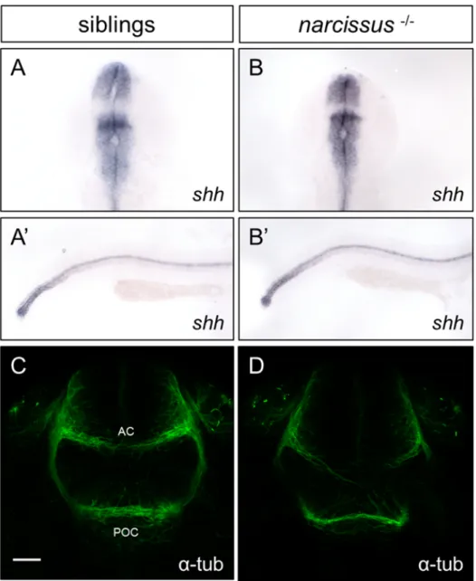
Indeed, an intact midline is required for early asymmetric markers to be cor-rectly expressed in only one side of the brain (Concha et al., 2000). To assess whether the IPN phenotype observed in nss mutants could be partly due to midline defects, we probed for the expression of the floor plate and notochord marker sonic hedgehog (shh). At 24 hpf we could not detect any differences in the expression domain of shh in wild-type siblings (Figure 3.2A-A’) and nss
-/-(Figure 3.2B-B’), suggesting that there are no major defects in midline tissues development in these mutants.
Additionally, guidance cues such as Slits, Ephrins and Semaphorins are re-quired to repel commissural axons to the contralateral side once they have reached the floor plate (Nawabi and Castellani, 2011). To address if commissural axons are being correctly guided through the midline in nss mutants, we have anal-ysed the formation of the post-optic commissure (POC) and anterior commissure (AC) in the forebrain by acetylated α-tubulin staining at 28 hpf embryos. Al-though most POC and AC axons correctly crossed the midline in all wild-type siblings examined (Figure 3.2C), 6/10 nss mutant embryos displayed abnormal crossing of commissural axons, particularly in the AC (Figure 3.2D, white arrow heads), suggesting that midline-derived axonal guidance cues are disrupted in nss mutants.
Previous studies have demonstrated that the secreted Class III Semaphorin, Sema3D, and its Neuropilin family receptor, Nrp1a, act as guidance cues to direct dHbL axons to the dIPN (Kuan et al., 2007). Indeed, nrp1a was shown to be specifically expressed in the left habenula as early as 2 dpf, while sema3D is expressed in the midline between the dHb and the IPN at this timepoint. Also, Nrp1a and/or Sema3D depletion by morpholino (MO) injection leads to the majority of kctd12.1+neurons to project to the vIPN. It is thus possible that the failure of dHbL axons to project to the dIPN in nss mutants is caused by defective guidance modulated by Nrp1a/Sema3D signalling. To test this hypothesis, we checked nss mutants for the expression of nrp1a. As previously reported, nrp1a is specifically expressed in the left habenula in wild-type siblings at 4 dpf (Figure
3.1A). In nss mutants, however, this expression is greatly reduced (Figure 3.1B). Taken together, these results indicate that the failure to correctly express nrp1a in the dHbL and, consequently, to respond to Sema3D signalling in nss mu-tants might preclude dHbL axons from properly projecting to the dIPN, thereby converging with dHbM axons in the vIPN. Additionally, failure in Sema3D-mediated midline signalling might explain the observed L-R segregation of habe-nular axons in the IPN of nss mutants.
Figure 3.1: dHbL axons innervation pattern is affected in nss mutants. A, In situ hybridization of nrp1a in wild-type siblings and B, nss mutants. C,C’, Retrograte dye labelling reveals that left habenular (lHb) axons (green) mainly project to the dorsal interpeduncular nucleus (dIPN) while right habenular (rHb) axons (magenta) innervate almost exclusively the ventral interpeduncular nucleus (vIPN) in 4dpf wild-type siblings, but D,D’ both lHb and rHb axons converge to the vIPN in 4dpf nss mutants. lHb and rHb nuclei were labelled with DiO and DiI, respectively. E, Immunolabelling of Tg(Pku558:GFP) neurons reveal that these project to the dorsal IPN in wild-type siblings, while in nss mutants F, the pattern of projection is affected, with pku558+ neurons projecting mostly to the vIPN. White arrowheads indicate misdirected axons projecting posteriorly.
Figure 3.2: nss mutants have normal midline tissues development but display defects in commissure formation. A-B’, sonic hedgehog (shh) is correctly expressed in the floorplate and notochord in both wild-type siblings, A-A’ and nss mutants B-B’ at 24 hpf. C-D, acetylated α-tubulin immunostaining of the anterior commissure (AC) and post-optic commissure (POC) at 28 hpf reveals that C, they are normally formed in wild-type siblings (n = 12) but D, AC axons fail to cross the midline in 6/10 analysed nss mutants. Scale bar = 25 µm. Anterior is to the top in A and B and to the right in A’ and B’, while dorsal is to the top in C and D.
3.2
Parapineal and olfactory bulb efferent
pro-jections to the habenulae are affected in nss
mutants, and projections from the pallium
remain asymmetric
The parapineal axonal projections to the left habenula have a critical role in the acquisition of dHbL fate, as specific ablation of parapineal anlage prior to its leftward migration or inhibition of the latter leads both habenulae to acquire a right-sided fate (Gamse et al.,2003). Considering this, it is possible that the left-type isomerism observed in nss habenular nuclei might be a result of defective parapineal-mediated induction of habenular fate.
We have previously observed that the parapineal organ correctly migrates to the left side in nss mutants (Figure 1.5A-B). To clearly visualize axonal trajec-tory of parapineal neurons and to assess whether the pattern of innervation is affected in mutants, we have carefully analysed axonal processes in confocal opti-cal sections of 4dpf Tg(foxD3:GFP) embryos. We found that, unlike in wild-type sibling (Figure3.3A) where axonal terminations tend to accumulate closer to the parapineal, these seem to extend onto a broader region of the left habenula in nss-/- (Figure 3.3B). In addition, we observed some axonal projections directing
toward the midline in mutants. These observations were further confirmed by quantification of the total area of projections in relation to their distance to the parapineal (Figure 3.3E).
More recently, Miyasaka and colleagues have described another asymmetric afferent projection specific to the right habenula, originating from a population of mitral cells in the zebrafish olfactory bulb (OB) and labelled by gap-YFP expression in Tg(lhx2a:gap-YFP) embryos (Miyasaka et al., 2009). Analysis of Tg(lhx2a:gap-YFP) embryos revealed that lhx2atg+ neurons fail to extend neu-ronal processes to the right habenula nucleus in nss mutants at 5 dpf (Figure
3.3D-D’), in contrast to wild-type siblings and consistent with the bilateral left-sided identity of habenular neurons (Figure3.3C-C’).
Cells labelled by transgenic expression in Tg(ClnB:GFP), probably originat-ing in the pallium, were identified by our lab to specifically project to the left
habenula. If the left-sided isomerism observed in the habenulae of nss mutants is complete, we would expect to see these neurons projecting to the rHb as well as to the lHb. An analysis of these projections in nss mutants, however, revealed that ClnBTg+ neurons still project asymmetrically to the left habenula (Figure
3.3E-F’).
Taken together, these results show that in nss mutants there is an overall disruption in habenular afferent projections, non-concordantly with a complete bilateral specification of the habenulae.
Figure 3.3: Asymmetric afferent projections to the dHb are disrupted in nss mutants. A-B, Immunolabelling in of Tg(foxD3:GFP) at 4 dpf reveals that the parapineal (pp) innervation to the left habenula nucleus (lHb) extends towards a broader region in nss mutants (B) than in wild-type siblings (A). C-D’, Immunolabelling of lhx2atg+ (green) olfactory bulb asymmetric projections
Figure 3.3: E-F’, Immunolabelling of ClnBtg+ (green) pallium asymmetric pro-jections to the left habenula (lHb) coupled with fluorescent in situ hybridization of kctd12.1 (magenta) shows that these remain asymmetric in nss mutants. G, Quantification of the parapineal projections pattern by correlation of the average area of the projections with the distance to the parapineal (n = 19). Positive values represent distances to the left while negative values represent distances to the right of the parapineal. White dotted boxes represent the zoomed region in C’,D’,E’ and F’. Scale bar = 25 µm. P, pineal.
3.3
Axonal projections in Tg(lhx2a:gap-YFP)
are not required for dHbM fate induction
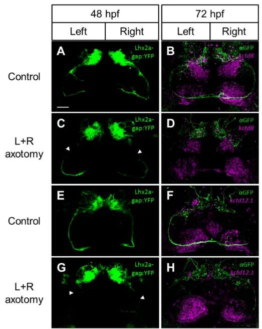
As kctd12.1 bilateral expression in nss mutant habenulae only develops from 48 to 72 hpf onwards, and no asymmetric disruption is seen before 48 hpf, we have hypothesised that what mediates this disruption might act during or from that time period onwards. We performed a time-course analysis revealing that the asymmetrical projection of lhx2atg+ OB neurons into the right habenula nucleusis first apparent at 52 hpf (Supplementary Figure B.1C, white arrowhead). The complete lack of these efferent projections in nss mutants, together with the fact that these innervate the right habenula nucleus at a seemingly critical time period in the development of the asymmetric defects observed in this mutant background led us to hypothesise that asymmetrical epithalamic innervation by OB mitral cells play a role in the development of right habenula identity. Much like what has been described for the inductive role of the parapineal in lHb development, mitral cells innervation to the right habenula progenitors could directly or indirectly promote dHbM or repress dHbL fates in this nucleus. As such, a failure to innervate the rHb in nss mutants would account for the failure to induce a right-type phenoright-type in the right epithalamus. To test this hypothesis, we conducted a laser-mediated axotomy of the axonal fibre bundles in Tg(lhx2a:gap-YFP). Using a multi-photon laser coupled with a confocal microscope, both left and right fibre bundles were selectively severed at around 48 hpf, before they had reached the epithalamic region. After the axotomy, embryos were left to develop until 72 hpf and then labelled for both the transgenic and either kctd8 or kctd12.1
expression as markers of dHbM and dHbL fates, respectively. Control embryos were subjected to the same laser pulses but in a YFP negative region close to the fibre bundles (Figure 3.4A-H). Time-lapse analysis after the axotomies and immunodetection of YFP in fixed embryos revealed that, unlike expected, the severed axons did not grow back, but rather regressed to their cell bodies in the OB (Supplementary Figure B.2). Despite this, kctd8 and kctd12.1 asymmetric expression domains were comparable in both axotomised and control embryos at 72 hpf (Figure 3.4A-H). These results indicate that, although present at the correct timing, OB mitral cells asymmetrical projections to the right habenula are not required to promote dHbM fates in the right epithalamus, and so, the observed lack of these projections in nss mutants are most likely a consequence and not a cause of the bilateral left-sided character in the habenulae of these mutants.
Figure 3.4: Asymmetric afferent projections from the OB in Tg(lhx2a:gap-YFP) are not required to specify dHbM neurons. A,E, Left and right lhx2aTg+ lateral fasciculi axotomised and C,G, non-axotomised controls display normal expression of both the B,D, dHbM marker kctd8 and F,H, dHbL marker kctd12.1. Images in A, C, E and G represent the endogenous in vivo transgenic expression of Tg(lhx2a:gap-YFP) after the administration of the laser pulses at 48hpf, while embryos in B,D,F and H represent these same embryos fixed at 72 hpf, immunolabelled against GFP (green) and probed for ei-ther kctd8 or kctd12.1 expression by fluorescent in situ hybridization (magenta). Arrowheads in C and G indicate where the axotomy pulses were administred. Scale bar = 25 µm.
3.4
Habenular neurogenesis is affected in nss
mutants
One of the mechanisms through which is possible to explain the development of an asymmetric habenula is the asymmetric onset of neurogenesis in left versus right epithalamus. dHbL and dHbM neurons are born at different time points during development: while the majority of dHbL neurons are born at around 32 hpf on the left habenula, most dHbM neurons are specified around 50 hpf on the right habenula (Aizawa et al., 2007). One possible explanation for the double left-sided phenotype in nss mutants is a disturbance in the asymmetric onset of neurogenesis, leading to both left and right dHb neurons being born during the first wave of neurogenesis and thereby acquiring a dHbL fate. Another explanation is that both left and right-sided neurons are correctly born during the first and second neurogenic waves, respectively, but a failure to induce dHbM character in neurons born during the second wave would result in the ectopic specification of a dHbL fate. To distinguish between these two hypotheses and to identify when ectopic right-sided kctd12.1+ neurons are born in nss mutants, we
conducted a birth-date analysis of habenular neurons by BrdU pulse labelling at both 32 hpf and 50 hpf (Figure3.5C-F,J). After BrdU pulse, embryos were chased until 5 dpf, a time point where both asymmetric gene expression and laterotopic projections are already established, and probed for kctd12.1 expression to mark dHbL (kctd12.1+) and dHbM (kctd12.2-) domains.
In both nss mutants and wild-type siblings, BrdU cells labelled at 32 hpf acquired a more dorsal and lateral position in the both habenulae, while cells la-belled at 50 hpf typically acquired a ventral and medial position (Figure3.5C,E), as previously described (Aizawa et al.,2007). In respect to their L-R disposition, no significant differences in the number of proliferating (BrdU+) cells were found between left and right habenular nuclei in wild-type siblings when the pulse was administered at 32 hpf, though significantly more of these cells acquired a dHbL fate (kctd12.1+) on the left side compared to the right (Figure 3.5C,G,J). In
contrast, no L-R differences in either the total number of BrdU+ or kctd12.1+
cells were found in nss mutants at this timepoint (Figure3.5D,H,J). BrdU pulse at 50 hpf revealed that, as expected for this time point, significantly more cells