October 2012
Escola de Engenharia
Alexandra Maria Rodrigues
Development and optimization of
production and purification of the
human protein BMP-2 in Escherichia
coli for biomedical applications
UMinho|20 12 Ale xandr a Maria Rodrigues De velopment and op
timization ofproduction and purification of t
he human pro
tein BMP-2 in
Escherichia coli
Dissertation thesis for the Master
degree in Biomedical Engineering
Supervisor:
Dr. Lucília Domingues
Co- Supervisor:
Dr. Miguel Gama
October 2012
Alexandra Maria Rodrigues
Development and optimization of
production and purification of the
human protein BMP-2 in Escherichia
COMPROMETE;
Universidade do Minho, ___/___/______
i
Acknowledgments
The present work was carried out in Biological Engineering Department of University of Minho, under the supervision of Dr. Lucília Domingues and Dr. Miguel Gama.
I am especially grateful to Dr. Lucília for the opportunity to perform this work, for all availability to constant discussions, advices and guidance for this project.
I would like to thank Dr. Miguel Gama for his availability and support with this work.
I particularly wish to thank my colleague Sofia Costa for her enormous support, excellent coordination of my work, and also for her great help for my everyday life in the laboratory.
I want to thank to my colleagues of Ecology and Molecular Microbiology Laboratory for being present and ready to help at anytime.
To my sister and my parents I would like to thank all the support, comprehension, motivation, courage and all the conditions which they gave me for achieving the objectives of my work. You are the most important people in my life.
Finally, I would like to address my thanks to all the other people, who have not been mentioned here by names, but who helped me during my thesis work and who made my stage in Biological Engineering Department a great experience.
iii
Development and optimization of production and purification of
the human protein BMP-2 in Escherichia coli for biomedical
applications
Abstract
Bone morphogenetic protein 2 (BMP-2) is one of the main representatives osteoinductive protein of a group of the transforming growth factor-β (TGF-β) superfamily of multifunctional cytokines. BMP-2 plays a critical role in cartilage and bone formation during skeletal development and repair. Several clinical studies have proven the clinical benefit of BMP-2 in the treatment of bone defects, being an effective alternative to bone grafts. Nowadays, BMP-2 is one of the two bone morphogenetic proteins approved as biological method to stimulate bone repair in humans. However, these commercial solutions are expensive, the protein purification is a laborious process and the obtained yields are low.
A landmark occurred with the cloning of the human BMP-2 gene and its production by DNA recombinant technology. Production of recombinant human BMP-2, using prokaryotic systems is the preferred method, being Escherichia coli one of the most popular systems for protein production. The bacterium E. coli is a well studied expression system, which provides a fast and economical production of recombinant proteins. Various studies on the production and purification of BMP-2 protein using E.
coli as expression system have been reported; however BMP-2 is difficult to express in
soluble form and its purification under native conditions remains a challenge.
In this work, the soluble expression of the recombinant BMP-2 protein in E. coli was studied using the fusion protein technology, with the novel FH8 tag.
The production of soluble recombinant fusion protein was initially achieved using as backbone the pQE-30FH8 vector, with the study of different culture conditions, in order to determine the conditions that maximize this soluble expression. Then, in order to increase soluble expression of FH8BMP-2, the cloning of bmp-2 gene was performed in two other different vectors: the pETMFH8 and pStaby1.2FH8, using different E. coli strains. Purification strategy of FH8BMP-2 was performed by the following chromatography techniques: using the FH8 as purification tag by hydrophobic interaction (HIC); affinity chromatography with nickel (using the His tag as purification tag) and ion exchange.
iv
The results presented in this work showed that recombinant fusion protein was successfully soluble expressed in the three vectors used. The pETM system was the one that showed the highest soluble expression. In relation to purification, the FH8-HIC technique, when combined with calcium addition to soluble fraction, shows good results in purification profile of target protein and a high yield of purified protein. Calcium ions and the own conformation of FH8 can have an important role on the aggregation of FH8BMP-2 into a dimer.
Overall, the BMP-2 was efficiently expressed in E. coli as a soluble protein and an optimized purification strategy was developed to obtain this bone morphogenetic protein under native conditions.
This work may be important for further steps for an in vitro production and biomedical application of recombinant BMP-2 protein.
v
Desenvolvimento e otimização da produção e purificação da
proteína humana BMP-2 em Escherichia coli para aplicações
biomédicas
Sumário
A proteína morfogenética do osso tipo 2 (BMP-2) é uma das proteínas osteoindutoras mais representativas da superfamília do fator de transformação do crescimento β (TGF-β) que agrupa um conjunto de citoquinas multifuncionais. A proteína BMP-2 desempenha um papel crítico na formação e regeneração do osso e cartilagem, durante o desenvolvimento do esqueleto. Vários estudos clínicos têm demonstrado o benefício clínico da BMP-2 no tratamento de defeitos ósseos, sendo uma alternativa eficaz aos enxertos do osso. Atualmente, a BMP-2 é uma das duas proteínas morfogenéticas do osso que se encontra aprovada como método biológico para estimular a reparação óssea em seres humanos. No entanto, estas soluções comerciais são dispendiosas, a purificação da proteína é um processo laborioso e os rendimentos obtidos são baixos.
Um marco importante ocorreu com a clonagem do gene humano da proteína BMP-2 e a sua produção através da tecnologia de DNA recombinante. A produção desta proteína humana recombinante, utilizando os sistemas procarióticos é o método preferido, sendo a bactéria Escherichia coli um dos sistemas mais populares de produção proteica. A bactéria E. coli é um sistema de expressão bem estudado, que proporciona uma produção rápida e económica de proteínas recombinantes de interesse. Vários estudos têm reportado a produção e purificação da proteína BMP-2, usando como sistema de expressão a E. coli; contudo, a proteína BMP-2 é difícil de expressar na forma solúvel e sua purificação em condições nativas continua a ser um desafio.
Neste trabalho, a expressão solúvel da proteína BMP-2 recombinante em E.coli foi estudada através da tecnologia de proteínas de fusão, com o novo tag de fusão FH8.
A produção solúvel da proteína de fusão recombinante foi conseguida usando como vetor inicial o pQE-30FH8, com o estudo das diferentes condições de cultura, com o objetivo de determinar as condições que maximizam essa expressão solúvel. Em seguida, com a finalidade de aumentar a expressão solúvel da FH8BMP-2, foi realizada a clonagem do gene da BMP-2 em dois vetores diferentes: o pETMFH8 e pStaby1.2FH8, usando diferentes estirpes de E. coli. A estratégia de purificação da
vi
proteína FH8BMP-2 envolveu a realização das seguintes técnicas de cromatografia: interação hidrofóbica (HIC), usando o tag FH8 como tag de purificação; cromatografia de afinidade com níquel (usando o His tag como tag de purificação) e troca iónica.
Os resultados apresentados neste trabalho mostraram que a proteína de fusão recombinante foi expressa solúvel com sucesso nos três vetores utilizados. O sistema pETM foi aquele que apresentou um maior nível de expressão solúvel. Em relação à purificação, a técnica de FH8-HIC, quando combinada com a adição de cálcio à fração solúvel, apresenta bons resultados em relação ao perfil de purificação da proteína alvo e um elevado rendimento de proteína purificada foi obtido. Os iões de cálcio e a própria conformação do tag FH8 podem ter um importante papel na agregação da FH8BMP-2 na forma dimérica.
Em geral, a BMP-2 foi eficientemente expressa no sistema E. coli como proteína solúvel e uma estratégia de purificação otimizada foi delineada para obter esta proteína morfogenética do osso em condições nativas.
Este trabalho pode ser importante para novas etapas de uma produção in vitro e aplicação biomédica da proteína BMP-2 recombinante.
vii
Table of Contents
Acknowledgments ... i Abstract ... iii Sumário ... v List of Figures ... xiList of Tables ... xiii
Abbreviations ... xv
Motivation and aim of the project... 1
Chapter 1: Review Literature ... 3
1.1 Bone morphogenetic proteins ... 3
1.1.1 Bone morphogenetic protein 2 (BMP-2) ... 4
1.2 Commercial BMP2 ... 5
1.3 Recombinant production of BMP-2 ... 6
1.3.1 E. coli as a host ... 7
1.4 Production of soluble recombinant BMP-2 ... 8
1.4.1 Fusion Protein Technology ... 9
1.4.1.1 Fusion system – FH8 tag ... 10
Chapter 2: Materials and Methods ... 13
2.1 Sterilization of material, culture media and solutions ... 13
2.2 Chemical products, solutions and reagentes ... 13
2.3 Vectors and Bacterial strains ... 13
2.3.1 pQE-30 QIAexpress system ... 14
2.3.2 pETM vector ... 14
2.3.3 pStaby1.2 Express System ... 15
2.4 Molecular Biological Methods ... 16
2.4.1 Extraction of plasmid DNA and template sequence of BMP-2 from E. coli . 16 2.4.2 PCR of insert fragment – bmp-2 gene ... 16
2.4.3 Agarose Gel Electrophoresis ... 18
2.4.4 DNA purification... 18
2.4.5 Digestion of DNA with restriction enzymes ... 18
2.4.6 Dephosphorylation of Plasmid DNA ... 19
2.4.7 DNA quantification ... 19
2.4.8 Ligation of DNA insert fragment to the vector ... 19
2.4.9 Preparation of chemical competent E. coli cells ... 20
2.4.10 Transformation of recombinant DNA to competent E. coli cells ... 20
2.4.10.1 Transformation of chemically competent DH5α E. coli cells ... 20
2.4.10.2 Transformation of chemically competent Top 10 E. coli cells ... 21
viii
2.4.12 DNA Sequencing... 22
2.4.13 Transformation of chemically competent BL21 Codon Plus Ril and Rosetta 2 E. coli cells ... 22
2.4.14 Transformation of chemically competent SE1 E. coli cells ... 22
2.5 Soluble Expression of recombinant FH8BMP-2 protein in E. coli ... 22
2.5.1 Study of the soluble expression conditions of recombinant FH8BMP-2 protein in E. coli M15/pQE-30 ... 23
2.5.2 Soluble expression of recombinant FH8BMP-2 protein in E.coli M15 /pQE30 ... 24
2.5.3 Study of the soluble expression conditions of recombinant FH8BMP-2 protein in pETM: E. coli BL21 Codon Plus Ril and Rosetta2 ... 24
2.5.4 Soluble expression of recombinant FH8BMP-2 protein in E. coli BL21 Ril /pETM10 ... 25
2.5.5 Conditions study of soluble expression of recombinant FH8BMP-2 protein in E. coli SE1/pStaby1.2... 25
2.6 Electrophoresis under denaturing conditions: SDS-PAGE... 26
2.7 Purification of recombinant FH8BMP-2 protein ... 27
2.7.1 Hydrophobic Interaction Chromatography (HIC) ... 27
2.7.1.1 Small scale screenings protocol ... 30
2.7.1.2 0.5-1L Purification Assays protocol ... 31
2.7.2 Immobilized metal ion Affinity Chromatography (IMAC) ... 32
2.7.2.1 Small scale screenings protocol ... 33
2.7.2.2 0.5-1L Purification Assays protocol ... 34
2.7.3 Ion Exchange (IEX) chromatography ... 34
2.8 Protein dialysis ... 36
2.9 Protein quantification ... 36
2.10 Dynamic light scattering (DLS) ... 37
Chapter 3: Results and Discussion ... 39
3.1 Soluble expression and purification of recombinant FH8BMP-2 in E. coli using the pQE-30 system ... 39
3.1.1 Soluble test expression of recombinant FH8BMP-2 in E. coli M15 using the pQE-30 system ... 39
3.1.2 Small screening purification test of soluble recombinant FH8BMP-2 produced in E. coli M15 using the pQE-30 system... 43
3.1.3Larger scale purification test of soluble recombinant FH8BMP-2 in E. coli M15 using the pQE-30 system ... 46
3.2 Cloning of bmp-2 gene into pETMFH8 and pStaby1.2FH8 plasmids ... 49
3.3 Soluble expression and purification of recombinant FH8BMP-2 in E. coli using pETMFH8 and pStaby1.2FH8 systems ... 53
3.3.1 Small scale screening ... 53
3.4 Larger scale production and purification tests of soluble recombinant FH8BMP-2 in E. coli using pETM system ... 57
ix
3.4.2 Hydrophobic interaction chromatography ... 58
3.4.2.1 Optimization of HIC wash step ... 62
3.4.3 Ion exchange chromatography ... 65
3.5 Larger scale production and purification tests of soluble recombinant FH8BMP-2 in pStaby1.2 system ... 67
Chapter 4: Main Conclusions and suggestions for forthcoming work ... 69
Chapter 5: Reference List ... 71
Chapter 6: Appendixes ... 77
Appendix 1: Detailed composition of chemical products, solutions and reagents ... 77
Appendix 2: Hidrophobicity analysis of FH8BMP-2 using Kyte and Doolittle scale 78 Appendix 3: Neb-Cutter analysis ... 79
Appendix 4: BLASTN analysis of the sequencing results of pETM/FH8BMP-2 and pStaby1.2/FH8BMP-2 clones ... 80
xi
List of Figures
Figure 1: Activation of BMP dimer ... 4
Figure 2: Gene transcription by BMP-Smad activation ... 4
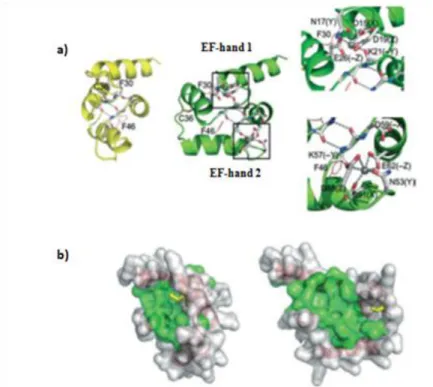
Figure 3: a) Model structure of FH8 in the closed conformation (yellow). After calcium ligation occur a change to open conformation (green) and on the right is possible to see in detail the calcium binding loops, where calcium ions are represented by grey spheres. b) Molecular surface representation of FH8 in closed (left) and open (right) conformations ... 11
Figure 4: Graphic of plasmid map of pQE-30, Qiagen ... 14
Figure 5: Graphic of plasmid map of pETMFH8, EMBL ... 15
Figure 6: Graphic of plasmid map of pStaby1.2FH8, Delphi ... 16
Figure 7: Soluble expression conditions of FH8BMP-2 in pQE system ... 23
Figure 8: Variation of surface net charge according to pH of the medium ... 34
Figure 9: a) 15%-4% SDS-PAGE solubility analysis of BMP-2 in pQE-30 system; b) 15%-4% SDS-PAGE solubility analysis of HBMP-2 in pQE-30 system ... 40
Figure 10: 15%-4% SDS PAGE analysis of expression level of recombinant FH8BMP-2 in E. coli M15/pQE-30 ... 41
Figure 11: SDS-PAGE Solubility analysis of FH8BMP-2 in pQE-30 system of 250 ml cultures ... 42
Figure 12: 15%-4% SDS-PAGE analysis of samples obtained from HIC small scale purification assays of FH8BMP-2: ... 44
Figure 13: 15%-4% SDS-PAGE analysis of samples obtained from IMAC-Ni small scale purification assay ... 45
Figure 14: HIC purification assay of 1L E. coli M15/pQE-30 culture of FH8BMP-2 .. 47
Figure 15: Second HIC purification of eluted samples of first HIC assay containing recombinant FH8BMP-2 ... 48
Figure 16: Cloning strategy to pETM/FH8BMP-2 and pStaby1.2/FH8BMP-2 constructs ... 50
Figure 17: 1% Agarose electrophoresis analysis of bmp-2 PCR products ... 51
Figure 18: 1% Agarose electrophoresis analysis of DNA plasmids ... 52
Figure 19: 1.2% Agarose electrophoresis analysis of transformants of recombinant vectors: a) Transformants of pETMFH8BMP-2; b) Transformants of pStaby1.2FH8BMP-2 ... 53
xii
Figure 20: 15%-4% SDS-PAGE solubility analysis of FH8BMP-2 in pETM system in 20 ml cultures: a) E. coli BL21 Codon plus Ril culture; b) E. coli Rosetta 2 culture .... 54 Figure 21: 15%-4% SDS-PAGE solubility analysis of pETM/FH8BMP-2; pETM/HisBMP-2; pETM/FH8 and pETM/His samples ... 55 Figure 22: 15%-4% SDS-PAGE Solubility analysis of FH8BMP-2 in pStaby1.2 system in E. coli SE1 20 ml cultures in comparison with pStaby1.2/FH8 (negative control – without protein) ... 56 Figure 23: 15%-4% SDS-PAGE analysis of samples obtained from IMAC-Ni small scale purification assay: ... 58 Figure 24: 15%-4% SDS-PAGE gel with samples of 1L of E. coli BL21 Codon plus Ril culture of FH8BMP-2 and HIC purification samples ... 59 Figure 25: 15%-4% SDS-PAGE gel with HIC purification samples from 1L of E. coli BL21Codon plus Ril culture of FH8BMP-2, with lower calcium concentration ... 60 Figure 26: 15%-4% SDS-PAGE gel with samples of second HIC purification performed with eluted samples obtained from first HIC purification assay ... 61 Figure 27: 15%-4% SDS-PAGE gel with samples of HIC purification performed with different wash buffers ... 64 Figure 28: 15%-4% SDS-PAGE gel with samples of HIC purification performed with wash buffers W0 and W1 ... 64 Figure 29: Illustrative diagram of steps performed in IEX procedure. ... 65
Figure A1: Analysis of the hydrophobicity of pQE-30/FH8BMP-2, according to the Kyte and Doolittle scale. ... 78 Figure A2: Analysis of the hydrophobicity of pETM/FH8BMP-2, according to the Kyte and Doolittle scale ... 79 Figure A3: BLASTN analysis of pETM/FH8BMP-2 and pStaby1.2/FH8BMP-2 clones ... 80 Figure A4: Results of the analysis of protein domains in fusion conserved domain by NCBI software. ... 81
xiii
List of Tables
Table 1: Selected works reporting the production of rhBMP-2 in different expression
systems ... 6
Table 2: Studies of the production and purification of rhBMP-2 in E.coli. ... 8
Table 3: Commonly used solubility fusion tags ... 10
Table 4: Plasmid vectors and bacterial strains used in the present work. ... 13
Table 5: Sequence of primers used in PCR reaction. ... 17
Table 6: PCR program. ... 17
Table 7: Description of restriction enzymes used. ... 19
Table 8: Colony PCR program. ... 22
Table 9: SDS-PAGE gels composition. ... 26
Table 10: Buffers recipe for FH8-HIC small screenings. ... 29
Table 11: Buffers recipe for FH8-HIC 0.5-1L purification assays... 29
Table 12: Buffers recipe for IMAC small screenings. ... 33
Table 13: Buffers recipe for IMAC 0.5-1L purification assays. ... 33
Table 14: Buffers recipes for IEX chromatography. ... 36
Table 15: Studied culture conditions for soluble expression of FH8BMP-2. ... 41
Table 16: Recipe of buffers of HIC purification step of recombinant FH8BMP-2 protein in E. coli M15/pQE-30. ... 45
Table 17: Induction conditions of screening tests for soluble expression of recombinante FH8BMP-2. ... 54
Table 18: Recipe of buffers used in IMAC-Ni purification step of recombinant FH8BMP-2 protein in E. coli BL21 Ril/pETM. ... 57
Table 19: Recipe of buffers used in first HIC purification step of recombinant FH8BMP-2 protein in E. coli BL21 Ril/pETM. ... 58
Table 20: Washing buffers tested in HIC purification. ... 62
Table A1: Composition of solutions used in the project. ... 77
xv
Abbreviations
APS – Ammonium Persulfate;
BMP-2 – Bone Morphogenetic Protein 2; bp – base pair;
BSA – Bovine Serum Albumin; CV – Column Volumes;
Da – Dalton;
DNA – deoxyribonucleic acid;
dNTPs – deoxyribonucleoside triphosphates; IPTG – Isopropyl β-D-1-thiogalactopyranoside; LB – Luria Broth;
MCS – Multiple Cloning Site; min – minute(s);
OD – optical density;
PBS – Phosphate Buffer Saline; pI – isoelectric point;
rpm – revolutions per minute; RT – Room Temperature; SDS – sodium dodecyl sulfate;
SDS-PAGE – Sodium Dodecyl Sulfate Polyacrylamide Gel Electrophoresis; TEMED – N,N,N’,N’-tetramethylethylenediamine;
TGF-β – Transforming Growth Factor β; UV – Ultraviolet light.
1
Motivation and aim of the project
The research on bone regeneration began decades ago with intensive studies on bone growth and healing. Most researches have been conducted to identify proteins capable to induce new bone formation and methods to perform biological applications that can lead to decrease or elimination of bone graft [1,2].
It was in 1965 that a landmark on bone regeneration began with the work developed by Urist [3]. The pioneering work of Urist showed that demineralized bone matrix had the capacity to induce endochondral bone formation and he named the mixture of proteins present in matrix as bone morphogenetic proteins (BMPs) [1,4]. During the decade of 80s and 90s many investigators developed research in this area with the cloning of BMPs genes, demonstrating that these proteins are responsible to initiate a cascade of events, in which stem and mesenchymal cells are differentiated into osteogenic linage capable of producing bone tissue. Since then, several BMPs have been identified and purified from bone of different species, including human, for clinical applications [1,2,4].
Bone morphogenetic protein 2 (BMP-2) is one of the main morphogenetic protein implicated in growth and regeneration of bone and cartilage [5,6]. The expensive production of BMP-2 is a limitation of using this protein in therapeutic applications. A landmark step was made with the use of recombinant gene technology, which allowed the production of large quantities of recombinant human BMPs (rhBMPs) [7,8]. Over the years has been reported the production of recombinant human bone morphogenetic 2 (rhBMP-2) in prokaryotic system. However, to obtain BMP-2 protein in soluble form and in native conditions remains a challenge.
In this work, the expression system chosen for the production of recombinant BMP-2 is the E. coli system and for soluble expression, the fusion protein technology with the novel FH8 tag is studied. Beyond the pQE-30 vector, it is used other two different vectors - pETM and pStaby1.2 - to express the fusion protein FH8BMP-2. The main purpose is to get an increase of soluble expression and purity level of target protein. The pET system is a powerful system for the cloning and expression of recombinant proteins in E. coli. In relation to pStaby1.2 system, the selection
2
mechanism is an important feature of this plasmid, because instead of antibiotics, this system uses antidote genes present naturally in plasmids. It is an advantage for commercialization systems, as it is described that contamination of the product by antibiotics is unacceptable by regulatory institutions.
The main objectives of the project are the following:
Production of soluble recombinant BMP-2 protein fused to the FH8 tag, using the pQE-30 expression system;
Purification of the fusion protein via the FH8 tag by hydrophobic interaction chromatography and via affinity chromatography with nickel, using the His-tag in the system pQE-30;
Cloning of the gene coding for BMP-2 and its insertion into two other expression systems (pETM and pStaby1.2) containing the His-FH8 tag;
Production of soluble recombinant fusion protein in both expression systems;
Purification of fusion proteins obtained in both systems (pETM and pStaby) by nickel affinity chromatography (His-tag) and hydrophobic interaction chromatography (FH8-tag);
3
Chapter 1: Review Literature
1.1 Bone morphogenetic proteins
Discovered in 1965, BMPs belong to a group of cytokines from the transforming growth factor-β (TGF-β) superfamily. To date, at least fifteen BMPs have been identified and characterized, and the members of BMPs family have been subdivide into subsets according to their amino acid sequence homology and similarity in protein structure. The main biological functions of BMPs are based especially on bone and cartilage formation [1,9]. BMPs are homo or heterodimers, which chains are connected via disulphide bridges. In general, BMPs are biological active as both homo or heterodimer conformation. The structure of these proteins are based on conserved motif of seven cysteines, that are involved in the formation of six intrachain disulphide bonds and a single interchain bond, to promote the dimer formation [1,4,9]. However, to form active dimer, BMP molecules are dissociated between the propeptide and mature region after proteolytical cleavage (Figure 1). These proteins are produced as large precursor molecules containing a hydrophobic signal sequence, a long and poorly conserved N-terminal pro-region sequence, a mature domain with a highly conserved C-N-terminal region and an N-terminal region that varies among the different BMPs [1,9]. The molecular signalling of BMPs is based on binding of these proteins to serine-threonine kinase receptors, which are present in the cells surface. This binding promotes important intracellular events that are responsible for activation of gene transcription, leading to cell proliferation and differentiation.
In particular, there are three types of receptors for TGF-β superfamily members, but only receptors type I and II are involved in the signalling of BMPs. That is, the cascade of intracellular events that are involved with BMPs signalling occurs when the binding of these proteins to type I and II receptors (BMPRI, BMPRII) triggers a signal transduction cascade via Smad family proteins. As a consequence, BMP-Smad pathway will activate direct or indirectly target genes, responsible for osteoblast differentiation (Figure 2) [1,4,10].
4 Figure 1: Activation of BMP dimer. Adapted from [9]
Figure 2: Gene transcription by BMP-Smad activation. Adapted from [4]
1.1.1 Bone morphogenetic protein 2 (BMP-2)
BMP-2 is one of the main BMPs member implicated in growth and regeneration of bone and cartilage, and therefore is one of the most intensely investigated growth
5
factor. BMP-2 is localized in bone tissue and is released in response to bone damage, stimulating differentiation of mesenchymal cells into osteoblasts and inducing cell proliferation via the Smad pathway demonstrated in Figure 2. In terms of structure, human BMP-2 consists of a long precursor protein of 396 amino acids, which is glycosylated, proteolytically cleaved and dimerized to form the mature homodimeric protein consisting of two 114 residue subunits [11]. Other feature related to the structure of BMP-2 is a heparin-binding domain located in the N-terminal region of the mature polypeptide, which can cause alterations on its biological activity [1]. The surface of the BMP-2 dimer is very hydrophobic causing its low solubility in aqueous solutions. Its osteoinduction properties have been subject of numerous preclinical and clinical experiments, described in literature (McKay and Sandhu (2002,2007); Govender et al. (2002); Raiche and Puleo (2004)), showing various therapeutic applications of BMP-2 [2,5,12–15]
.
1.2 Commercial BMP2
As a result of its therapeutic potential, BMP-2 has been studied as an alternative to autologous bone grafting in many clinical situations. These include spinal fusion, osteoporosis, treatment of bone defects, non-union fractures and root canal surgery [1,2,16–18]
. The approval commercial systems involving this protein in human application occurred in 2002, when the US Food and Drug Administration (FDA) and the European Medicines Agency (EMEA) have approved the clinical use of 2. Currently, BMP-2 is commercially available under the implant kit name Inductos™ (Medtronic Sofamor Danek and Wyeth Pharmaceuticals). This kit contains the BMP-2 protein as a lyophilized powder, dissolved in sterile water and is applied to an absorbable collagen matrix made of type 1 bovine collagen, for the treatment of acute open tibial fracture. Another similar product is the Infuse™ bone graft (Medtronic Sofamor Danek), indicated to the treatment of degenerative lumbar disc disease [2,5,12–15]. These commercial preparations of BMP-2 are, however, produced abroad and are expensive [1,2,5]
.
To overcome this bottleneck, during the last years, the investigation has been focused in strategies to increase production of BMP-2 at lower cost taking advantage of the advances in recombinant technology.
6
1.3 Recombinant production of BMP-2
As with the majority of BMPs, reduced yields are obtained when native BMP-2 is isolated from bone (around 1-2 µg/kg bone). Since the production and purification of this native protein presents difficulties and its clinical applications are limited by the potential health risks associated with its isolation from allogeneic bone donor, researchers have been encouraged to express BMP-2 protein, as well as the other BMPs by DNA recombinant technology [1,4,5,19,20].
Recombinant human BMP-2 (rhBMP-2) was first obtained using mammalian cultures of chinese hamster ovary cells and the recombinant protein promoted ectopic bone and cartilage formation after two weeks of implantation in rats. After this pioneering work, other investigations were performed, to obtain rhBMP-2 using different expression systems (Table 1) [1,2,4].
Table 1: Selected works reporting the production of rhBMP-2 in different expression systems. Adapted from [1]
BMP Expression system Novelty References
BMP-2 CHO cells
Bone formation and characterisation of
expressed BMP-2
Wang et al – 1990 Israel et al - 1992
BMP-2 Insect cells Alternative expression
system Maruoka et al – 1995
BMP-2 E.coli
The heparin binding domain reduces ALP and
specific in vitro biological activity
Ruppert et al - 1996
BMP-2 E.coli High density expression
in bacteria Li et al - 1998
BMP-2 E.coli, pCYTEXP3 Optimisation of
refolding conditions
Vallejo et al – 2002 Vallejo and Rinas -
2004
BMP-2 E.coli
Additional heparin binding domains enhance in vivo bone
formation
Wurzler et al - 2004
BMP-2 E.coli, pET-11a Comparison with BMP-2
propeptide Hillger et al - 2205
BMP-2 E.coli, pET-21a Use of different
refolding buffers Long et al – 2006
BMP-2 E.coli, pET-25b Bioactivity in human
7
Nowadays, recombinant human BMPs (rhBMPs) are produced mainly by two expression systems: in mammalian cells or in bacteria. However, with mammalian cells expensive cultivation and poor yields render this procedure cost-intensive, especially at the industrial scale. Production in prokaryotic hosts is usually the preferred method, because these offer important advantages, such as, high yield, low cost cultivation/production and high bio-safety. One of prokaryotic systems widely used for the rapid and economical production of recombinant proteins is Escherichia coli (E.
coli) [1,12,16,17,19].
1.3.1 E. coli as a host
Recombinant protein expression has revolutionized all aspects of the biological sciences, namely by the dramatically expansion of the number of proteins that can be studied both biochemically and structurally. In spite of the development of multiple nonbacterial recombinant expression systems over the last three decades (yeast, baculovirus, mammalian cell, cell free systems), E. coli is still the preferred host for recombinant protein expression, being widely used in industry and in academic research for this purpose [21–24]. The vast advantages of E. coli turn it into a valuable organism for the high-level production of recombinant proteins. Among the important features, one can find: rapid growth, and expression, its well-characterized genetics, high product yields, easy of genetically manipulation, and it is relatively inexpensive to culture. The ability of this bacterial host to accumulate recombinant proteins up to 80% of its dry weight and to survive a variety of environmental conditions represent also important advantages for a wide range of downstream applications . Actually, the expression of human proteins in E. coli can reach a high success rate of 75% [21,22,24–26].
However, common drawbacks can occur when using E. coli as an expression host. E. coli is not capable of producing eukaryotic post-translational modifications, such as glycosylation, which can be critical for the production of folded and active protein. Equally important is the fact that many proteins of biomedical interest have proved difficult to express properly in this host system because they are easily turned into insoluble and instable protein aggregates, identified as inclusion bodies. This aggregation of recombinant proteins in bacterial cells is a result of accumulation of high concentrations of folding intermediates or from inefficient processing by molecular chaperones [24,27]. The main disadvantages of obtaining proteins from inclusion bodies
8
are related with solubilisation and refolding time-consuming steps and with the use of denaturing agents to extract protein of interest. This can cause problems with protein native conformation and consequently its activity. In the case of BMP-2, it is a protein that is biologically active in homodimeric form, so refolding steps are necessary after recovery from inclusion bodies [5,18,27].
During the last years some investigators have performed important scientific work in this field (in Table 2 is possible to observe some of these publications) in order to produce and purify rhBMP-2 in E. coli, from the recovery of inclusion bodies.
To overcome limitations associated to the production of recombinant protein in the form of inclusions bodies, several strategies have been described in literature to achieve soluble protein expression in E. coli. In following section it will be described some of these strategies, in which fusion expression technology has an important focus [23,28–30]
.
Table 2: Studies of the production and purification of rhBMP-2 in E.coli. Author/date
Study
Zhang et al. /
2011 Expression, purification, and refolding of a recombinant rhBMP-2
Zhang et al. /
2010 Optimized procedure for expression and renaturation of rhBMP-2 at high protein concentrations
Sharapova et al.
/2011 Production of the rhBMP-2 in E. coli and Testing of Its Biological Activity in vitro and in vivo
Bessa et al. /
2007 Osteoinduction in human fat-derived stem cells by rhBMP-2 produced in E. coli
Long et al. /
2006 Expression, purification, and renaturation of rhBMP-2 from E. coli
1.4 Production of soluble recombinant BMP-2
According to recent studies of structural genomics centres, more than half of all recombinant proteins accumulate in the form of insoluble aggregates when they are
9
overproduced in E. coli. However, in many biomedical applications, the objective is to obtain a product that is soluble, folded and active [21,31].
Many methodologies that have been applied to increase the solubility of recombinant proteins in E. coli consist on the manipulation of culture conditions, and altering the temperature at which the target protein is being produced [31,32]. Also, changes in the E. coli expression strains, the use of different promoters and co-expression methodology may contribute to improve the solubility of recombinant proteins in E. coli.
In relation to BMP-2, there is only a study by Ihm et al in which is reported the soluble expression of BMP-2 in E. coli, using co-expression of thioredoxin gene in a different expression vector [18].
As referred above, in most cases, strategies previously described do not solve completely the problem and other technologies are been widely explored as, for instance, the use of solubility fusion tags [31,32].
1.4.1 Fusion Protein Technology
Fusion protein technology is widely used for a rapid, efficient and cost-effective protein expression and purification process [33]. Fusion tags are described to be proteins or peptides that are fused to the protein of interest and the main functions of these partners are to increase target protein production yields, promote its solubility and help on its purification [29,31,32]. Over the years several fusion partners had already been described in literature (Table 3), but none of them is ideal with respect to all parameters referred above.
Generally, it is difficult to choose the best fusion system for a specific protein of interest, due to different factors such as the expression system, the target protein itself (its stability or hydrophobicity) and the application of the purified protein. Thus, to determine the “best” tag for a specific target protein remains a challenging.
Additionally, there are some factors related to the fusion tags that can affect the soluble protein expression levels and its purification, for example the placement of the tag, either in N-terminal or C-terminal. On the other hand, as many of affinity tags are large proteins, they can affect important characteristics like the structure of the protein to be studied. Also, biological activity of the recombinant expressed protein can be affected by the presence of a fusion tag and thus, in these cases, it is necessary to
10
remove the tag after the purification of the fusion protein [21,31,32,34]. The most commonly used solution to remove the tag is to place a protease cleavage site between the solubility tag and the partner protein, allowing an in vitro reaction after purification to remove the fusion tag [32]. The commonly proteases used are: enterokinase, tobacco etch virus (TEV), thrombin, and factor Xa. The recovery process of target protein depends on the cleavage efficiency [35].
Table 3: Commonly used solubility fusion tags. Adapted from [32]
1.4.1.1 Fusion system – FH8 tag
The Hitag® fusion system consists of two novel fusion tags, the H and FH8 tags, which have demonstrated to increase protein expression levels in E. coli [36,37].
The FH8 tag is an 8 kDa protein with 69 aminoacids and it is a homologous antigen secreted by the parasite Fasciola hepatica in the early stages of infection. Previous studies demonstrated that FH8 is a binding calcium protein by the presence of two hands, which contain motifs involved in calcium coordination. Structurally, EF-hands proteins are organized in functional domains which form stable helical bundles. The binding calcium properties of FH8 can cause conformational changes on its structure: when calcium binds to EF-hands domains, a switch from a closed to an open
Tag Protein name Source organism
MBP Maltose-binding protein E. coli
GST Glutathione-S-transferase Schistosoma japonicum
Trx Thioredoxin E. coli
NusA Utilization substance E. coli
SUMO Small ubiquitin-modifier Homo sapiens
SET Solubility-enhancing tag Synthetic
DsbC Disulfide bond C E. coli
Skp Seventeen kilodalton protein E. coli
T7PK Phage T7 protein kinase Bacteriophage T7
GB1 Protein G B1 domain Streptococcus sp
ZZ Protein A IgG ZZ repeat domain
Staphylococcus aureus
11
conformation occurs. This reorientation of the protein conformation leads to the exposure of hydrophobic regions, which act as a target binding surface [36,38]. Figure 3 shows in detail this change of conformation after calcium binding (Figure 3 a)) and also the conformational change of hydrophobic area of FH8, which becomes larger in calcium loaded state (Figure 3 b)).
As other members of the EF-hand family, FH8 tag demonstrates an unusual stability at high temperatures, and in the presence of calcium it is even more stable in other denaturant conditions, such as in the presence of high urea concentrations [38,39].
Another interesting feature as a result of calcium interaction is a dimmerization of FH8 protein [38,39]. A study conducted of FH8 shows the analysis, in non-denaturing conditions, of higher molecular weight band, suggesting the formation of dimers and pentamers. The pentamers are considered the most stable structure of the antigen and its formation is dependent on calcium [38].
Currently, FH8 tag has been tested as solubility and purification tag with proteins difficult to express in E. coli [29,37,39,40]. The fusion of these proteins to the FH8 tag resulted in an increase of soluble expression and due to this positive effect, it will be studied the FH8 tag application with BMP-2 protein.
Figure 3: a) Model structure of FH8 in the closed conformation (yellow). After calcium ligation occur a change to open conformation (green) and on the right is possible to see in detail the calcium binding loops, where calcium ions are represented by grey spheres. b) Molecular surface representation of FH8 in closed (left) and open (right) conformations. Adapted from [38]
13
Chapter 2: Materials and Methods
2.1
Sterilization of material, culture media and solutionsThe sterilization of materials, solutions and culture media was carried out by autoclaving at 121°C for 20 minutes. Thermolabile solutions were sterilized by filtration with sterile filters of 0.2 micrometers.
2.2 Chemical products, solutions and reagentes
Composition of all solutions, loading samples and reagents used in this work are described in Table A1 of appendix 1.
2.3 Vectors and Bacterial strains
The plasmid vectors and the corresponding E. coli strains used to produce the recombinant fusion protein in this study are presented in Table 4. It is important to note that only the construction of pQE-30/FH8BMP-2 was already available. The other two constructions were conducted in the scope of this work. A more detailed description of each plasmid vector will be presented in this section.
Table 4: Plasmid vectors and bacterial strains used in the present work.
Backbone plasmid Constructed plasmid E.coli strain Phenotype
pQE-30 (QIAexpress system – Quiagen)
pQE-30/FH8BMP2 M15[pREP4]
NaIS, StrS, RifS, Thi–, Lac–, Ara+, Gal+, Mtl– , F–, RecA+, Uvr+,
Lon+
pETM (EMBL) pETM/FH8BMP2
BL21 CodonPlusRil
B F–ompT hsdS(rB– mB–) dcm+ Tetr gal endA Hte [argU ileY
leuWCamr] Rosetta 2 F – ompT hsdSB(rB–mB– ) gal dcm pRARE23 (CamR) pStaby1.2 (StabyExpress system - Delphi) pStaby1.2 /FH8BMP2 SE1
gal, dcm, DE3 (lacI, T7 polymerase under the control of the lacUV5
14
2.3.1 pQE-30 QIAexpress system
This expression system, distributed by Qiagen, consists of pQE-30 vector and it was used to express BMP-2 protein with and without the FH8 fusion tag. The pQE-30 plasmid, represented in Figure 4, uses a transcription–translation system based in T5 promoter/lac operator, which allows high expression levels of recombinant proteins in
E. coli [41]. This system has other important feature: the presence of a 6xHis-tag
codifying sequence in the N-terminal, which when genetically bound to the protein of interest, can facilitate the purification process by affinity chromatography.
Figure 4: Graphic of plasmid map of pQE-30, Qiagen. Adapted from [41]
2.3.2 pETM vector
The pETM vectors derive from the pET vector series initially developed by Studier and colleagues, which represent nowadays a powerful system to cloning and expression of recombinant proteins in the E. coli host. The pETM plasmids use the same transcription-translation system as the pET collection based in T7 promoter to clone the genes of interest. Some important features of pETM vectors are: the presence
15
of two 6xHis-tags (one before and other after the MCS), a conserved multiple cloning site (MCS) and a TEV protease recognition site.
In this particular study it was used the pETMFH8 plasmid, containing the FH8 tag and a TEV recognition site between the fusion tag and protein. This expression vector has also the presence of a gene responsible to confer kanamycin resistance. The MCS is located between TEV site and the second 6xHis-tags. A schematic diagram of pETMFH8 vector is shown in Figure 5.
Figure 5: Graphic of plasmid map of pETMFH8, EMBL.
2.3.3 pStaby1.2 Express System
This expression system, commercialized by Delphi Genetics, was also used to clone bmp-2 gene and produce FH8BMP-2 protein. The plasmid used in this work was the pStaby1.2FH8, which has the following features: T7 promoter, 6xHis-tag at the C-terminal end of the protein; the plasmid is stable without the use of antibiotics. As in the case of pETMFH8 vector, the gene of codifying for BMP-2 was cloned into the pStaby1.2FH8 plasmid, but in this vector FH8 tag and the 6xHis residues are in opposite ends, which may improve the purification process. Plasmid scheme of pStaby1.2FH8 is displayed in Figure 6.
16 Figure 6: Graphic of plasmid map of pStaby1.2FH8, Delphi.
2.4 Molecular Biological Methods
2.4.1 Extraction of plasmid DNA and template sequence of BMP-2 from E. coli
The pQE-30/BMP-2 plasmid, already available in the lab, was used as a template for amplifying the codifying sequence of bmp-2 gene. E. coli M15 pQE-30/BMP-2 cells were incubated at 37 ºC o/n from a plate containing LB/amplicillin/kanamycin and plasmid DNA was extracted. The DNA from bacterial cultures containing the pETMFH8 (LB/kanamycin) and pStaby1.2FH8 (LB/ amplicillin) plasmids were prepared in the same way. The DNA extraction was performed using the NZYMiniprep (Nzytech) kit, according to manufacturer’s instructions [42]. At the end of the protocol, fractions of 30-50 µl were recovered, containing DNA of template sequence and plasmid DNA.
2.4.2 PCR of insert fragment – bmp-2 gene
To obtain the desired constructs, the bmp-2 gene was amplified by PCR using specific primers accordingly to the destination vector (pETMFH8 or pStaby1.2FH8). The first step before proceeding to the amplification reaction was primer design, in
17
which specific pairs of primers were constructed, each pair related to the corresponding vector. Template sequence was amplified using the primers presented in Table 5.
Table 5: Sequence of primers used in PCR reaction. Cloning bmp-2
gene into
Primer forward – 5’- 3’ Primer reverse – 5’- 3’
pETMFH8 TCTATTCCATGGGATCCACTTTCGGCCACGA TGGTAAAGG AATAGACTCGAGCTAGCGACA GCCACAACCCTCCACAAC pStaby1.2FH8 TCTATTGAGCTCGAGAATCTTTATTTTCAGG GCATGACTTTCGGCCACGATGGTAAAG AATAGACTCGAGGTAGCGACA GCCACAACCCTCCACAAC
The forward primers contain the recognition sequences (nucleotides underlined) for NcoI and SacI to be used in the pETMFH8 and pStaby1.2FH8 plasmids, respectively; the reverse primers contain the same restriction site for XhoI.
PCR was performed in 50 µl of reaction mixture containing 1 µl of DNA template, 1 µl of each primer, 1 µl of dNTP’s mixture, 10 µl 5x Phusion HF buffer, 0.5 µl of Phusion DNA Polymerase (Fermentas) and ultrapure water to complete final volume. The amplification reaction was held on a thermal cycler My CyclerTM Thermal
Cycler (Biorad) with program described in Table 6.
After visualization on agarose gel, the two products of amplification (bmp-2 for pETMFH8 and bmp-2 for pStaby1.2FH8) were purified to be further cloned into the final vectors.
Table 6: PCR program.
Step Temperatute, Time Cycles
Initial Denaturation 98 ºC, 30 seconds 1 Denaturation 98 ºC, 10 seconds
30-35 Annealing 68 ºC, 30 seconds
Elongation 72 ºC, 30 seconds
Final extension 72 ºC, 10 minutes 1
18
2.4.3 Agarose Gel Electrophoresis
For DNA analysis, electrophoresis procedure was performed using agarose gel stained with GreenSafe Premium (Nzytech). GreenSafe Premium is a new nucleic acid stain which can be used as a safer alternative to the traditional ethidium bromide stain for detecting nucleic acids in agarose gels [43]. The agarose was dissolved in TAE buffer and the percentages of agarose gels varied between 1% and 1.2 %, according to DNA fragment that is visualized. The DNA samples were mixed with 5x sample loading dye and then were loaded into the gel. The DNA molecular weight used in this project was NZYDNA Ladder III (Nzytech). Electrophoresis running occurred at 90 V in horizontal cells (Biorad) with TAE buffer. The gels photos were taken by a transilluminator Gel
Doc 2000 (BioRad).
2.4.4 DNA purification
After 1% agarose gel electrophoresis (technique described in section 2.4.3), the DNA bands of PCR products were cut off using UV light panel of a transilluminator
Gel Doc 2000 (BioRad) and were treated following standard protocol of QIAEX II Gel
Extraction Kit (QIAGEN) [44].
2.4.5 Digestion of DNA with restriction enzymes
For the digestion of DNA fragments, 2 µg of DNA was mixed with corresponding enzymes in a reaction volume of 50 µl, in the presence of the recommended 10x reaction buffer and BSA 100x solution. The reaction mix was incubated at 37 ºC o/n and stopped by incubation at 65ºC for 20 min. The incubation time of reaction mix and concentration of restriction endonuclease were applied according to manufacturer’s instructions. Description of restriction enzymes used during this procedure is presented in Table 7.
19 Table 7: Description of restriction enzymes used.
Restriction Enzyme Cleavage site Source
NcoI 5'-C^C A T G G-3'
3'-G G T A C^C -5' New England Biolabs
SacI 5'-G A G C T^C-3'
3'-C^T C G A G-5' New England Biolabs
XhoI 5'-C^T C G A G-3'
3'-G A G C T^C -5' New England Biolabs
2.4.6 Dephosphorylation of Plasmid DNA
Dephosphorylation of cloning vectors pETMFH8 and pStaby1.2FH8 was carried out with Shrimp Alkaline Phosphatase (SAP) of Fermentas. This enzyme is used to prevent re-circularization and re-ligation of linearized vector DNA. Dephosphorylation procedure was performed in 55 µl of reaction mixture, containing linear DNA, 5.5 µl of 10x reaction buffer, 1 µl of SAP and ultrapure water to complete final volume. The mixture was incubated at 37 ºC, during 1 h and stopped at by incubated at 65ºC for 15 min. After SAP inactivation, plasmid DNA was purified according to the procedure described in section 2.4.4.
2.4.7 DNA quantification
The concentration of nucleic acids was measured by spectrophotometry with NanoDrop™ 1000 (Thermo Scientific), in which 2 µl of DNA sample was used.
2.4.8 Ligation of DNA insert fragment to the vector
After purification of the insert and vector DNAs, it was estimated the amount of insert required at a specific molar ratio of vector:insert for DNA ligation. In this work, for ligation between insert fragment bmp-2 and pETMFH8 (ligation a) a molar ratio of vector:insert of 1:3 was used; the ligation between insert fragment bmp-2 and pStaby1.2FH8 (ligation b) involved a molar ratio of vector:insert of 1:2. Apart from insert fragment and vector, ligation reactions implicated the mixture of T4 DNA Ligase (Promega) and the recommended buffer. In this way, ligation reactions of 10 µl and 20 µl to ligation a and b, respectively, were prepared. To ligation a, it was mixed 2 µl of
20
vector DNA, 2.4 µl of insert DNA, 1 µl of 10x ligase buffer, 0.5 µl of T4 DNA ligase 3u/µl and ultrapure water to complete final volume. Reaction mixture of ligation b containing 15 µl of DNA vector, 0.5 µl of insert DNA, 2 µl of 10x ligase buffer, 1 µl of T4 DNA ligase 3u/µl (Promega) and ultrapure water up to final volume. Both reactions were incubated o/n at 4 ºC.
E. coli DH5α and Top 10 cells were transformed with the resulting DNA from
ligation of bmp-2 insert and the corresponding plasmids (methodology described in section 2.4.10).
2.4.9 Preparation of chemical competent E. coli cells
A pre culture of 10 ml of E. coli DH5α or Top 10 cells was prepared and incubated at 37 ºC with shaking o/n. Then 5 ml of pre culture was added to 250 ml LB medium with the corresponding antibiotics and cells grew at 37 ºC with vigorous shaking (200-250 rpm) until OD600 reaches 0.5. The culture was kept on ice for 10 min and then transferred to sterilized 50 ml falcon tubes, followed by a centrifugation at 4000 g (4 ºC) for 10 min. For each falcon tube, the pellet was resuspended carefully in 20 ml of ice-cold TB solution and kept on ice for another 10 min. Then, the suspension cells were recovered by centrifugation at 4000 g (4 ºC) for 10 min and the pellet was resuspended in equivalent volume of TB solution and DMSO was added for each tube to a final concentration of 7%. The competent cell suspension was dispensed into microtubes (200 µl/tube) and stored at -80 ºC.
2.4.10 Transformation of recombinant DNA to competent E. coli cells 2.4.10.1 Transformation of chemically competent DH5α E. coli cells
This protocol was made to transform DNA of ligation a, according to the following steps: 8 µl of DNA solution were added to 50 µl aliquot of DH5α competent cell suspension and this mixture was incubated on ice for 20 min. A heat-shock was made at 42 ºC for 45 seconds and after cell suspension was kept on ice for 2 min. Then, 800 µl of LB medium was added and the mixture was continually incubated at 37 ºC for 1h. At the end, transformed cell suspension was plated onto LB/kanamycin plate.
21
Separately and following the same procedure, it was prepared a DH5α cells control (without ligation reaction) and a negative control (ligation without insert fragment). The plates were incubated at 37 ºC overnight to develop colonies of the transformed cells.
2.4.10.2 Transformation of chemically competent Top 10 E. coli cells
To transform DNA of ligation b it was performed a similar transformation protocol to the previous one, but in this case using competent Top 10 E. coli cells. In this way, 20 µl of DNA solution were added to 200 µl aliquot of Top 10 competent cell suspension and this mixture was incubated on ice for 30 min. A heat-shock was made at 42 ºC for 30 seconds and after cell suspension was kept on ice for 10 min. Then, 800 µl of SOC medium was added and the mixture was continually incubated at 37 ºC for 1h. At the end, transformed cell suspension was plated onto LB/ ampicillin plate. Separately and following the same procedure, it was prepared a Top 10 cells control (without ligation reaction) and a negative control (ligation without insert fragment). The plates were incubated at 37 ℃ overnight to develop colonies of the transformed cells.
2.4.11 Screening of Transformants
To determine if the insert fragment of bmp-2 gene was successfully cloned into pETMFH8 and pStaby1.2FH8 vectors, colony PCR methodology was used for screening the transformed bacteria. Colony PCR protocol is presented as follows: about half of a isolated colony (from transformed cells plate) was picked using a sterile toothpick and resuspended in a 25 µl amplification reaction, which containing 1 µl of MgCl2 2 mM, 0.5 µl of dNTP’s mixture, 0.5 µl of T7 forward primer, 0.5 µl of T7 reverse primer, 2.5 µl of 10x reaction buffer, 0.5 µl of DNA Taq polymerase 5 u/µl (Nzytech) and ultrapure water up to the final volume. Target DNA was amplified using cycling conditions appropriate for screening primers used (universal T7 forward and T7 reverse primers) and size of amplifier product. The amplification reaction was held on a thermal cycler My CyclerTM Thermal Cycler (Biorad) with program described in Table 8. An aliquot of the completed PCR was recovered and analysed by agarose gel electrophoresis to identify the product length, which indicates if the correct insert is present in the clone.
22 Table 8: Colony PCR program.
2.4.12 DNA Sequencing
All constructions made with bmp-2 insertion in pETMFH8 and pStaby1.2FH8 vectors were confirmed by sequencing using the Eurofins MWG Operon (Germany) service.
2.4.13 Transformation of chemically competent BL21 Codon Plus Ril and Rosetta 2 E. coli cells
The general protocol followed in this transformation step was the same to the described in section 2.4.10.1.
2.4.14 Transformation of chemically competent SE1 E. coli cells
The general protocol followed in this transformation step was the same to the described in section 2.4.10.1.
2.5 Soluble Expression of recombinant FH8BMP-2 protein in E. coli
This section will describe the main stages of cell growth, protein induction and recovery of soluble fraction of target protein, FH8BMP2, for the different E. coli strains used in this work.
Step Temperatute, Time Cycles
Initial Denaturation 94 ºC, 7 minutes 1 Denaturation 94 ºC, 30 seconds
30 Annealing 55 ºC, 30 seconds
Elongation 72 ºC, 2 minutes
Final extension 72 ºC, 10 minutes 1
23
2.5.1 Study of the soluble expression conditions of recombinant FH8BMP-2 protein in E. coli M15/pQE-30
These experiments were carried out to evaluate the effect of culture conditions for soluble expression of FH8BMP-2 protein produced in E. coli M15/pQE-30.
From a freshly bacterial biomass of E. coli M15[pREP4] pQE-30/FH8BMP-2, a pre culture of 25 ml LB/ampicilin/kanamycin was prepared and it was allowed to grow at 37 ºC, o/n with shaking (200 rpm). The next day, four erlenmeyer flasks were prepared as follows: in each one, containing 250 ml LB/amplicilin/kanamycin medium, was inoculated 1/50 ratio of the pre culture. Cell cultures were incubated at 37 ºC and 200-250 rpm until the OD600 reach a value between 0.4-0.6. At this point, the expression of FH8BMP-2 was induced by the addition of IPTG to a final concentration of 0.1 mM and incubated at 18-20 ºC at two different induction times: two erlenmeyer flasks were incubated for 16h and the other two were induced for 24h. At the end of induction times, cells were harvested by centrifugation at 4000 g (4 ºC) for 20 min and the resulting cell pellets were stored at -20 ºC.
To study the optimal conditions of soluble expression of FH8BMP-2, cell pellets were resuspended in two different lysis buffers: Buffer A – 50 mM Tris-HCl, 250 mM NaCl, pH 8 and Buffer B – 50 mM Sodium Phosphate, 300 mM NaCl, pH 8. Thus, the four pellets obtained (each one corresponding to 250 ml of culture), were resuspended in 10 ml of buffer A and buffer B with 1 mM PMSF (phenylmethylsulfonyl fluoride (serine protease inhibitor)) as proteases inhibitor, according to their induction time culture, as shown in the scheme of Figure 7.
Figure 7: Soluble expression conditions of FH8BMP-2 in pQE system.
Then bacterial cells were incubated at room temperature for 15 min and then transferred to ice and further sonicated (Branson sonifier – 30 seconds on and 30 seconds off for 6
24
cycles, Duty cycle 50 % and Output control 5) and soluble fraction was separated from its insoluble part by centrifugation at 16000 g (4 ºC), for 30 min. Samples of soluble fractions and total lysates (the sample of total lysate corresponds to the total extract of
E. coli immediately after cell lysis) of the four cultures studied were further analyzed by
electrophoresis under denaturing conditions: SDS-PAGE (methodology described in section 2.6).
2.5.2 Soluble expression of recombinant FH8BMP-2 protein in E.coli M15 /pQE30
After being determined the conditions that maximize the soluble production of recombinant FH8BMP-2, its expression was performed through total cultures of 1L resulting from small cultures of 250 ml. The soluble production of target protein was made according to the process described in section 2.5.1.
2.5.3 Study of the soluble expression conditions of recombinant FH8BMP-2 protein in pETM: E. coli BL21 Codon Plus Ril and Rosetta2
In the extent of these experiments, two main goals were established: to determine the most favourable conditions of soluble expression of FH8BMP-2 in pETM system and to select the E. coli strain that maximize FH8BMP-2 production. To accomplish these goals, small scale screenings using cultures of 20 ml were performed. 3 ml of LB medium and specific antibiotics (final concentrations of kanamycin: 50 ug/ml and chloroamphenicol: 33 ug/ml) were added to 15 ml falcon tubes and single colonies were picked from corresponding bacterial biomass and inoculated in the medium. Pre-cultures had grown at 37 ºC, o/n with shaking (200 rpm). The next day, 20 ml LB/kanamycin/chloroamphenicol medium was inoculated with 1/50 ratio of the each pre culture (400 µl) using 100 ml erlenmeyer flasks. Cell cultures were incubated at 37 ºC and 200-250 rpm until the OD600 reach a value between 0.4-0.6 (2-3h of incubation). At this point, liquid cultures of 20 ml were divided into 2×10 ml cultures in 50 ml falcons to study different inductor concentrations and time of induction. In this way, cultures were placed as follows: the first falcons (2× 50 mL falcons, corresponding to BL21 Codon Plus Ril and Rosetta2 cultures) at 18ºC, o/n with IPTG to a final concentration of 0.2 mM and the second group of falcons (2 × 50 mL falcons,
25
corresponding to BL21 Codon Plus Ril and Rosetta2 cultures) were incubated at 30ºC for 3h with IPTG 0.5 mM.
After the induction period, cells were harvested by centrifugation at 4000 g, 4 ºC, for 10 min and resulting cell pellets were stored at -20 ºC, o/n.
The next day, bacterial pellets were resuspended in 5 ml of lysis buffer - 50 mM Tris-HCl, 250 mM NaCl, pH 7.4 with 1 mg/ml lysozyme and 1 mM of PMSF; incubated at room temperature for 10 min and then transferred to ice and sonicated (Branson sonifier – 30 seconds on and 30 seconds off for 6 cycles in position 8, Duty
cycle 50 % and Output control 5). Soluble fraction was separated from its insoluble part
by centrifugation at 16000 g (4 ºC), for 30 min. Aliquots of soluble fractions and total lysates of all cultures studied were further analyzed by electrophoresis under denaturing conditions: SDS-PAGE (methodology described in section 2.6).
2.5.4 Soluble expression of recombinant FH8BMP-2 protein in E. coli BL21 Ril /pETM10
After being determined the conditions that maximize the production of recombinant FH8BMP-2, its expression was performed through total cultures of 1L, as a result of four cultures of 250 ml. The soluble production of target protein was made according to the process described in section 2.5.3.
2.5.5 Conditions study of soluble expression of recombinant FH8BMP-2 protein in E. coli SE1/pStaby1.2
The process followed to solubility analysis of FH8BMP-2 protein in pStaby1.2 vector using SE1 E. coli strain was identical to that described in section 2.5.3, with the exception of certain aspects: it was studied another induction condition at 37 ºC for 3h and cultures were prepared without antibiotics due to the special feature of pStaby1.2 system, described previously.
26
2.6 Electrophoresis under denaturing conditions: SDS-PAGE
This methodology was used to analyze all the samples of protein extracts and fractions obtained before, during and after the purification process. This type of electrophoresis is based on Laemmli system [45]. This system uses discontinuous polyacrylamide gel to separate proteins based on molecular weight; an electric field is applied across the gel, causing the negatively-charged proteins to migrate across the gel towards the positive electrode (anode). Depending on their size, each protein will move differently through the gel matrix: short proteins will more easily fit through the pores in the gel, while larger ones will have more difficulty. The polyacrylamide gel is cast as a separating gel (usually known as resolving or running gel) topped by a stacking gel and secured in an electrophoresis apparatus. In this system, denaturation of proteins occurs in the presence of β-mercaptoethanol to reduce disulfide bonds and SDS reagent, which confers negative charge to molecules making the running uniquely dependent on
differences in molecular weight. Ammonium Persulfate (APS) and
Tetramethylethylenediamine (TEMED) catalyze the polymerization of acrylamide solutions into gel matrices. SDS-PAGE gels composition is presented in Table 9.
Table 9: SDS-PAGE gels composition.
Reagents Resolving gel Stacking gel 40% Acrilamid/Bis 3 ml 0.5 ml 0.5M Tris-HCl, pH 6.8 - 1.25 ml 0.5M Tris-HCl, pH 8.8 2.5 ml - 10% SDS 100 µl 50 µl 10% APS 50 µl 25.5 µl TEMED 5 µl 2.5 µl H2O 4.35 ml 3.17 ml Final volume 10 ml 5 ml
Before application into the gel, samples were treated by adding 5x loading sample buffer and by incubating in a bath at 100 ºC for 5-10 min. If samples were not immediately loaded into the gel, they were kept on ice or at -20 ºC. The protein molecular weight marker used in SDS-PAGE electrophoresis was PageRuler™ Unstained Broad Range Protein Ladder (5-250 kDa) from Fermentas.
![Figure 2: Gene transcription by BMP-Smad activation. Adapted from [ 4 ]](https://thumb-eu.123doks.com/thumbv2/123dok_br/17803011.840967/23.892.230.663.142.445/figure-gene-transcription-bmp-smad-activation-adapted.webp)


![Figure 4: Graphic of plasmid map of pQE-30, Qiagen. Adapted from [41]](https://thumb-eu.123doks.com/thumbv2/123dok_br/17803011.840967/33.892.170.728.416.836/figure-graphic-plasmid-map-pqe-qiagen-adapted.webp)


![Figure 8: Variation of surface net charge according to pH of the medium [46] .](https://thumb-eu.123doks.com/thumbv2/123dok_br/17803011.840967/53.892.280.669.681.952/figure-variation-surface-net-charge-according-ph-medium.webp)
