Development of a new ultra-fast freezing procedure for
zebrafish sperm cryopreservation
Sandra Raquel de Melo Vieira Martins Rebocho
Development of a new ultra-fast freezing procedure for
zebrafish sperm cryopreservation
Sandra Raquel de Melo Vieira Martins Rebocho
Dissertação para obtenção do Grau de Mestre em Biotecnologia dos Recursos
Marinhos
Dissertação realizada sob a orientação da Doutora Ana Catarina Certal e coorientação da Doutora Clélia Afonso
iii Development of a new ultra-fast freezing procedure for zebrafish sperm cryopreservation
Copyright Sandra Raquel de Melo Vieira Martins Rebocho, ESTM, IPL
A Escola Superior de Turismo e Tecnologia do Mar e o Instituto Politécnico de Leiria têm o direito, perpétuo e sem limites geográficos, de arquivar e publicar esta dissertação/trabalho de projeto/relatório de estágio através de exemplares impressos reproduzidos em papel ou de forma digital, ou por qualquer outro meio conhecido ou que venha a ser inventado, e de a divulgar através de repositórios científicos e de admitir a sua cópia e distribuição com objetivos educacionais ou de investigação, não comerciais, desde que seja dado crédito ao autor e editor.
v Acknowledgements
This work was partially funded by the Champalimaud Foundation and developed with the support from the research infrastructure Congento, co-financed by Lisboa Regional Operational Programme (Lisboa2020), under the PORTUGAL 2020 Partnership Agreement, through the European Regional Development Fund (ERDF) and Fundação para a Ciência e Tecnologia (Portugal) under the project LISBOA-01-0145-FEDER-022170.
The author wishes to thank the Fish Platform staff for their support and for the continuous development and implementation of rigorous protocols. The researcher Ana Raquel Tomás was an important help for the development of part of the work and Georgios Strimpakos, besides being a wonderful person full of joy and good mood, was the one who shared the principles used in rodents for the development of the main method described in this thesis. For the elucidation of doubts and sharing capacity the author thanks to Toshinobu Tokumoto.
Special thanks to Bruno Rebocho for the motivation and constant vitality in all the work progress. For our little being who was about to meet us, but meanwhile decided to give us an early visit…never give up and always follow your dreams, whatever they may be, little Raquel.
Toutes les grandes personnes ont d’abord été des enfants. (Mais peu d’entre elles s’en souviennent.)
Antoine de Saint-Exupéry in LE PETIT PRINCE
vi
vii Resumo
À medida que novas linhas novas geneticamente modificadas vão sendo geradas, aumenta o desafio de manter este vasto número de peixes-zebra. Desta forma, a criopreservação de esperma tem sido considerada uma ótima opção para o armazenamento a longo prazo de material genético reduzindo assim custos inerentes à sua manutenção.
Os métodos tradicionais de criopreservação e vitrificação são os mais utilizados para criopreservar células. A vitrificação apresenta inúmeras vantagens sobre o método tradicional, entre as quais a não formação de cristais de gelo através de elevadas taxas de arrefecimento. Desta forma, a concentração de agentes crioprotetores utilizados pode ser menor, diminuindo o seu efeito tóxico nas células.
A falta de padronização e resultados coesos em estudos anteriores foram as principais razões para o desenvolvimento de um método simples e consistente na Plataforma de Peixes da Fundação Champalimaud. Desta forma, o objetivo principal desta tese foi o desenvolvimento de um protocolo fácil, económico e coerente, designado por congelamento ultra-rápido. Este método otimizado para a criopreservação de esperma de peixe-zebra terá um impacto muito importante na comunidade científica.
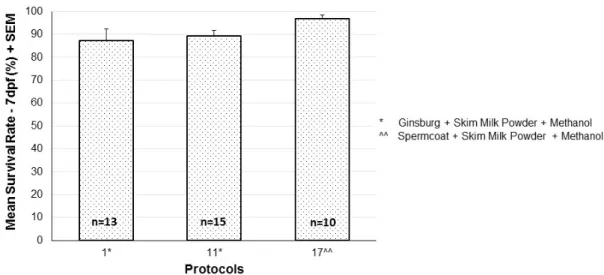
De 23 protocolos testados (n = 201), foram escolhidos para criopreservação de esperma de peixe-zebra os que apresentaram melhores resultados tendo em conta a percentagem de recuperação de linhas, a taxa média de fertilização, a taxa média de sobrevivência das larvas e a taxa média de malformações. Desta forma, foram selecionados dois protocolos com combinações de extender e crioprotector distintas.
Em relação aos métodos complementares utilizados na otimização do protocolo de criopreservação, a estimulação hormonal das fêmeas com 17α,20β-DHP traduziu-se claramente numa melhoria na qualidade e quantidade dos oócitos, sendo um passo muito importante na otimização deste protocolo; a quantificação da concentração de esperma é útil apenas com amostras translúcidas, no entanto indicou que a concentração de esperma não pode ser correlacionada com a taxa de fertilização; o protocolo otimizado de FIV (fertilização in vitro) é, no momento, um serviço prestado pela Plataforma de Peixes da Fundação Champalimaud.
Paralelamente à otimização da criopreservação de esperma, foi otimizado um protocolo de transgénese usando o sistema de transposão Tol2 com microinjeção in vitro de
viii oócitos seguido pela técnica de FIV otimizada anteriormente. Com este método, a percentagem de fundadores transgénicos de uma linha transgénica estável foi de 66.67%, muito superior à observada em microinjeção no estádio de zigoto. Foi ainda realizada uma análise da viabilidade do esperma em peixes alimentados com duas dietas comerciais (Skretting Gemma® e SparosZebrafeed®) tendo como conclusão que ambas as dietas são semelhantes.
ix Summary
As new genetically modified lines are being generated the challenge of maintaining this vast number of zebrafish increases. Therefore, cryopreservation of sperm has been considered a good option for the long-term storage of genetic material, thus reducing costs inherent to its maintenance.
The traditional cryopreservation method and vitrification are the most used methods to cryopreserve cells. Vitrification has many primary advantages and benefits over the other methods, such as no ice crystal formation through increased cooling rates. This way, the concentration of cryoprotectant agents used can be less, decreasing its toxic effect in the cells.
The lack of standardization and coherent expectable results in previous studies were the major causes for the development of a simple and consistent method at the Champalimaud Fish Platform. This way, as the main goal of this thesis, an easy, cheap and reliable protocol procedure was developed and optimized, designated by ultra-fast freezing. This optimized method for zebrafish sperm cryopreservation will have a very important impact in the zebrafish community.
The best protocols of 23 tested (n=201) were chosen for zebrafish sperm cryopreservation according to the percentage of line recovery, mean fertilization rate, mean larvae survival rate, and mean malformation rate. This way, two protocols with different combinations of extender and cryoprotectant were selected.
Regarding the complementary methods for the ultra-fast freezing method, female stimulation with 17α,20β-DHP was clearly an improvement in the oocyte quality and quantity and a very important step in the optimization of this protocol; quantification of sperm concentration is useful only with transparent samples but indicated that sperm concentration can´t be correlated with the fertilization rate; IVF (in vitro fertilization) optimized protocol is at the moment a state-of-the-art service in the Champalimaud Fish Facility.
In parallel with sperm cryopreservation optimization, a transgenic protocol was optimized using the Tol2 transposon system with oocyte in vitro microinjection followed by the optimized IVF technique previously optimized. With this method the percentage of germline transgenic founders was 66.67%, a higher percentage than the one observed in
x one-cell stage microinjection. It was also performed a sperm viability analysis using fish fed with two dietary regimens currently commercialized (Skretting Gemma® and SparosZebrafeed®), concluding that both feedings are equal.
xi Index of contents Acknowledgments ... V Resumo ……….………... VII Summary ………... IX I. Introduction ………... 1 II. Aims ……... 3
III. Literature review ...………... 5
1. Gamete production ...……….………... 5
2. Biophysics of zebrafish sperm …...……… 6
3. Cryopreservation ………...……….. 7
3.1. Embryo, oocyte and primordial germ cells cryopreservation ...………… 8
3.2. Sperm cryopreservation ………... 10
3.2.1. Cryopreservation solutions ………... 11
3.2.2. Freezing……….. 12
3.2.3. Thawing and activation……….………...…………. 14
3.2.4. In vitro fertilization ……… 14
IV. Methods ………...……….. 17
1. Zebrafish and husbandry procedures ………... 17
2. Techniques directly involved in sperm cryopreservation ………... 17
2.1. In vitro fertilization ………... 17
2.2. Female hormonal stimulation ... 19
2.3. Sperm ultra-fast freezing ……… 20
2.4. Sperm thawing and reconstitution...………. 21
2.5. Determination of sperm concentration ………. 22
3. Technological procedures performed in parallel with sperm cryopreservation optimization……… 22 3.1. In vitro oocyte injection ………... 23
3.1.1. Microinjection ………... 23
3.1.2. Transient expression ………... 24
3.1.3. Screening for stable expression ……… 24
3.1.4. Establishment of stable transgenic lines ……….. 24
3.2. Sperm viability analysis taking into account two dietary regimens ……….. 24
xii
3.2.2. Statistical analysis ……… 25
3.2.3. Feeding regimens ……… 25
V. Results………. 27
1. Techniques directly involved in sperm cryopreservation ………... 27
1.1. In vitro fertilization ……… 27
1.2. Females hormonal stimulation ……….. 28
1.3. Sperm ultra-fast freezing ……… 29
2. Technological procedures performed in parallel with sperm cryopreservation optimization ………... 34 2.1. In vitro oocyte injection ………... 34
2.1.1. Microinjection ……….... 34
2.1.2. Primary screen ………. 35
2.1.3. Germline transmission ………. 37
2.2. Sperm viability analysis taking into account two dietary regimens ……….. 38
VI. Discussion and conclusions ………... 39
1. In vitro oocyte injection ……… 39
2. Sperm viability taking into account different dietary regimens ……….. 41
3. Importance of complementary procedures for sperm cryopreservation optimization ………... 41 4. Sperm ultra-fast freezing .……… 42
Bibliography ………...………. 49
xiii Index of figures Figure III.1……… 6 Figure IV.1………... 17 Figure IV.2………... 17 Figure IV.3………... 17 Figure IV.4………... 20 Figure IV.5………... 20 Figure IV.6………... 21 Figure IV.7….………... 22 Figure IV.8………... 23 Figure V.1……….. 27 Figure V.2……….. 28 Figure V.3……….. 29 Figure V.4……….. 30 Figure V.5……….. 31 Figure V.6……….. 32 Figure V.7……….. 32 Figure V.8……….. 33 Figure V.9……….. 36 Figure V.10……… 37 Figure V.11……… 38 Figure V.12……… 38
xv Index of tables
Table IV.1……… 26 Table V.1……… 35
xvii List of abbreviations, acronyms and symbols
17α,20β-DHP - 17α,20β-Dihydroxy-4-pregnen-3-one BSMIS - Buffered sperm motility inhibiting solution CPA - Cryo Preservation Agent
DMA - N,N-dimethylacetamide DMF - N,N-Dimethylformamide dpf – Days post fertilization E3 - Embryo medium FIV - Fertilização in vitro
FTIR - Fourier transform infrared spectroscopy GOI - Gene of interest
HBSS300 - Hanks’ balanced salt solution at an osmolality of 300 mOsmol/kg hpf – Hours post fertilization
IVF – In vitro fertilization LN2 – Liquid nitrogen Lp - Water permeability N2- Nitrogen
PGCs - Primordial germ cells RFP - Red fluorescent protein RT – Room temperature SG – Spermatogonia
SOP – Standard operations procedure UAS - Upstream Activating Sequence Vb - Osmotically inactive component
1 I. Introduction
Sperm cryopreservation is a technique involving many steps including sample collection, sperm extension, cryoprotectant selection, cooling, storage, thawing, and viability detection (Tiersch 2000). Its success can be assessed by in vitro fertilization and production of live offspring. Protocols for sperm cryopreservation can vary due to species-specific differences in sperm size, shape, and biochemical characteristics. In sperm cryopreservation, Cryo Preservation Agents (CPAs) are additives necessary for protection against freezing damage due to intracellular ice crystal formation and excessive dehydration (Yang et al. 2007).
Using genome manipulation techniques, biomedical research has been creating thousands of new mutant strains of mice and zebrafish, the two main vertebrate animal models. For mice, the number of strains is so big that it has become impossible in terms of cost and space to maintain more than a fraction as breeding colonies. Consequently, an increasing number and proportion of strains are being maintained by cryopreservation of their germplasm (Mazur et al. 2008). Zebrafish is by now the second most used animal model in biomedical research and the increasingly fast generation of new transgenic and mutant fish lines in recent years (Clark et al. 2011, Howe et al. 2013, Varshney et al. 2013, Ata et al. 2016) urges for simple and efficient cryopreservation programs. The development of more effective, reproducible, easier and cheaper methods of sperm cryopreservation not only guarantees safe preservation of the genotypes but also addresses the inevitable space limitation to maintain live strains in fish facilities thus limiting research.
With recent improvements in the methodology, cryopreservation of spermatozoa in zebrafish is quickly becoming the favoured method for archiving animal lines, leading to much less ‘‘front-end’’ work required for safely storage. One disadvantage of sperm cryopreservation is that only one haploid gamete is preserved. However, the gamete cryopreservation remains the most used technique for the archiving and shipping of valuable animal models (Du et al. 2010).
There are two main cryopreservation techniques used for sperm conservation, the slow equilibrium freezing cryopreservation (traditional method) and vitrification. The most commonly used cryopreservation method relies in sperm being slowly frozen and then stored in liquid nitrogen. However, as in humans and rodents, this technique has drawbacks, including loss of motility and vitality, and membrane damage. For zebrafish, published
2 protocols are ponderous, with multiple steps rendering them error-prone. More recent studies have demonstrated that the use of French straws can improve this method (Yang et al. 2009), however a programmable freezer is very expensive which can be a limitation to many zebrafish facilities. Vitrification on the other hand involves suspending cells in sufficiently high concentrations of mixtures of CPAs that, in combination with sufficiently high cooling and warming rates, prevent the cells and the surrounding medium from undergoing ice formation during cooling or warming (Mazur et al. 2008). Vitrification is a freezing method with several advantages over the traditional cryopreservation method, including a significant increase in sperm motility in humans and rodents (Kasai & Mukaida 2004). In the latest tests in zebrafish, vitrification consisted of freezing primordial germ cells, however this method had very low success rates and entailed quite laborious steps. Simultaneously, there were suggestions that cryopreservation of these cells can have negative consequences in gametogenesis due to hypermethylation (Riesco & Robles 2013).
3 II. Aims
Due to the inherent problems associated to the slow equilibrium freezing in cryopreservation (traditional method) and vitrification, it is thus becoming urgent to have a better, easier and functional method for zebrafish sperm cryopreservation. To achieve this goal it was decided to develop an ultra-fast freezing method. This is an intermediate freezing method that, to the best of our knowledge, had not been adopted yet in zebrafish. This new method for zebrafish sperm cryopreservation was developed based on previous murine and zebrafish protocols. Therefore, the main goal of this project was to develop and optimize a new sperm cryopreservation protocol that would be easily reproducible not requiring excessive training nor specific skills, not too laborious and as cheap as possible making this procedure accessible for all the zebrafish community. More importantly, we wanted a protocol as more independent as possible from individual sperm quality in order to have more balanced success rates.
To assess the success of the procedure, fertilization rate was used as a quality check. In order to have a reliable fertilization rate, the in vitro fertilization technique itself was optimized using several approaches.
5 III. Literature review
1. Gamete production
In contrast to other vertebrate groups, reproduction in fishes exhibits great diversity and many original features. Reproductive strategies are as diverse as the adaptations to numerous aquatic environments that are found in fishes. This diversity may concern sexuality, spawning, and parental behavior, sensitivity to environmental factors, and specific features of gametogenesis (Jalabert, 2005 in Bone & Moore 2008). Knowledge of fish reproduction and life history is important. The prospects of breeding fish requires an understanding of reproductive physiology, breeding behavior, and genetics (Purdom, 1993 in Bone & Moore 2008). Because fish have evolved to live in diverse environments, there are substantial differences in fish morphofunctional characteristics. Fish have had to develop adaptation mechanisms to survive in diverse environmental conditions, and as a result spermatozoa from fish species demonstrate significant differences in their reactions to cryopreservation protocols. For example, there is a striking difference in post-thaw survival of reproductive cells of marine and freshwater species. Sperm of marine species were successfully cryopreserved and reported (Blaxter 1953 in Stoss 1983 and in Agarwal 2011, Tsai & Lin 2012) soon after the discovery of the first cryoprotectant, whereas the cryopreservation of freshwater fish gametes was more challenging and took longer to achieve (Graybill & Horton 1969, Moczarski 1977, Stein & Bayrle 1978 in Stoss 1983, Tsai & Lin 2012).
The fish male yields several hundreds of billions of spermatozoa per year per kg of body weight, or more than 100x106/g of testis per day, which is 10 times higher than production recorded in mammals. Sperm concentration, also very high, is between 10 and 40x109 spermatozoa/ml of sperm in trout and pike, 7x10⁹ in coregones, 14x109 in carp, and 10x109 and 30x109 in the perch. However, only a part of these spermatozoa can be collected in some species during the reproductive period, the rest remain in the testis where they are gradually reabsorbed. Two original features characterize the spermatozoa physiology of most of the studied species: immotility in the genital tract and extremely short lifespan after motility is triggered. Immotility can be due to the presence of a specific ion in the seminal fluid but other factors, such as elevation of osmotic pressure or sucrose may also inhibit motility (Bellard 1988 in Alavi & Cosson 2006). Female fecundity is generally high, depending on the species and the mode of reproduction. Fecundity or egg size in the same species may also depend on the time of reproduction, which, in turn, depends on seasonal differences in food availability for the parents (Bagenal 1971 in Demartini & Sikkel 2006). After ovulation,
6 the fertilization ability of the ova remaining in the female genital tract declines more or less rapidly (varying from hours to weeks), depending on the species (Bellard 1988 in Alavi & Cosson 2006).
2. Biophysics of zebrafish sperm
Cell cryopreservation cannot be improved without proper basic physiological knowledge. Successful cryopreservation of germplasm must consider intrinsic biophysical properties (e.g., water and cryoprotectant permeability, osmotic tolerance limits, intracellular ice nucleation) to maximize survival (Rall 1993 in Hagedorn et al. 2009). It is important to understand and avoid the mechanisms by which sperm is damaged or destroyed during cryopreservation.
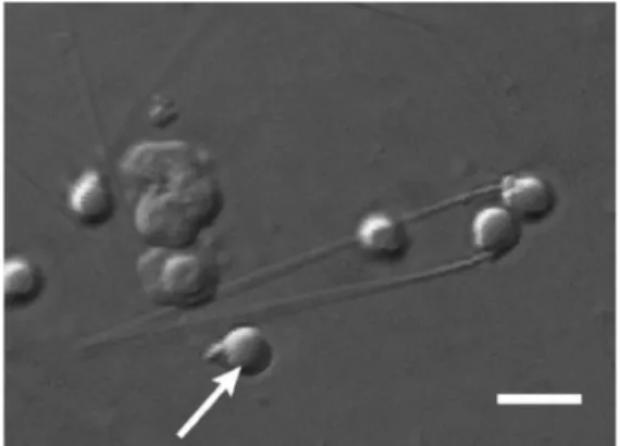
Zebrafish sperm have small, round heads and a smaller midpiece (Figure III.1) that together are approximated as a prolate spheroid with an average major and minor axes of the combined head and mid-piece of 2.2± <0.1 µm (SEM) and 1.9 µm ± <0.1 m, respectively, an average tail length of 27.6 ± 0.5 µm with an average tail thickness of 0.4 ± <0.1 µm in diameter (Hagedorn et al. 2009).
Figure III.1: Zebrafish sperm morphology. Zebrafish sperm display a prolate head and mid-piece (arrow), and a tail ~30 µm in length. Bar = 4 µm (in Hagedorn et al. 2009).
Sperm volume measured in the Coulter electronic particle counter is 12.1 µm3. The optimal osmotic range for these cells is from approximately 200 to 600 mOsm/kg (Hagedorn et al. 2009).
Zebrafish sperm have a low osmotically-inactive component (Vb) (determines how much osmotically-active water is in the cell) compared to that of sperm from most mammals. Vb of zebrafish is 0.37 compared to 0.61 for mouse, for example. Water permeability (Lp) for zebrafish sperm is low, approximately 30-fold lower than in mammalian sperm, as might be
7 predicted for a cell that must function in a hypotonic environment (e.g., fresh water) when fertilizing an egg (Hagedorn et al. 2009).
3. Cryopreservation
The preservation of biological material in a stable state is a fundamental requirement in biological/medical science, agriculture, and biotechnology. It has enabled standardization of experimental work over time, has secured lifesaving banks of cells and tissue ready for transplantation and transfusion at the time of need, and has assured the survival of critical germ plasm in support of programs for the conservation of species. Cryopreservation is one of the widely accepted and preferred techniques for achieving long-term storage, and has been applied to an increasingly diverse range of biological materials. (Day & Stacey 2007). Advantage of cryopreserving the fish semen is well established. It is not only a useful management tool, it offers several benefits such as stock protection due to outbreak of diseases, natural disaster, or over exploitation. Other application of cryopreservation includes stable supply of sperm for optimal utilization in hatchery production, and easy stock transportation among hatcheries, with stocks being maintained more economically and effectively, and for laboratory experiments providing experimental material for advanced studies such as gene transfer (Agarwal 2011).
Although the basis for many methodologies is common, many laboratories lack expertise in applying correct preservation and storage procedures and many apply out-dated or inappropriate protocols for storing samples or cultures (Day & Stacey 2007).
Cryopreservation is the use of very low temperatures to preserve structurally intact living cells and tissues (Agarwal 2011, Pegg 2007). This technique allows virtually indefinite storage of biological material without deterioration over a time scale of at least several thousands of years (Agarwal 2011, Mazur 1985 in Hiemstra et al. 2005), but probably much longer. Important progress in cryobiology was achieved in the second half of the previous century (Hiemstra et al. 2005).
In cryopreservation, cells are suspended in a suitable solution, cooled, stored in liquid nitrogen, warmed to room temperature, and returned to a physiological solution. During each step of this process, cells are at risk for various types of damage. The primary injury is that caused by the formation of intracellular ice during cooling and warming (Kasai &
Mukaida 2004). The biological effects of cooling are dominated by the freezing of water,
which results in the concentration of the solutes that are dissolved in the remaining liquid phase. Rival theories of freezing injury have envisaged either that ice crystals pierce or tease apart the cells, destroying them by direct mechanical action, or that damage is from secondary effects via changes in the composition of the liquid phase. Cryoprotectants, simply
8 by increasing the total concentration of all solutes in the system, reduce the amount of ice formed at any given temperature, but to be biologically acceptable they must be able to penetrate into the cells and have low toxicity. Both damaging mechanisms are important, their relative contributions depending on the cell type, cooling rate, and warming rate (Pegg 2007).
Whether freezing is permitted (conventional cryopreservation) or prevented (vitrification), the cryoprotectant has to gain access to all parts of the system. However, there are numerous barriers to the free diffusion of solutes (e.g., membranes), and these can result in transient, and sometimes equilibrium, changes in compartment volumes, which can be damaging. Hence, the processes of diffusion and osmosis have important effects during the introduction of cryoprotectants, the removal of cryoprotectants, the freezing process, and during thawing (Pegg 2007).
It was not until 1948 that a general method was discovered allowing for the freezing of many types of animal cells with subsequent restoration of structure and function (Agarwal 2011). In 1949, Polge et al. published their landmark paper in which they showed that the inclusion of 10–20% of glycerol enabled the spermatozoa of the cock to survive prolonged freezing at –80°C. Regarding fish, several different approaches were initially tested including storage of fish sperm in medium saturated with different gases (Holtz et al 1976), preservation of sperm at temperatures above zero (Ginzburg 1968), as well as in the frozen state (Blaxter 1953) and drying (Zell 1978). However, to date, low-temperature preservation has proven to be the most effective approach, with the first successful cryopreservation of fish sperm being reported by Blaxter in 1953 (Agarwal 2011).
3.1. Embryo, oocyte and primordial germ cell cryopreservation
Teleost primordial germ cells (PGCs), as the embryonic precursors of gametes, have tremendous importance in the fields of developmental biology and aquaculture. They are an optimal cell type to be cryopreserved because they conserve both paternal and maternal genomes. Moreover, recent studies have demonstrated the competence and suitability of these cells for surrogate production. The implementation of these technologies provides precise control over many relevant reproductive aspects, for example, PGCs or spermatogonias (SGs) xenotransplantation that could offer a solution for the management of species with reproductive failures, or for those species with long maturation periods (Robles et al. 2017).
In zebrafish, PGCs have been cultured and marked by using a transgenic line that expresses red fluorescent protein (RFP) under the PGC-specific vasa promoter (optimizing
9 the culture conditions by counting the number of fluorescent cells) (Fan et al. 2008). The possibility of generating PGCs in vitro would represent a powerful tool in biotechnological research and aquaculture. The number of PGCs is limited in embryos and cell proliferation is difficult to achieve once they are cultured in vitro. Generation of these cells in vitro would provide the means to increase the number of cells per embryo which could be important for germplasm banking purposes, it would be a source of cells for surrogate production and it would increase the possibility to genetically manipulate embryonic cells (easier to transfer than PGCs) in teleosts (Robles et al. 2017).
Both embryo and oocyte cryopreservation have not been successful in fish yet. Most of the cryopreservation protocols have been developed for sperm, which disregards the female genome (Robles et al. 2017). Teleost oocytes and embryos have intrinsic biophysical properties that make their cryopreservation difficult. To minimize cryodamage and maximize survival rates, water exchange and cryoprotectant influx have to be studied and tested, taking into account that both these factors are influenced by membrane permeability, osmotic tolerance limits, surface-volume ratio, and yolk amount. (Hagedorn et al 1997). All previous attempts to cryopreserve fish embryos have been unsuccessful so far. The analysis of the permeability parameters of the zebrafish embryo predicted that a major site for lethal cryodamage would occur within the yolk compartment (Robles et al. 2017). Presumably, without sufficient cryoprotectant entering the yolk, damaging ice-crystals will form (Hagedorn et al 1997 in Robles et al. 2017). Therefore, protocols for fish embryo vitrification with removal of some yolk have been studied (Higaki et al. 2013). Regarding oocytes, after cryopreservation, these cells require post-thaw in vitro maturation and fertilization, thus a functional protocol for germ cell survival would not necessarily guarantee successful production of zygotes. Therefore, cryopreservation of PGCs and spermotogonia represent an important tool in gene banking until fish embryo cryopreservation is successfully achieved (Robles et al. 2017).
Besides the cryoprotectant exposure time (Higaki et al. 2009), combination of external and internal CPAs at lower doses (Robles et al. 2007), microencapsulation with dissociated cells (Kasai & Mukaida 2004), or incorporation of antifreeze protein effects, other strategies, such as yolk removal (Higaki et al. 2013) have been used to examine the effects of this partial removal and cryoprotectant mixtures on the viability and the differentiation ability of cryopreserved zebrafish PGCs. All of these studies have provided important advancements for PGC cryobanking and have established a basis for future improvements.
10 3.2. Sperm cryopreservation
The low-temperature preservation method has been applied widely and has become not only a routine tool in aquaculture for fish hybridization and selective breeding, but also an important tool in programs of biodiversity and preservation of endangered species. Gamete banks of rare or almost extinct species were created (Harvey et al. 1998) with the objective of protecting endangered species. The technique has also found applications in research programs for maintaining laboratory animals and sperm of more than 200 species of fish have been successfully cryopreserved (Agarwal 2011, Blesbois & Labbé 2003 in Hiemstra et al. 2005, Rana & Gilmour 1996). However, despite the extensive number of studies that have been undertaken there is still ambiguity (great variability and poor reproducibility) in the data reported in the literature, primarily because of lack/poor standardization of methodology and data analysis. Zebrafish sperm cryopreservation protocols are far from optimized and further improvement is necessary.
Because oocyte or embryo cryopreservation has not yet been successful in zebrafish (Guan et al. 2010, Lin et al. 2009, Robles et al. 2017), sperm freezing is currently the best option for genetic resource banking. Although there are many protocols available for low-temperature storage of sperm of freshwater fish (Agarwal 2011, Kopeika & Novikov 1983, Tiersch & Mazik 2003) there is much work still to be done to improve this technology. Most of the events associated with freezing are a result of the osmotic properties of cells. The cellular damage during the freezing process is all due to the osmotic shock, intracellular ice formation, increased intracellular concentration of solutes and solution effects (Agarwal 2011). In general, approximately 40–90% of spermatozoa from freshwater species are usually damaged after cryopreservation, whereas only 10–20% of spermatozoa are damaged in marine species (Tiersch & Mazik 2003). Post-thaw survival of fish sperm is strongly predetermined by their sensitivity to osmotic changes in extracellular media (Tiersch & Mazik 2003), leading to a generally low (0–30%) average post-thaw motility (Morris et al. 2003, Yang et al. 2007). Decrystallization is pointed out as the main cause for cryodamage, and rewarming is the critical step for post-thaw survival (Mohammad et al. 1997, Medrano et al. 2002).
To develop reliable protocols of cryopreservation for fish spermatozoa, individual fish and species-specific properties must be taken into consideration (Agarwal 2011). A cryopreservation protocol needs to have several optimized steps such as gamete collection, stimulation of maturation (used in specific cases), gamete storage and equilibration, freezing, storage in liquid nitrogen, thawing, and fertilization. Due to the multiple steps and their
11 interactions, errors at each step can accumulate and lead to considerable losses of viable cells. Thus, careful attention should be given to the numerous details at each step, and care should be taken to reduce or eliminate sources of uncontrolled variation (Leibo, 2000).
3.2.1. Cryopreservation solutions
The solutions used for sperm cryopreservation include:
(1) Extender to storage the gametes and retain the functional capability and fertilizing ability of sperm by controlling pH, osmolality, ion concentration, and in some cases, the supply of energy (Stoss & Holtz 1981 in Yang & Tiersch 2009). The choice of appropriate extender depends on the species. The osmolality of the extender solution is one of the most important factors in preparation of an appropriate extender (Kopeika et al. 2007). Specifically, for zebrafish the most common extenders are Ginsburg Ringer’s fish solution with skim milk powder, Hanks’ balanced salt solution (HBSS300) and buffered sperm motility inhibiting solution (BSMIS) and all of them generally function well to retain fertility of post-thaw sperm (Harvey et al. 1982, Morris et al. 2003, Draper et al. 2004, Yang et al. 2007).
(2) Cryoprotectant solution. The absence of an ideal cryoprotectant, makes selection of a common single cryoprotectant difficult for different species. However, the optimal cryoprotectant can be determined empirically. The addition of cryoprotectants interacts with the membranes to make them more flexible and thus reduces damage due to solution effects. Thus, the basic principle of cryopreservation is to cause cell dehydration and eventually concentrate the cytosol with minimum injury so that ice crystallization in the cytosol is minimized during cooling in liquid nitrogen (Agarwal 2011). The concentration of cryoprotectant usually varies in the range between 5 and 12% (v/v) (Kopeika et al. 2007). Better cell protection can be achieved by employing higher concentrations, but this has to be balanced with toxicity effects of the cryoprotectant (Yang & Tiersch 2009). The addition of non-penetrating agents, such as sucrose, is generally considered to be beneficial. However, direct mixing of fish sperm with cryoprotectants inevitably leads to the death of all cells (Scott & Baynes 1980 in Gwo et al. 2009). The level of dilution of the cryoprotectant medium is equally important and it is species sensitive (Agarwal 2011, Lahnsteiner 2000). The most commonly used cryoprotectants for fish sperm cryopreservation are permeating ones, such as dimethyl sulfoxide (DMSO), ethylene glycol, methanol, ethanol, glycerol, and N,N-dimethylacetamide (DMA) and non-permeating ones, such as egg yolk, milk, and proteins (Kopeika et al. 2007, Yang & Tiersch 2009). For zebrafish, the toxicity of DMSO, N,N-dimethyl acetamide, methanol, and glycerol at concentrations of 5, 10, and 15% have been evaluated with sperm cells. Glycerol was the most toxic, and was eliminated for sperm cryopreservation. The other three chemicals have been used for sperm cryopreservation,
12 and analysis of post-thaw motility have shown that methanol at a concentration of 8% was the best choice (Yang et al. 2007). This was also the choice in two earlier studies (Harvey et al. 1982, Draper et al. 2004). In addition, DMA (10%) was used as a cryoprotectant for zebrafish sperm (Morris et al 2003), but the fertilization level after thawing (9–14%) was lower than that observed (28–51%) when methanol was used (Harvey et al. 1982, Draper et al. 2004, Yang et al. 2007). Cryoprotectant permeabilities are in the range expected for most sperm (~10-4 cm/min). Sperm suffers changes in cell volume caused by dimethylsulfoxide, however 10% methanol and 10% N,N-dimethylformamide do not cause any changes in cell volume as they enter and exit the cell (Hagedorn et al. 2009).
Hagedorn et al. 2009 analysed sperm membranes with Fourier transform infrared spectroscopy (FTIR), which is an established tool for biophysical characterization of cell membranes (Crowe et al. 1989) extremely sensitive to changes in lipid conformational order, and allows for measurement of membrane fluidity and lipid organization in intact cell membranes. The FTIR data suggest that freezing zebrafish sperm without cryoprotectant causes membrane damage and large-scale lipid reorganization.
Cold shock damage has been directly linked to lipid phase transitions that cause the sperm membrane to become transiently leaky, thereby compromising membrane integrity (Agca et al. 2005, Arav et al. 2000, Drobnis et al. 1993 in Hagedorn et al. 2009). Ice formation and changes in osmotic pressure are the major causes of spermatozoa damage during cryopreservation, and the ability of sperm plasma membrane to resist structural damage during cryopreservation may be related to the type of fatty acids in the spermatozoa plasma membrane and the strength of the bonds between membrane components (Agarwal 2011) causing irreversible phase separation (clustering) and rearrangement of membrane components in sperm (DeLeew et al 1990, Hotl & North 1984 in Hagedorn et al. 2009). During chilling, the key is to minimize the number and cooperativity, or sharpness, of lipid phase transitions, thus keeping the membrane fluid and structurally intact (Hagedorn et al. 2009).
3.2.2. Freezing
The freezing step can be achieved using different methods:
(1) Freezing in vapour-phase liquid nitrogen, which implies placing vials or straws above the liquid nitrogen horizontally on a rack at a predetermined position. The position of the sample and the time of exposure at that position depend on the sample volume, type of container, and temperature at that position;
(2) Freezing in alcohol baths. Similar results can be obtained by freezing sperm in cold baths that are capable of maintaining a set temperature;
13 (3) Freezing in dry-ice using Falcon type tubes as a support placed deep in dry ice; (4) Controlled-rate cooling using programmable freezers.
Freezing in liquid nitrogen vapour or in dry ice are more practical methods compared to a controlled-rate freezer and are also the closest easily achieved approximation to an exponential cooling regime (Agarwal 2011, Harvey et al. 1982, Kopeika et al. 2007).
Cooling rate is a crucial factor in sperm cryopreservation because it affects the osmotic and pH balance of intracellular and extracellular solutions during freezing. Theoretically, with an excessively slow cooling rate, osmotic equilibrium is maintained, and much of the freezable water leaves the cell resulting in excessive dehydration; with an excessively fast cooling rate, little or no freezable water leaves the cell, and thus large intracellular crystals can form, causing damage to the cell. Ideally, a balanced situation allows survival when the cooling rate is fast enough to minimize the time of exposure to concentrated solutions and yet is slow enough to minimize the amount of intracellular ice formation. Optimum cooling rates vary with different cryoprotectants and the physiology of sperm cells from different species (Yang & Tiersch 2009).
The packaging of samples for freezing and storage is also important to standardize the cooling rate, and to assure proper sample identification. Currently, several different kinds of containers have been used, such as plastic cryovials, glass tubes and ampules, and plastic straws. The different materials and shapes of these containers result in different heat transfer properties during freezing and thawing. Even for the same style of container, differences can exist with products from different manufacturers, which can result in variation of cooling or thawing rates (Yang & Tiersch 2009).
Currently, reported sperm cryopreservation protocols on zebrafish include:
(1) Freezing in glass capillary tubes on dry ice using methanol and powdered skim milk as cryoprotectants (Harvey et al. 1982) and various adaptations of this method (Westerfield 1995, Ransom & Zon 1999, Brand et al. 2002, Draper et al. 2004) (Agarwal 2011). Morris et al. 2003 were unable to reproduce the results reported by Harvey et al. 1982 and its updated protocols;
(2) Freezing in 1.5 mL cryotubes on dry ice using N,N-dimethylacetamide as cryoprotectant (Morris et al. 2003, Berghmans et al. 2004) or methanol (Draper & Moens 2009);
(3) Freezing in 0.25 mL French straws with a programmable freezer using methanol as cryoprotectant (Yang et al. 2007). Bai et al. 2013 were unable to repeat the success with
14 methanol when samples were frozen in 0.25 mL French straws with a programmable freezer. DMA was found to be worse than methanol in the straw freezing method (Yang et al. 2007). (4) Cryomicroscopy which allows real time observation of the entire freezing and thawing process, tracking throughout all temperature regions events such as cell motility, membrane integrity, and ice formation status. Cryomicroscopy yields a two-step freezing protocol that employs a faster cooling rate of 25°C/min initially from 4 to 30°C, and then a slower cooling rate of 5°C/min from 30 to 80°C before plunging into liquid nitrogen for permanent storage. For freezing, the equipment is a controlled-rate freezer and sperm is suspended in 8% DMSO in 0.25 ml French straws (Bai et al. 2013). Bai et al. 2013 tested the efficiency of this method through sperm motility observation.
3.2.3. Thawing and activation
By the time sperm is thawed and ready to be used for fertilization, it has gone through a series of stresses. Therefore, special care has to be taken during handling of sperm after thawing and pure water should not be used as an activator for cryopreserved-thawed sperm during fertilization (it affects functional activity of weak sperm cells post-thaw). Improved activation will be attained in activation media that have higher osmolality than pure water. However, the increase in osmolality in the activating medium has to be within the range that is safe for the eggs (Kopeika et al. 2007). In zebrafish, once activated by
hypotonic osmolality, sperm have a short burst of motility (30 s to 5 min) (Yang et al. 2007).
Theoretically, the process of thawing is the reverse of freezing, and thus the damage that can occur during cooling can also occur during warming, primarily through formation of ice crystallization between −40 and 0°C (Leung 1991, Til et al. 2016). Therefore, it is usually desirable to rapidly thaw cryopreserved samples to minimize the period of crystal propagation (termed “recrystallization”) (Yang & Tiersch 2009).Studies on optimization of the thawing regime have demonstrated that the best thawing regime for 1–2 mL vials is using a water bath between 33 and 40°C (Kopeika et al. 2007, Draper & Moens 2009).
3.2.4. In vitro fertilization
In vitro fertilization (the collection of spermatozoa and ova and their mixing together in various media that keep spermatozoa motile) is commonly carried out in several freshwater species, such as salmonids, cyprinids and acipenserids. The eggs of most teleosts are fertilized externally, which means that after passing through the micropyle, the spermatozoon penetrates the cytoplasm. Traditionally, fresh water (or sea water for marine species) is used as the medium in which the male and female gametes are mixed. However,
15 fresh water is not a very favourable medium because hypotonic shock causes the sperm structure to deteriorate in several minutes and the egg is activated quickly. These problems can be avoided by using as media various saline solutions of different composition, depending on the species. These media prevent sperm deterioration, prolong slightly the duration of motility or limit it, and prevent or defer the cortical reaction. The length of gamete survival is an important factor to consider in carrying out artificial reproduction (Agarwal 2011, Bellard 1988 in Alavi & Cosson 2006).
For zebrafish, artificial fertilization protocols have been established with fresh sperm, and can be directly modified to provide fertilization analysis of cryopreserved sperm (Westerfield 1995 in Yang & Tiersch 2009). Eggs can be collected by squeezing of females, held in isotonic buffer to retain fertility, and then be mixed with a sperm suspension for fertilization. After mixing of sperm and eggs, fresh water needs to be added to activate gametes for fertilization. Fertilization and hatching are determined by assessing the percentage of developing embryos or hatched fry (Yang & Tiersch 2009).
17 IV. Methods
1. Zebrafish and husbandry procedures
Several strains of zebrafish that are widely used in biomedical research: wild-type AB and TU, the Nacre (mitfa-/-) mutant and several transgenic lines were used. Housing and husbandry of all animals were performed according to Martins et al. 2016. Fish were housed in 3.5L tanks at a maximum density of 10 fish per liter or housed in 3.5L tanks with a divider dividing males and females at a maximum density of 4 fish per liter (depending on the purpose). Fish did not stay divided for more than two consecutive weeks.
The feeding regimen implemented consisted of feeding the fish 3 times per day (one time with live Artemia nauplius and two times with powder Skretting® Gemma Micro 500).
2. Techniques directly involved in sperm cryopreservation 2.1. In vitro fertilization
Spawning trials are necessary to test the fertility of cryopreserved sperm. This process includes a series of steps: egg collection, holding of eggs prior to fertilization, thawing of cryopreserved sperm, mixing of the sperm and eggs, gamete activation, fertilization confirmation, hatching of fertilized eggs, and offspring harvest (Yang & Tiersch 2009).
Large numbers of synchronously developing embryos can be obtained by in vitro fertilization (IVF). IVF can be performed when experiments depend on synchronized embryos, when natural mating doesn´t occur or for line recovery of a cryopreserved sperm sample. Fertilization with cryopreserved sperm requires slightly more time and equipment that fertilizion using fresh sperm.
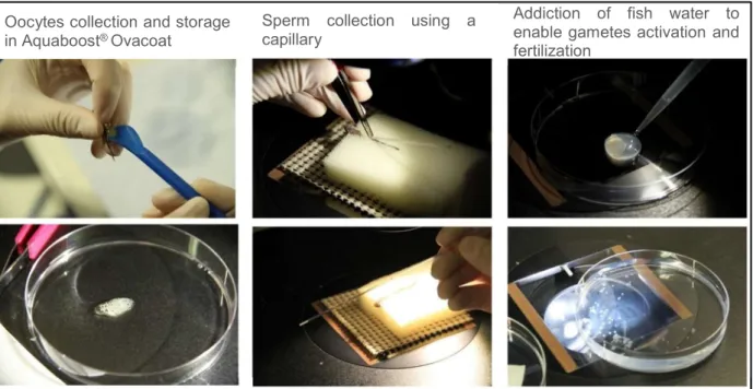
Gametes were collected from breeding adults by gentle pressure and stored in commercial solutions (Aquaboost® Ovacoat for oocytes and Aquaboost® Spermcoat for sperm) in order to retain fertility (figure IV.1). These commercial solutions can maintain sperm inactive for 24 hours on ice and oocytes for 30 minutes at room temperature (CUG 11/13 2015), allowing for the collection of several or pooled samples and the time optimization of the procedure.
Briefly, after collection, the gametes were mixed together in a petri dish, fish water was added to the egg-sperm mixture and fertilization took place very rapidly in 20 to 60 seconds (figure IV.1). After 1 minute, the sperm is no longer active. Embryos were then placed in the
18 incubator at 28°C with a photoperiod of 14h:10h/light:dark. Between 15 and 24hpf the success of fertilization was checked by observing on the stereoscope the development of the embryos (figures IV.2 and IV.3, appendix A).
Figure IV.1: Zebrafish in vitro fertilization main steps. Procedure currently in use at the Champalimaud Fish Facility.
Figure IV.2: Zebrafish embryo development at 6hpf, 80x magnification. F – fertilized embryo; NF – egg not fertilized; NA – egg not activated.
Figure IV.3: Zebrafish embryo development at 24hpf, 40x magnification. NF – eggs not fertilized; NA – eggs not activated.
F NF
NF NA
Oocytes collection and storage in Aquaboost® Ovacoat
Sperm collection using a capillary
Addiction of fish water to enable gametes activation and fertilization
19 The Champalimaud IVF protocol was optimized based on several established protocols (ZFIN, ZIRC, UCL and Cryogenetics). The standard operations procedure (SOP) is described in appendix B.
For the IVF tests, good breeders were selected. Four month-old females and males were incrossed at least three times in a weekly basis and housed in pairs in 1.1L tanks. The couples that produced, during the three mating episodes, at least 50 to 200 fertilized embryos were considered good breeders and transferred to a 3.5L tank with a divider dividing males and females at a maximum density of 4 fish per liter.
Males were squeezed every two weeks, females were squeezed every two/three weeks. Fish were used in a rotating system as being separated for too long reduces their productivity and can trigger inflammation caused by egg accumulation.
IVF was performed in the first three hours after the room lights turn on. Alternatively, when female hormonal stimulation was used, IVF was performed approximately 6-7 hours after the room lights turn on.
2.2. Female hormonal stimulation (adapted from Tokumoto et al. 2009).
Female egg quality is an important factor in successful IVF but it can sometimes be a challenge to obtain good usable eggs. Not all females are fecund but 1/3 of squeezed females can have good quality eggs (whereas males will give sperm >90% of the times) (Pegg 2007).
After the first trials, in vitro fertilization started to be hampered by the quantity and quality of oocytes. In a first attempt to understand how many females were needed to guarantee enough oocytes to perform IVF and to reduce costs and human resources, the percentage of high quality oocyte clutches (yellow oocytes, with no white debris indicative of degradation, dry and sticking together) within two wild type strains was determined for four months. For this study, only data from wildtype AB and TU females was collected.
In order to trigger zebrafish oocyte maturation and ovulation, to make sure the female population has higher quality clutches, the natural teleost maturation-inducing hormone, 17alpha,20beta-dihydroxy-4-pregnen-3-one (17α,20β-DHP) was used as a tool for artificially inducing ovulation in zebrafish (adapted from Tokumoto et al. 2009). 100nM 17α,20β-DHP was administered directly to the water where zebrafish were housed. This direct administration allows the steroid hormone to penetrate the fish body, causing an effect upon oocyte maturation. Besides being an enhancer of good egg clutches, this technique allows IVF procedures to have an extra daily working window of 2-3 hours, as fertilizable
20 oocytes can be obtained up to 4-5 hours upon addition of 17α,20β-DHP. The respective SOP is described in appendix C.
2.3. Sperm ultra-fast freezing
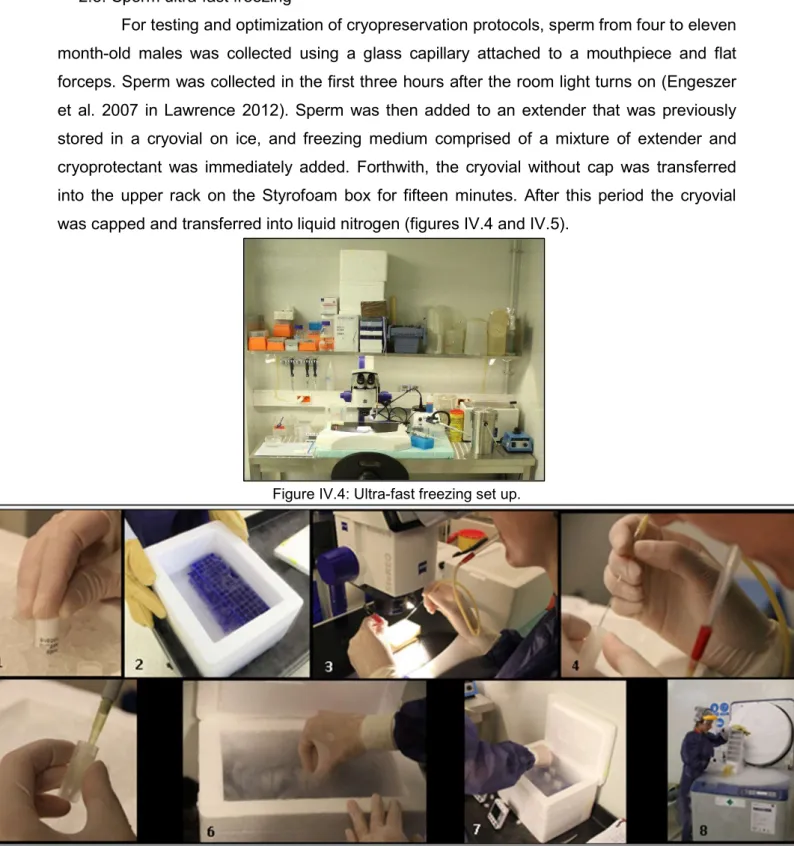
For testing and optimization of cryopreservation protocols, sperm from four to eleven month-old males was collected using a glass capillary attached to a mouthpiece and flat forceps. Sperm was collected in the first three hours after the room light turns on (Engeszer et al. 2007 in Lawrence 2012). Sperm was then added to an extender that was previously stored in a cryovial on ice, and freezing medium comprised of a mixture of extender and cryoprotectant was immediately added. Forthwith, the cryovial without cap was transferred into the upper rack on the Styrofoam box for fifteen minutes. After this period the cryovial was capped and transferred into liquid nitrogen (figures IV.4 and IV.5).
Figure IV.4: Ultra-fast freezing set up.
Figure IV.5: Zebrafish sperm ultra-fast freezing main steps currently in use at the Champalimaud Fish Facility. 1 - Extender storage on ice; 2 - Styrofoam box with LN2; 3 – Sperm collection; 4 – Sperm addition to extender; 5 – Freezing medium addition; 6 – Cryovial transfer without cap; 15 minutes in N2 vapour; 7 – Capping of cryovial and transfer to LN2; 8 – Storage in N2 chamber at ~-180°C.
21 The container used in the cryopreservation procedure was not, as in classical protocols, an expensive metal container. A Styrofoam box filled with 5000 to 6500cm3 of liquid nitrogen was used as the cryopreservation main set up. The ultra-fast freezing set up was comprised of the Styrofoam box and two centrifuge tube racks - one placed at the bottom of the box completely immersed with liquid nitrogen and the other one for cryovials placed on the top of the other rack. In general, the total volume of solutions and sperm cryopreserved was 23µL per cryovial. The respective SOP is described in appendix D.
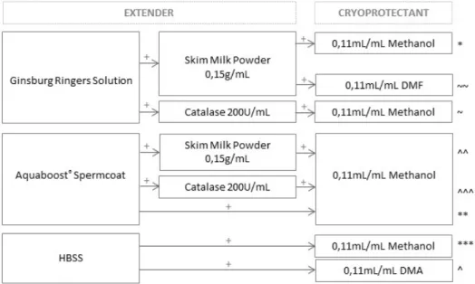
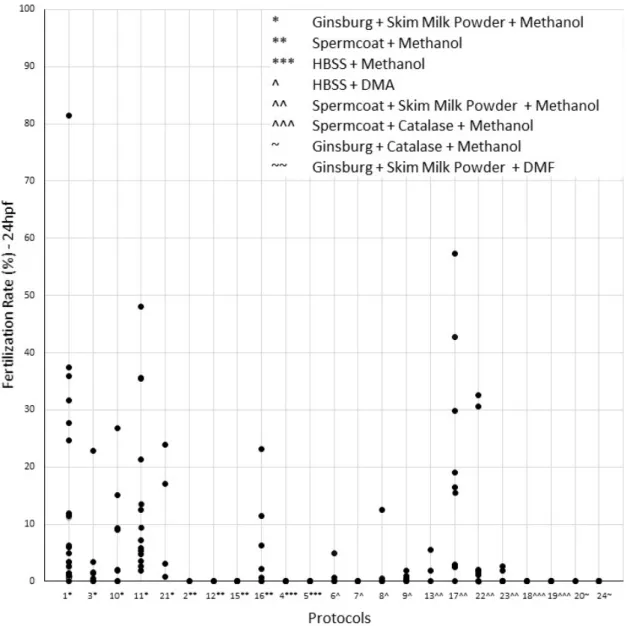
All tested cryopreservation solutions (extender and cryoprotectant) are schematized in figure IV.6. The freezing medium was a mixture of extender and cryoprotectant.
Figure IV.6: Tested combinations of extenders and cryoprotectants used in the sperm ultra-fast freezing tests. Symbols *, **, ***, ^, ^^, ^^^, ~ and ~~ identify each combination.
2.4. Sperm thawing and reconstitution
Examination of sperm viability generally includes evaluation of morphology, membrane integrity, motility, ability to bind oocytes, and fertilization. Motility is the most widely used assay, but fertilization is considered to be the most informative (Yang & Tiersch 2009). Fertilization rate was achieved by performing in vitro fertilization and was determined by assessing the percentage of developing embryos or hatched fries at 24hpf. Survival and malformations rates (defined as larvae viability rate) were evaluated between 6 and 7dpf.
A water bath was set at 33°C in order to pre-heat the extender and to use this temperature to thaw the sperm samples. Meanwhile, oocytes were collected from selected
22 females into petri dishes and stabilized in the extender Aquaboost® Ovacoat in order to prevent their activation.
Each cryovial was removed from liquid nitrogen, the cap opened, the liquid nitrogen tipped out and quickly immersed ~1/2 way into 33°C water bath for 15 seconds. 70µL of pre-heated extender was immediately added and mixed by gently pipetting up and down 2-3 times. A 200µL pipette tip was used with the tip cut off to prevent spermatozoa damage (figure IV.7). Before adding the sperm to the oocytes, Aquaboost® Ovacoat had to be gently removed from the oocytes to ensure proper contact between the two gametes. Immediately, 750μL of fish water was added and incubated 5 minutes at room temperature. After incubation, the petri dishes were filled with fish water and transferred to an incubator at 28°C with a photoperiod of 14h:10h/light:dark. The respective SOP is described in appendix D.
Figure IV.7: Sperm thawing main steps. 1. Cryovial removal from N2; 2 – Immersion of ~1/2 cryovial into a 33°C water bath; 3 – Addition of pre-heated extender and mix.
2.5. Determination of sperm concentration
A sperm suspension for analysis was obtained sampling 2 to 3μL of sperm with extender. Sperm concentration was estimated with a microspectrophotometer (NanoDrop®, Thermo Scientific, Wilmington, DE). The protocol for microspectrophotometry analysis was previously established by Tan et al. 2010. The standard equation used was:
Y = (3x10⁸) X - 3x10⁷
with “X” being defined as the absorbance measured at 400nm. Briefly, 1μL sample of sperm suspension was loaded onto the lower pedestal of the NanoDrop, and absorbance was measured at 400 nm.
3. Technological procedures performed in parallel with sperm cryopreservation optimization Despite sperm cryopreservation being the main goal of this study, other zebrafish procedures that needed optimization of the IVF technique were performed, showing the general importance of this technique. Therefore, those methods and results were included as part of this project.
23 3.1. In vitro oocyte injection
This subchapter is part of an on-going collaboration for the development of new transgenic procedures between the Fish Facility and CR researchers, in this particular case, the Orger Lab. A newly developed method involving reproduction techniques, namely IVF, was tested.
3.1.1. Microinjection
In vitro oocyte injection to improve transgenesis was performed based on the protocol described by Xie et al. 2016. Low efficiencies of genome editing and germline transmission result in time-intensive and laborious screening work, thus the optimization of strategies to minimize screening is crucial.
The standard method of introducing foreign genomic material into zebrafish is by microinjecting it in fish embryos immediately after fertilization, at one-cell stage. This new method consists on injecting oocytes instead of embryos (figure IV.8) and incubating them in a specific oocyte storage medium to significantly improve efficiencies of genome editing and germline transmission. According to Xie et al. 2016, micro-injecting zebrafish oocytes substantially improved genome editing efficiency, especially for sgRNAs with low targeting efficiency, providing an efficient alternative to decrease the time frame of generating heritable mutants in zebrafish by using the CRISPR/Cas9 system.
Figure IV.8: Comparison between oocyte injection (OI), 20x magnification, and one-cell stage injection (OCSI), 60x magnification.
The method developed in this study had slight differences to the original published method (Xie et al. 2016), namely the type of construct injected (the Tol2 system was used to generate random insertions of a transgene as opposed of using the CRISPR/Cas9 system to generate gene-specific mutants) and some variations in the storage medium. Usually, the agarose plates used for embryo injections are prepared with E3 medium but for oocyte injection this this would cause the oocytes to be activated immediately which is not
24 compatible with the procedure. Therefore, the medium in which the agarose is melted needs to be an extender, in this case Ginsburg Ringers Solution (also used in the sperm cryopreservation process described in subchapter 2.3.) or 90% Leibovitz’s L-15 medium with L-glutamine and 0.5mg mL 1 bovine serum albumin, pH 9.0 (Xie et al. 2016).
The constructs injected - HuC:GFF 10xUAS:GCaMP6sEF05 and HuC:GCaMP6sEF05 - were chosen by the Orger Lab.
The SOP is described in appendix F. Injection was done in a TU zebrafish strain with Nacre (mitfa+/-) background. At 24hpf plates were cleaned off of dead embryos and the medium was replaced with fresh E3.
3.1.2. Transient expression
At 48hpf all injected larvae were screened for pan-neuronal transgenic transient expression on a PentaFluor-equipped V8 stereoscope (Zeiss) using a blue filter with a spectrum range from 400–460nm.
Positive transgenic larvae (stringently selected: enough labelled cells and expected panneuronal expression pattern for the HuC/elav3 promoter) were raised according to the conditions published by Martins et al. 2016.
3.1.3. Screening for stable expression
Fish were raised until adulthood (~3 months or when sexual maturity was observed). Fish were then individually crossed with the driver line Isl3:Gal4(+/+) and the offspring was screened on a PentaFluor-equipped V8 stereoscope (Zeiss) using a blue filter with a spectrum range from 400–460nm. Animals with positive progeny were kept. From the positive progeny (F0, founders), stable transgenic lines (F1) were established.
3.1.4. Establishment of stable transgenic lines
F1 stable lines were generated crossing each Tg(10xUAS:GCaMP6sEF05) F0 (founder) fish with the driver line HuC:GFF. At 48-72hpf the expression pattern was checked (strong fluorescence in the brain tectum) and the positive larvae raised.
3.2. Sperm viability analysis taking into account two dietary regimens
This subchapter is part of a parallel study performed by the Champalimaud Fish Facility in which the impact of different feeding regimens on zebrafish survival, growth and reproductive performance was studied. Two feeding regimens were created using combinations of two commercial dry feeds - Skretting® Gemma Micro and Sparos®
25 Zebrafeed - and one live feed (rotifers). Results from this study were submitted for publication.
3.2.1. Sperm viability
To evaluate the influence of the dietary regimen on reproduction, both embryo development and sperm viability were studied.
In order to have more information to analyse, fertilization rate assays were performed. Thus fertilization was achieved by performing in vitro fertilization and was determined by assessing the percentage of developing embryos or hatched fries 24hpf. Oocytes from 5-10 month-old wild type fish (AB and TU strains) were collected by female squeezing. Clutches from each female were divided in two petri dishes and held in an extender Aquaboost® Ovacoat to retain fertility. In order to calculate the fertilization rate depending on the diet on which each male had been fed, sperm from two individual regimens (dietary groups 2 and 5; Table IV.1) was mixed to the half clutch for in vitro fertilization. After mixing, fresh fish water was added to activate gametes in order for fertilization to occur. Males were squeezed during four months every two weeks (6-10 month-old fish). Survival and malformation rates (defined as larvae viability rate) were evaluated between 6 and 7dpf.
3.2.2. Statistical analysis
All data was analysed with the IBM SPSS Statistics software (v. 23, IBM Corp.,
Chicago, IL). As assumptions were not verified
(normality and homoscedasticity), the Wilcoxon-Mann-Whitney test was used followed by pairwise comparison. Results were considered statistically significant for p-values <0.05.
3.2.3. Feeding Regimens
Skretting® Gemma®Micro 150, 300 or 500 was provided to animals with <30 dpf, 30-90 dpf, and >30-90 dpf, respectively. Similarly, Sparos® Zebrafeed® 200-400 and 400-600 was used to feed fish with ages 30-90 dpf, and >90 dpf, respectively. Regardless of the dietary regimen, all fish were fed 4x/day between 8 dpf and 60 dpf, 3x/day from 60 to 90 dpf and 2x/day from 90 dpf onwards. On weekends and holidays, they were fed 1x/day with the dry feed of the corresponding experimental group. All tanks were given a similar volume of rotifer solution and a similar amount of dry feed. The density of each tank was readjusted at 30 dpf, to ensure uniformity and reduce the influence of density on the results. Two experimental dietary groups were designed using different combinations of dry feeds (Skretting® Gemma® Micro or Sparos® Zebrafeed®) and a live feed (type “L” saltwater rotifers) (table IV.1).
26
Table IV.1: Feeding regimens for dietary groups 2 and 5
Dietary Group
6 - 30dpf 30 - 60dpf 60dpf - end of study
Live Feeding Dry Feeding Live Feeding Dry Feeding Dry Feeding
2 2x Rotifers 2x Gemma Micro 150 1x Rotifers 2x Gemma Micro 300 2x Gemma Micro 500
27 V. Results
1. Techniques directly involved in sperm cryopreservation 1.1. In vitro fertilization (IVF)
The first IVF test was performed using two strains of wild type zebrafish, AB and TU, one mutant strain, Nacre (mitfa -/-), and one transgenic strain, Tg(Isl3:Gal4). AB, TU and Nacre fish had been previously selected as good breeders and were four to six months old. The transgenic fish were nine months old and were randomly selected. Before using the gametes for IVF, a quality assessment was done. Only yellow, with no white debris indicative of degradation, dry and sticking together oocytes and bright white sperm were used.
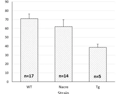
The mean fertilization rate at 24hpf was 69.58±22.42%, 70.72±27.30% and 38.66±8.10% for wild type, Nacre and Isl3:Gal4, respectively (figure V.1).
Figure V.1: Mean fertilization rate ± standard error at 24hpf using the IVF technique, according to the strain. x̄(WT)=69.58±22.42%, n=17; x̄(Nacre)=70.72±27.30%, n=14; x̄(Tg)=38.66±8.10%, n=5. WT is a pool of AB and TU lines.
Eight IVF trials were performed using oocytes in phase I and II of maturation and with some percentage of white debris or using sperm that had low motility (<50% of spermatozoa without motility observed after activating the gametes with fish water). All the experiments resulted in no fertilized embryos (data not shown), further emphasizing that samples with these characteristics should never be used. Indispensable characteristics for success in IVF are oocytes with a transparent to yellow colour, a firm and rounded chorion and clutches should form an aggregation of cells within a transparent fluid. Sperm should be opaque white and present at least with 80% of motility.
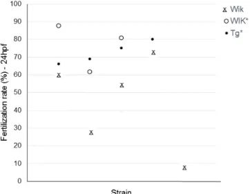
28 Some of the fish lines existing at the Champalimaud Fish Facility lost their natural mating behaviour due to either inbreeding through many generations (a requirement for the particular type of behavioural research for which they are used) or to aging. Therefore, in these circumstances, IVF has to be performed to maintain the line. One extra step in the initial optimized IVF procedure was added, in which the natural teleost maturation-inducing hormone (17α,20β-DHP) was used. Figure V.2 demonstrates that IVFs performed with oocytes collected from females stimulated with the hormone 17α,20β-DHP have higher fertilization rates.
Figure V.2: Percentages of fertilization success for every IVF performed due to loss of natural mating behaviour. Wik lines lost the natural mating behaviour due to inbreeding; Tg line (HuC:GFF UAS:mCherry) lost the natural mating behaviour due to fish aging. * refers to IVFs performed with oocytes from females stimulated with the hormone 17α,20β-DHP.
1.2. Female hormonal stimulation
In order to understand how many females would be sufficient to have always enough oocytes to perform IVF, a study with a total number of 473 females was performed. The mean of females with good clutches (yellow, with no white debris indicative of degradation, dry and sticking together oocytes) after squeezing was 33.23±27.51% and 19.50±24.29% for AB and TU, respectively (figure V.3A). Similar data from females stimulated with 17α,20β-DHP, shows a much higher number of females producing good quality clutches, 66.34±32.53% and 50.82±26.54% for AB and TU, respectively (figure V.3B).