Article
0103 - 5053 $6.00+0.00
*e-mail: [email protected], [email protected]
Spectroscopic Evidence of Photodegradation by Ultraviolet Exposure of
Tris(8-hydroxyquinoline) Aluminum (Alq
3) Thin Films
W. R. Brito,*,a G. Aráujo,a W. G. Quirino,b C. Legnani,b Y. Angulo,c
M. Cremonab,c and M. L. M. Rocco*,a
aInstituto de Química, Universidade Federal do Rio de Janeiro, 21949-900 Rio de Janeiro-RJ, Brazil
bDivisão de Metrologia de Materiais (Dimat), Inmetro, 25250-020 Duque de Caxias-RJ, Brazil
cDepartamento de Física, Pontifícia Universidade Católica do Rio de Janeiro,
22453-970 Rio de Janeiro-RJ, Brazil
A degradação extrínseca de diodos orgânicos emissores de luz (OLEDs) permanece como uma questão crucial, especialmente no que se refere à degradação, devido à exposição à luz. Poucos estudos existem e faltam dados a respeito dos produtos de fotodegradação e respectivos mecanismos, responsáveis pela extinção da luminescência. Com o objetivo de investigar os mecanismos de degradação causados pela exposição à luz de ilmes inos de Alq3 usado com
sucesso como camada transportadora de elétrons e material emissivo na fabricação de OLEDs, a fotodegradação foi monitorada em função da irradiação com luz UV através das técnicas de fotoabsorção e de fotoemissão nas bordas 1s do carbono, do nitrogênio e do oxigênio bem como nas bordas 2s e 2p do alumínio. A inluência da exposição à luz foi simulada com o emprego de três comprimentos de onda diferentes: 254, 365 e 307 nm, os dois primeiros correspondendo aproximadamente a máximos de absorção no espectro de UV do Alq3. Após exposição, todos os espectros apresentam um decréscimo nos sinais de fotoabsorção e de fotoemissão. Entretanto, enquanto a radiação de 307 nm causa mudanças menores nos espectros de NEXAFS, modiicações mais acentuadas são observadas para a exposição a 254 e 365 nm. Medidas de fotoemissão para ilmes inos de Alq3 não-irradiado e irradiado com luz de 307 nm também foram conduzidas.
Embora o sinal correspondente ao alumínio mantém-se praticamente intacto, mudanças mais signiicativas são observadas nas intensidades e deslocamentos dos picos nas bordas 1s do carbono, do nitrogênio e do oxigênio.
The extrinsic degradation of organic light emitting diodes (OLEDs) remains a critical issue especially concerning degradation due to exposure to light. Very few studies exist and little is known about the related photodegradation products and mechanisms, responsible for quenching of luminescence. In order to gain insight into the degradation mechanisms caused by light exposure of Alq3 thin ilms, used successfully as electron transport layer and emissive material in the
fabrication of OLEDs, we have monitored UV photodegradation through synchrotron radiation-based photoabsorption and photoemission techniques at the carbon, nitrogen, and oxygen 1s edges as well as at the aluminum 2s and 2p edges. The inluence of light exposure was simulated using three different wavelengths, namely 254, 365 and 307 nm, the irst two nearly corresponding to absorption maxima in the UV spectrum of Alq3. After exposure all spectra show decrease of the
photoabsorption and photoemission signals. However, while irradiation at 307 nm causes lesser changes in the total electron yield NEXAFS spectra, strong spectral modiications are observed for 254 and 365 nm exposures. Core level photoemission measurements from non-irradiated and irradiated Alq3 thin ilms at 307 nm were also performed. While the Al peaks maintained almost
intact, changes in peak intensities as well as shifts for carbon, nitrogen and oxygen are much more dramatic.
Introduction
Due to their applications in many different areas and manufacturing simplicity, organic light emitting diodes (OLEDs) represent a promising research line in the
development of new optoelectronic devices.1 However, the
degradation of the materials that are commonly used in the OLED fabrication is still the principal weakness of these devices. Many efforts have been made in order to understand the factors that have inluenced the different degradation mechanisms of the OLEDs. One of the principal focus of these studies was to investigate their stability against radiation originated from the environment, such as intense sunlight. To better understand the degradation processes, which occur when organic materials used in OLEDs are submitted to intense radiation a series of investigations
using spectroscopic techniques were carried out.2-9
Among the materials used in OLED fabrication,
tris-(8-hydroxyquinoline) aluminum (Alq3) is one of the most
attractive electroluminescent material, used successfully as electron transport and emissive material in organic devices, and therefore has stimulated many studies focusing in a better comprehension of the degradation processes due to
light exposure. Thangaraju et al.10 studied the inluence of
light in the photoluminescence from Alq3 thin ilms. They
conirmed that the photoluminescence (PL) originates from its two geometrical isomers, namely, facial and meridional,
which coexist in amorphous Alq3 thin ilms. PL from both
isomers decreases for increasing light exposure time leading to the creation of luminescent quencher (carbonyl group)
in the Alq3 thin ilm. Kumar et al.11 employed ellipsometry,
photoluminescence and infrared spectroscopies to study the inluence of light exposure on the optical properties of
Alq3 ilms. Attenuation of the photoluminescence of Alq3
ilms exposed to light is discussed by Thangaraju et al.12
using luorescence, FT-IR, mass, MALDI-ToF-MS and 1H
and 13C NMR spectroscopy. They reported that the affected
molecule contains the carbonyl group, which acts as luorescence quencher. Recent theoretical and experimental
IR work pointed out to the degradation of Alq3 through
formation of carboxylate groups bound to aluminum after
UV irradiation at 307 nm.13 Comparative infrared studies
among exposed and non-exposed Alq3 ilm revealed a
band at 1697 cm-1 that corresponds to vibrational band of
carbonyl group.13
In the present work photoabsorption (NEXAFS, Near-edge X-ray absorption ine structure) and core-level photoemission (XPS, X-ray photoelectron spectroscopy) techniques were employed to investigate the degradation of
Alq3 thin ilms as a function of UV irradiation. Exposure at
254 nm and 365 nm causes larger degradation, as observed
by total electron yield NEXAFS results at the carbon and nitrogen 1s edges. Core level photoemission measurements at 307 nm show dramatic changes for nitrogen, oxygen and carbon photoelectric signals.
Experimental
We have employed the photoabsorption and photoemission techniques using synchrotron radiation provided by the SGM beamline from the Brazilian Synchrotron Light Source (LNLS), located at Campinas,
São Paulo in order to investigate the stability of Alq3 thin
ilms under UV irradiation. The beamline is mounted with a spherical grating monochromator for VUV and Soft X-ray spectroscopy (250-1000 eV), which may furnish an
energy resolution (E/∆E) better than 2000 and a spot size
on the sample of about 0.5 × 0.5 mm2. The experimental
set-up includes a sample manipulator and a concentric hemispherical electron energy analyzer housed in an UHV
chamber with a base pressure of 10-8 mbar. Alq
3 in form
of thin ilms was attached directly to the sample holder using a conducting double-side tape. In addition, silver glue was used in the corner of the sample to assure for a good electrical contact.
Films of Alq3 (Sigma Chem.) of 100 nm thickness were
thermally deposited onto silicon at room temperature in a high vacuum system (LEYBOLD, UNIVEX 300 model).
The base pressure was 9 × 10⁻⁶ mbar during evaporation and
the rate of deposition was in the range of 0.1 to 0.3 nm s-1.
By this procedure Alq3 is likely to form an amorphous ilm.
The ilms were exposed to UV light for 14-16 h on
non-controlled atmosphere using different UV lamps at λ = 254,
306, 365 nm wavelengths. The exposure conditions are as
follows: UV lamp at λ = 254 nm (ca. 85 µW cm-2) during
16 h; UV radiation using a He-Xe lamp at λ = 307 nm
(ca. 420 µW cm-2) during 14 h; and UV lamp at λ = 365 nm
(ca. 500 µW cm-2) during 14 h. The 254 and 365 nm
wavelengths nearly correspond to absorption maxima in the
UV spectrum of Alq3.14 The distance between source and
sample ranged from 0.5 to 30 cm. At the end of the irradiation periods no more luorescence emission could be observed. All irradiations were carried out in ambient atmosphere.
Acquisition of NEXAFS data was performed at the
carbon and nitrogen K-edges by measuring simultaneously
the total electron yield and the photon lux by an Au grid monitor, placed in the path of the incident beam. The energy calibration was performed by taking the values found in
the literature for Alq3.15 NEXAFS spectra were measured
sample charging was observed throughout the experiments. The energy calibration was performed by taking the values
from the literature.4 The incidence angle was about 40o
with respect to the sample surface. Photoabsorption and photoemission spectra were measured before and after UV exposure. Complementary studies for the 307 nm irradiated
sample were performed using an Al Kα X-ray source.
Results and Discussion
Near-edge X-ray absorption ine structure (NEXAFS)
spectra of Alq3 thin ilms, exposed and non-exposed to UV
light at three different wavelengths (254, 307 and 365 nm)
were obtained following carbon and nitrogen K-edges. These
spectra, covering from 280 to 310 and 390 to 430 eV photon energies, respectively, are depicted in Figures 1 and 2. NEXAFS spectra were obtained at normal incidence
measuring the drain current at the sample, i.e. in the total
electron yield mode (TEY), which from all detection modes using electrons is the least surface sensitive method.
At the C 1s edge the non-exposed Alq3 NEXAFS
spectrum depicted in Figure 1 shows four features, which corresponds to electronic transitions from the C 1s electrons
to unoccupied molecular orbitals of π* and σ* symmetries in
good agreement with previous studies.15 This spectrum was
not normalized to the reference current, which may account
for the higher intensity of the σ* resonances as compared
to the π* peaks. Due to the nine chemically inequivalent
carbon atoms, the C K-edge spectrum is broader with a
complex lineshape. As suggested by Yokoyama et al.16 the
irst two features are due to transitions from various C 1s levels to mixed states of LUMO and LUMO+1. The LUMO lies primarily on carbon (C1 and C3) and N atoms of the
pyridyl ring.4,17 LUMO+1 contains only carbon character.16-18
According to DeMasi et al.17 the irst absorption feature (at
285.2 eV) is primarily due to transitions to the C3 LUMO, which is in a simple C-H environment, located oppositely to the nitrogen atom, with some contribution from transitions to the LUMO+1 state on the C4 atom. The broad character of the second absorption peak (at 286.4 eV) can be accounted for by the fact that it contains not only transitions to the
LUMO+1 but also to the LUMO and LUMO+2 states.17
The third structure at 288.8 eV is assigned to transitions to
LUMO+2 and LUMO+3 followed by many σ* states. After
UV irradiation at 307 nm only a small reduction in peak intensity was observed. When irradiating using the 254 nm
UV lamp the spectrum changes drastically; we observe a
reduction of the second peak intensity, which has also strong
contribution from C 1s →π* (LUMO+1) transition. Since the
LUMO+1 is formed solely by carbon atoms,16-18 this suggests
that irradiation at 254 nm induces intense degradation at the carbon atoms moieties.
Figure 1. C K-edge NEXAFS spectra of Alq3 thin ilms before (solid line) and after 307 nm (dash line) and 254 nm (dot line) irradiation.
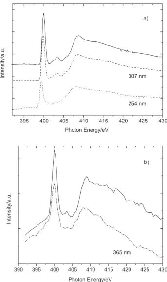
In Figure 2a we plotted the N 1s NEXAFS spectra of
as-prepared and light exposed at 307 and 254 nm Alq3
ilms. The spectrum shows basically three features and a shoulder at the lower energy side of the second structure, corresponding to excitation of localized 1s core level electron into unoccupied molecular orbitals, in good agreement with
earlier studies.15,16 The narrow character of the peaks in the
N K-edge spectrum as compared to the C K-edge spectrum
is related to the fact that the three nitrogen atoms from Alq3
are chemically equivalent. According to Yokoyama et al.16
the intense irst peak at 400.4 eV is due to transition of N 1s electron to the LUMO orbital, the second to LUMO+3 and the shoulder to LUMO+2 state. The broad band above
the π* transitions may be comprised of σ* states. Since the
nitrogen atom does not participate in the LUMO+1 orbital there is no transition to this orbital at the N 1s edge. While 307 nm irradiation causes again less inluence on the TEY NEXAFS spectrum, light exposure at 254 nm changes it drastically. The most important points here are the drastic
reduction of the irst π* peak and the complete loss of the
following two features. These results are also observed
when exposing the Alq3 ilm to 365 nm light as shown in
Figure 2b, obtained during a single-bunch run of the storage ring. As can be seen from Figures 2a and 2b the intensity of the peaks changes appreciably upon irradiation, showing evidence of strong degradation associated with the loss of the nitrogen atoms due to the incident radiation. This loss
disrupts the conjugated π system and therefore the charge
conduction through the molecule is interrupted, causing damage to the performance of the device. Similar results
were obtained when exposing Alq3 ilms during 30 min to
broadband synchrotron radiation in a simulation of intense
sunlight exposure.8
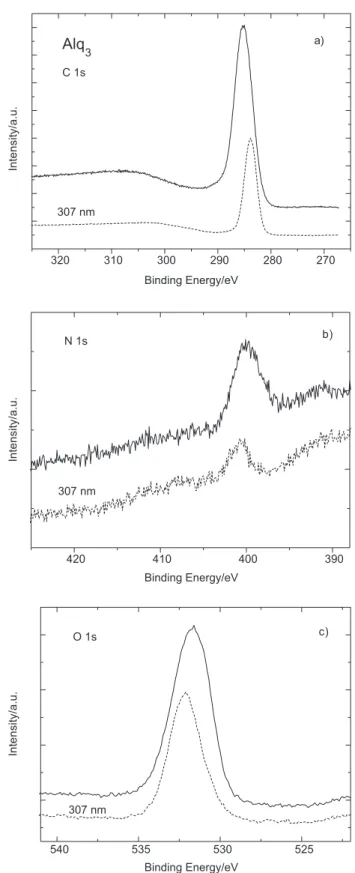
In order to further analyze evidence of degradation
presented at the Alq3 thin ilms upon UV irradiation
XPS studies were performed at the carbon, nitrogen, and oxygen 1s edges as well as at the aluminum 2s and
2p edges for Alq3 irradiated at 307 nm. XPS spectra are
displayed in Figures 3(a-c) and 4(a-b) before (solid line) and after (dash line) UV irradiation. The carbon 1s XPS spectrum shows basically a decrease in intensity together with a shift of the photoelectric peak to lower binding energy after irradiation. This behavior can be explained by the formation of new carbon containing species, as
observed before for Alq3 ilms after white light (i.e., full
non-monochromatized synchrotron radiation) exposure.4,8
The damage produced by the irradiation affects also the oxygen moieties (see Figure 3c), where besides the reduction of the photoelectric signal, a shift toward higher
binding energy is observed for exposed Alq3. Recent
theoretical and experimental IR work on exposed and
non-exposed Alq3 ilm revealed a band at 1697 cm-1 that
corresponds to vibrational band of the carbonyl group
after 307 nm irradiation.13
The N 1s XPS results show that the nitrogen signal decreases drastically after irradiation. A reduction of 80% was measured for the N 1s signal. Carbon and oxygen 1s photoemission signals were reduced about 60-65% of the original intensity. The 2s and 2p aluminum peaks presented a reduction less than 5% of the photoelectric signal. Similar
results were obtained by exposing Alq3 ilms to intense
broadband synchrotron radiation.4,8 The loss of nitrogen
followed by damage of the molecular structure may cause
disruption of the conjugated π system and consequently
interruption of the charge conduction through the molecule, degradating the performance of the device.
Conclusions
In order to gain insight into the degradation mechanisms
of Alq3 layer used as electroluminescent material in
OLEDs, synchrotron radiation-based photoabsorption and photoemission techniques at the carbon, nitrogen and
oxygen 1s edges and aluminum 2s and 2p edges were employed at the Brazilian Synchrotron Light Source (LNLS). The unoccupied molecular orbitals (LUMOs) of
Alq3 were probed using near-edge X-ray absorption ine
structure (NEXAFS) in the total electron yield (TEY) mode. NEXAFS results showed remarkable differences
between exposed and unexposed Alq3 when irradiated
by 254 and 365 nm UV lamps as compared to 307 nm irradiation. Attenuation of peak intensities was observed for all irradiations, although for 254 and 365 nm strong degradation was measured for the carbon and nitrogen 1s transitions. This suggests changes in the chemical
environment with damage of the π-system of the molecule
that may cause loss of electroluminescence and electron transport properties, and formation of dark emissive zones. XPS measurements for 307 nm exposed and non-exposed
Alq3 thin ilms were also performed. While the Al peaks
are less affected, changes for carbon, oxygen and especially nitrogen are much more dramatic, what may be related to the breakdown of charge conductivity.
Acknowledgments
Research partially supported by LNLS-Brazilian Synchrotron Light Laboratory, Brazil. The authors would like to acknowledge CNPq, CAPES, and FAPERJ for inancial support.
References
1. Sheats, J. R.; Antoniadis, H.; Hueschen, M.; Leonard, W.; Miller, J.; Moon, R.; Roitman, D.; Stocking, A.; Science1996, 273, 884. 2. Burrows, P. E.; Bulovic, V.; Forrest, S. R.; Sapochak, L. S.;
McCarty, D. M.; Thomson, M. E.; Appl. Phys. Lett.1994, 65, 2922.
3. Popovic, Z. D.; Aziz, H.; Hu, N.; Hor, A.; Xu, G.; Synth. Met. 2000, 111-112, 229.
4. Treusch, R.; Himpsel, F. J.; Kakar, S.; Terminello, L. J.; Heske, C.; van Buuren, T.; Dinh, V. V.; Lee, H. W.; Pakbaz, K.; Fox, G.; Jiménez, I.; J. Appl. Phys. 1999, 86, 88.
5. Curiori, A.; Andreoni, W.; Treusch, R.; Himpsel, F. J.; Haskal, E.; Seidler, P.; Heske, C.; Kakar, S.; van Buuren, T.; Terminello, L. J.; Appl. Phys. Lett. 1998, 72, 1575.
6. Liao, L. S.; Hung, L. S.; Chan, W. C.; Ding, X. M.; Sham, T. K.; Bello, I.; Lee, C. S.; Appl. Phys. Lett. 1999, 75, 1619. 7. Quirino, W. G.; Legnani, C.; Cremona, M.; Reyes, R.; Motta, G.
V.; Weibel, D. E.; Rocco, M. L. M.; J. Braz. Chem. Soc.2008, 19, 872.
8. Quirino, W. G.; Legnani, C.; Batista, L.; Cremona, M.; Motta, G. V.; Weibel, D. E.; Rocco, M. L. M.; De Sousa, E.; Braz. J. Vac. Appl.2006, 25, 1.
9. Chagas, M.; Quirino, W. G.; De Sousa, E.; Cremona, M.; Rocco, M. L. M.; Motta, G. V.; Thin Solid Films 2009, 517, 4461. 10. Thangaraju, K.; Kumar, J.; Amaladass, P.; Mohanakrishnan,
A. K.; Narayanan, V.; Appl. Phys. Lett.2006, 89, 082106, DOI:10.1063/1.2338566.
11. Kumar, S.; Shukia, V. K.; Tripathi, A.; Thin Solid Films2005, 477, 240.
12. Thangaraju, K.; Amaladass, P.; Shanmuga Bharathi, K.; Mohanakrishnan, A. K.; Narayanan, V.; Kumar, J.; Appl. Surf. Sci.2009, 255, 5760.
13. Rosselli, F. P.; Quirino, W. G.; Legnani, C.; Calil, V. L.; Teixeira, K. C.; Leitão, A. A.; Capaz, R. B.; Cremona, M.; Achete, C. A.; Org. Electron.2009, 10, 1417.
14. Garbuzov, D. Z.; Bulovic, V.; Burrows, P. E.; Forrest, S. R.; Chem. Phys. Lett. 1996, 249, 433.
15. Kim, P.-S. G.; Naftel, S. J.; Sham, T. K.; Coulthard, I.; Hu, Y.-F.; Moewes, A.; Freeland, J. W.; J. Electron Spectrosc. Relat. Phenom. 2005, 144-147, 901.
16. Yokoyama, T.; Ishii, H.; Matsuie, N.; Kanai, K.; Ito, E.; Fujimori, A.; Araki, T.; Ouchi, Y.; Seki, K.; Synth. Met.2005, 152, 277.
17. DeMasi, A.; Piper, L. F. J.; Zhang, Y.; Reid, I.; Wang, S.; Smith, K. E.; Downes, J. E.; Peltekis, N.; McGuinness, C.; Matsuura, A.; J. Chem. Phys. 2008, 129, 224705, DOI:10.1063/1.3030975. 18. Amati, M.; Lelj, F.; Chem. Phys. Lett. 2002, 358, 144.
Submitted: June 28, 2010