*e-mail: [email protected]
Chemsensor of NO
2Gas Based on Porphyrin of
5, 10, 15, 20-Tetraphenylporphyrin LB Films and LS Films
Nelício Faria de Sales, Herman Sander Mansur*
Department of Metallurgical and Materials Engineering,
Laboratory of Material and Surface Characterization, Federal University of Minas Gerais,
Belo Horizonte - MG, Brazil
Received: September 24, 2008; Revised: December 10, 2008
The sensitivity of 5, 10, 15, 20-tetraphenylporphyrin (H2TPP) to the presence of NO2 gas in diluted solutions and in Langmuir-Blodgett (LB) and Langmuir-Schaefer (LS) films was investigated by UV-visible spectroscopy. The shift of Soret and Q bands were analyzed and the energies involved were calculated. The exposure of LB porphyrin films deposited onto glass slides to NO2 has performed as an active chemsensor with 7000 ppm gas concentration. Furthermore, the UV-vis dichroism absorption results associated with the Soret bands have given evidence of the tilt angle of the macrocycle related to the substrate. H2TPP in LB film was tilted by an angle of 51 ±5° and in the LS film was tilted by an angle of 36° ±5° indicating the formation of a preferential organization of the molecular films depending on the deposition method.
Keywords: molecular film, Langmuir-Blodgett film, chemsensor, uv-vis spectroscopy
1. Introduction
Tetraphenyl porphyrins or H2TPP are the simplest porphyrins which have been investigated. These porphyrins yield isotherms with an extrapolated area of only 13-17 Å2 per molecule1. This contrasts
with an expected value of 225 Å2 per molecule, if the molecules
lie flat on the water surface, and with an expected value of 90 Å2
per molecule for a vertical packing array of molecules2.For these
macrocyclic molecules, organized supramolecular structures can be formed in monolayers. Such is the case in Langmuir-Blodgett (LB) monolayers and self-assembled monolayers (SAM), in which the macrocycles arrange themselves in a parallel alignment due to the inherent organization of the macrocyclic rings. The presence of double-conjugated bonds attributes the sp2 planar configuration
with several possibilities of π-π* transitions. These structures, referred to as a playing-card model, have been confirmed by AFM measurements of Langmuir and self-assembled monolayers of porphyrins and phthalocyanines2. The substantial attractive π-π
interaction between porphyrin macrocycles may cause significant pre-aggregation prior to the compression of the film forming domains2,3. Furthermore, the limitation of 3D conformation due
to the planar structure restricts the overall mobility on reaching a thermodynamic energy minimum.
Many free-base porphyrins have been studied as potential NO2 sensors because they possess distinctive UV–vis absorption spectra due to their highly conjugated π-electron systems 4.These sensors
reli-ant on changes in the optical properties of the active sensing material, generally operate at room temperature and remain relatively immune to interference effects5.The optical responses toward alcohol vapors
of H2TPP and its derivatives suggested different molecule organiza-tion between spin coated and vacuum evaporaorganiza-tion technique thin films6.Hence, supramolecular assemblies of porphyrins have been
extensively studied for two main kinds of applications: for mimick-ing natural photosynthesis process and as components in molecular devices. The porphyrin orientation and the distance between the rings in monolayer and films are two important parameters for the interactions between the rings7.
Based on the lower than expected area/molecule values, it can be concluded that in general the material must form multilayer stacks in which the structure and stability of the film (collapse 40 mNm–1) is
governed mainly by molecular packing rather than by hydrophilic/ hydrophobic interactions 1-3,7.The exposure of porphyrins to oxidizing
or strong reducing gases induce dramatic change in the UV-visible absorption spectrum (Soret and Q bands) that are associated with
π→π* transitions and provide the basis for a remarkably interesting gas sensing capability8,9.
The organization of supramolecular assemblies of H2TPP films and its derivatives are dependent of subphase temperature and sub-phase modifications2-3,7, such as the addition of different electrolytes7
or changes in pH10. Moreover, the interaction of central metal and
side-group substituents with weak and strong carboxylic acids by UV-vis spectroscopy were previously reported11. The UV-vis absorption
circular or linear dichroism can also be used to estimate the orientation angle of the porphyrin rings with respect to the normal angle of the substrates in Langmuir-Blodgett (LB), Langmuir-Schaefer (LS) and self-assembled films2,12-16.
The goal of the present study was to investigate the optical ab-sorbance behavior of H2TPP in solution and in LB and LS films by UV-vis spectroscopy when exposed to NO2 gas. The characteristics for gas sensing associated with sensitivity and response were analyzed by following the changes in the Soret and the Q bands. The concen-tration of the porphyrin and the subphase pH were used as major parameters for controlling the final aggregation state of molecules for chemsensor applications.
2. Materials and Methods
2.1. Materials
Hy-drochloric acid (Synth) and sodium hydroxide (Sigma Aldrich) were used for pH adjustment. Quartz plates (SPI Supplies,USA) were made hydrophilic by immersion in hydrogen peroxide (Cromato Produtos Químicos Ltda, 30%v/v).
2.2. Measurements of ð-A isotherms and preparation of LB
films at water interface.
The measurements of surface pressure-area (π-A isotherms) were carried out at a temperature of 23 ± 1 °C on a Nima model 311D Langmuir trough with a Wilhelmy plate balance. The trough and the barrier are made of Teflon®. The quality of the spreading solvent and
subphase was tested by compressing the interface after the spreading and evaporation of the solvent. The final surface pressure was always lower than 0.2 mN·m–1 assuring no contaminate residues. Distilled
water further purified by a Milli-Q system (>18.2 MΩ·c) was used as subphase after the pH adjustment.
H2TPP was dissolved in n-hexane/chloroform (in volumetric proportion of 6/4), and the concentration of H2TPP in the solu-tion was 30 µM. The solvents were allowed to evaporate for about 20 minutes and the barrier was compressed at constant rate of 7.8 Å2·min–1.molecule–1.
Langmuir-Blodgett (LB) films were prepared by the immersion of pre-treated hydrophilic quartz plates through the H2TPP monolayer-subphase interface maintained at 18 mN·m–1 for different solution
concentrations. In the sequence, LS films were deposited by manual immersion after extraction of LB film and surface pressure stabiliza-tion was reached. These samples were characterized by Ultraviolet-visible (UV-vis) spectroscopy measurements.
In order to obtain a hydrophilic surface, quartz plates (1.0 x 9.75 x 25.4 mm) were treated with hydrogen peroxide at 60 °C
for 30 minutes and then dried in an oven (at 60 °C for 24 hours). ACD/ChemSketch software (Advanced Chemistry Development, Inc, version 11.01) was used to estimate the value of area/molecule assuming a flat film formation (space-filled models).
2.3. Measurements of UV-vis spectra
UV-vis spectra were recorded on a Perkin Elmer Lambda EZ210 Spectrophotometer. Empty cuvette was used as the reference. The spectra were collected at 200 nm/min over a range of 300-900 nm, resolution within 2.0 nm.
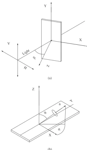
2.4. Molecular orientation of phorphyrin in LB and LS films
The orientation of porphyrin molecules in LB and in LS films was investigated by polarized UV-vis absorption spectroscopy. Figure 2a and Figure 2b show the space coordinates (x, y, z) for expressing the
porphyrin orientation and the direction of the incident polarized light on the substrate.
The optical absorption dichroism was used to study the orienta-tion of the porphyrin rings relative to the normal direcorienta-tion of the substrate by measuring the dichroic ratios Dβ,Dβ = Av /Ah; where
βis the angle of incident light regarding to the x-y plane, usually
β = 0° and 45°; the Av /Ah parameter is calculated through the ratio of absorbance values from the film submitted to polarized light with electric vectors parallel and perpendicular to the dipping direction, respectively2,12. The dipping direction for LB films is generally
con-sidered to be the y-axis.
2.5. Gas sensing test
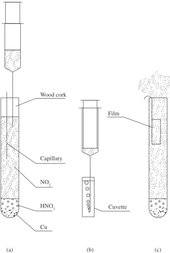
The configuration and setup for NO2 synthesis apparatus is il-lustrated in Figure 3a. Experiments involving the bubbling of gas were conducted using an assay tube (“reactor”) sealed with a wooden cork and two capillary, one for pressure equilibrium and other for the withdrawal of gas with a syringe. The just produced NO2 gas was then injected into a standard UV-vis cuvette of 10 mm path length (Figure 3b). Alternatively, LB and LS films were exposed to the NO2 gas at saturated concentration after opening the reactor and properly placing the film samples into the recipient (Figure 3c).
The production of NO2 starts with the reaction of copper (Cu0)
with concentrated nitric acid. Initially, the produced gas (NO) is colorless, that immediately reacts with the contained air inside the
N H
N N H
N R
R
R
R R
(b) Phenyl (a)
Figure 1. a) Free-base porphyrin macrocycle and b) H2TPP.
n V
Y
Light
H
X
(a)
(b)
X
Y
reactor producing the NO2 (brown color) according to the following reactions at 25 °C: (Equations 1, 2 and 3):
3Cu0 + 8HNO
3(aq) 3Cu(NO3)2(aq) + 2NO(g)↑ + 4H2O (1)
Cu0 + 4HNO
3(aq) 2H2O + Cu2+(s) + 2NO3-(aq) + 2.NO2(g)↑ (2)
2NO(g) + O2(g) 2NO2(g)↑ (3)
The relationship between NO2/NO was not of major concern in the experiment since the oxidative environment used (excess of oxygen) has driven the equilibrium to NO2 gas formation (reaction 3).
3. Results and Discussion
3.1.
π
-A isotherms and LB film deposition
Figure 4 shows the surface pressure–area isotherms for H2TPP with concentration of 30 µM and pH = 5 (Figure 4a) and pH = 8 (Figure 4b).
The surface pressure-area isotherms (π-A) for H2TPP films have shown typical curve profile associated with the presence of three main phases: G (gas phase), LE (liquid-expanded phase) and LC (liquid-condensed phase). The high value of projected area/molecule was obtained by using pH = 5.0 ± 0.1 with good range of control on film formation17. It can be understood that pH alters the charge
bal-ance in the macrocycle ring, mainly associated with the interaction between H+ and the non-bonding electronic pair in nitrogen. In the
porphyrin structure, the two pirrolenine nitrogen atoms bearing lone pairs of electrons (pKb ~ 9) can be easily protonated with acids such as trifluoroacetic acid11. Porphyrins can be considered amphoteric
compounds having both acidic (NH-acids) and basic (N-base) proper-ties at the same time. Nitrogen atoms of imine type (-N=) are able to accept surplus protons, whereas pyrrole type nitrogen atoms (N-H) are able to donate protons. In fact, the ionization and acid-base proper-ties of tetrapyrrolic compounds include the formation of anionic and cationic forms, i.e. processes that involve charge changes. Hence, the isotherm results are supported by considering a different packing of H2TPP films at the air-subphase interface. That means, H2TPP films are sensitive to acid (pH = 5) and alkaline (pH = 8) subphase solution due to the overall protonated-deprotonated charge balance17.
Porphyrin derivatives very usually form 3D aggregates or multi-layer with the porphyrin core tilted with respect to the water surface2.
The results have shown that it is possible to obtain high values of area/molecule (not shown), ranging from 160 to 300 Å2·molecule–1
for H2TPP concentrations below 60 µM which are equivalent to the area value of 225 Å2·molecule–1, calculated via simulation model
as-suming H2TPP molecules arranged in horizontal form (ChemSketch software). This result indicates the possibility of a flat film formation at air-water interface.
3.2. UV-vis absorption spectra of solution and LS film
The NO2 gas bubbling experiment was used to identify the po-tential for the H2TPP macromolecule to act as a chemical sensing material. The optical absorption spectrum of H2TPP solution, before bubbling with NO2, exhibits a sharp and intense absorption band around 417 nm (Soret band), and four bands of lower intensity and longer wavelength at 513, 548, 590 and 645 nm (Q bands) (Figure 5a). After NO2 exposure, the Soret band shifted to red (Figure 4b) with a decrease in the energy of about 0.15 eV (Table 1), and the bands at 513 and 548 nm decreased in intensity. The bands at 590 and 645 nm shifted to red and other bands developed at around 603 and 655 nm (Figure 5b inset). In this work the NO2 gas concentration was estimated to be about 7000 ppm based on stoichiometric calculation assuming 100% reaction conversion. In H2TPP solution, the Soret peak appears at 417 nm and after the bubbling experiment the Soret band has changed to 439 nm. It was possible to identify this iso-bestic point as the intersection between the spectra before and after exposure to the gas. Further studies need to be conducted in order to investigate the sensitivity of H2TPP films submitted to different NO2 concentrations and maybe its correlation. Analogous study from Dunbar and co-workers4 reported the kinetics response of Soret
band absorption for the 5,10,15,20-tetrakis[3,4-bis(2-ethylhexyloxy)
Figure 3. Schematic representation of bubbling experiments and the exposure of cast films to NO2.
Capillary
NO2
HNO3
Cu Wood cork
Cuvette Film
(a) (b) (c)
Figure 4. π-A isotherms of H2TPP, spreading solution concentration 30 µM: a) pH = 5 and b) pH = 8.
0 10 20 30 40 50
50 100 150 200 250 300 350 400 450 500 550 600 650 700
Area per molecule (Å2/molecule)
Surf
ace pressure (mN.m
-1)
LE LC
LE LC
phenyl]-21H,23H-porphine (EHO) films that improved dramatically when the samples were exposed to NO2.
The absorption Soret band of the film, presented a red shift in comparison with solution form (417 to 434 nm). The Soret band in the film is broader than in the solution with an average bandwidth of 168 nm compared to 142 nm in solution. This is attributed to the crystal field effect for the relatively closely packed porphyrin molecules in the thin film assembly9. Following the transference of
H2TPP onto a quartz plate, the Soret and Q bands were studied by exposing the samples (LB and LS) to the gas. The same sample of LS film was exposed to the NO2 in a test tube in steps of 4 seconds, totalizing 16 seconds of exposure. The effect of exposing the LS film of H2TPP (two layers, deposited at 30 µM concentration) to NO2 was a red shift of 33 nm of the Soret band (434 to 467 nm) (Figure 6), followed by a simultaneous disappearance of the Q bands (521, 553, 591 and 647 nm) and the development of a Q band at around 687 nm, when compared to a non-exposed H2TPP film (Figure 6 inset).
Both “red-shifts” (H2TPP solution → non-exposed H2TPP film → NO2 exposed H2TPP film) are attributed to the aggregation state of molecules in the film. These findings are partially endorsed by similar published research from Tonezzer and co-workers6
in-vestigated H2TPP and CoTPP films produced by spin coating and vacuum evaporated techniques. These systems were studied in dif-ferent alcohol atmospheres (methanol, ethanol and propanol) and their effect on optical absorbance was analyzed. Also, Richardson and co-workers9 have achieved a fast response time for calixarene/
porphyrin LB films which can be potentially applied as NO2 organic sensing material.
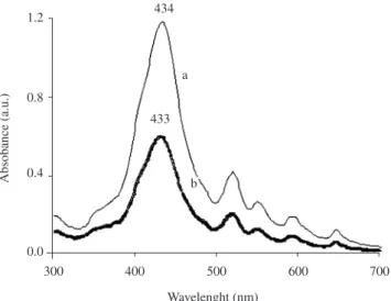
Figure 7 shows the difference between two films formed by LB and LS, indicating different intensities in absorbance and thereby
reflecting on the thickness of each formed film, by considering that absorption intensity is proportional to thickness – Beer’s law.
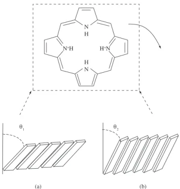
3.3. Molecular orientation of phorphyrin in LB and LS films
The orientation of H2TPP molecules in LB and LS films (two layers) was estimated by measuring the UV-vis absorption dichroism (Figure 8) at the Soret band from porphyrin (433 nm) and the polarized UV-vis spectra data are summarized in the Table 2. The orientation angles normal to the film and the dipping direction, respectively θ, φ were calculated18 by the following Equations 4 and 5:
cos2θ = [D
0 – (1 + D0 sin2β ) Dβ ] / [ (1 – 2 sin2β ) Dβ–
( 1 + Dβsin2β ) D
0 ] (4)
sin2θ cos2φ = (D
0 - cos2θ)/(1+D0) (5)
Considering the uncertainty of ±5° that comes from the polarized UV–vis method2, the results indicated that the macrocycle of H
2TPP in
LB film was tilted by an angle of 51 ±5° and in the LS film was tilted by an angle of 36 ±5° (Figure 9). Both values are in good agreement
Table 1. Electronic spectra of H2TPP film solutions before and after NO2 exposure.
Sample Soret band (nm)
Q(0.0) band (nm)
Q(0.1) band (nm)
Q(1.0) band (nm)
Q(1.1) band (nm)
Solution 417 513 548 590 645
LS film 439 N.A. N.A. 603 655
Δλ(nm)/
ΔE(eV)
22/-0.15 N.A. N.A. 13/-0.05 10/-0.03
N.A – Not applicable.
Figure 6. UV-vis spectra of LS H2TPP films (2 layers) deposited onto quartz substrate at pH = 5, exposed to NO2 gas at different times.
0.0 0.5 1.0 1.5
300 400 500 600 700 800 900
Wavelength (nm)
Absorbance (a.u.)
434 467
b a
c
0.0 0.5
500 600 700 800
520 552
595 648 687
a c b
1.2
0.8
0.4
0.0
300 400 500 600 700
Wavelenght (nm)
Absobance (a.u.)
434
433 a
b
Figure 7. UV-vis spectra of H2TPP films (2 layers) deposited onto quartz
substrate at pH = 5, a) LS film and b) LB film.
400 500 600 700
Absorbance (a.u.)
417
a
0 0.1 0.2 0.3 0.4 0.5
500 600 700
513
590
655
548 603
645 a
b 439
439
bb
Wavelength (nm)
Figure 5. Optical absorbance spectrum of 5 x 10⁻⁶ M solution of H2TPP, a) before
with the literature2. Hence, these results confirmed that the H 2TPP
molecules did not form flat LB and LS films and but the trend of a tilted molecular organization as preferential average conformation where the angle is dependent on the deposition method used. A more in-depth investigation is required to fully understand such behavior as far as the molecular film is transferred onto the substrate and its dynamic thermodynamic stabilization process.
The structures of the monolayers before and after being trans-ferred onto solid substrates may differ due to the different environ-ments. That is, the tilt angle of the porphyrin macrocycle normal to the substrates is slightly larger than that normal to air/liquid interface, arising from the elimination of the surface pressure19.According to
the playing-card model, porphyrin rings arrange parallel one to an-other, and the corresponding transition moments in adjacent rings are also parallel in the supramolecular structure formed at the air/water interface. For this kind of alignment of the transition moments, the exciton model developed by Kasha and others20 predicts that the shift
or splitting of absorption bands in weakly coupled electronic systems is caused by the interaction of localized transition dipole moments11.
This theory explains that for free-base porphyrins that have two or-thogonal transition moments, there are two anglesα1 and α2, related
to the center-center line between the adjacent rings. If α2=90° then α1
corresponds to the orientation angle of the ring θ related to the normal angle of substrate and the results for θ = α1 < 54.7º is the red shifted
of absorption band, indicating that J aggregates are formed3,7,21.
4. Conclusions
The response of 5, 10, 15, 20-tetraphenylporphyrin to the pres-ence of NO2 gas in diluted solutions and in Langmuir-Blodgett and Langmuir-Schaefer films was verified by UV-visible spectroscopy based upon the shift of Soret and Q bands. A relatively simple and inexpensive gas reactor was built for gas detection. The exposure of LB porphyrin films deposited onto glass slides to NO2 has performed as an active chemical detector with about 7000 ppm gas concentration. Additionally, the UV-vis dichroism absorption results associated with the spectroscopy characterization have given evidence of the tilt angle of the macrocycle deposited on the substrate. H2TPP LB film was tilted by an angle of 51 ±5° and the LS film was tilted by an angle of 36 ±5° indicating the formation of a preferential organization of the molecular films depending on the deposition method. In summary, the developed H2TPP molecular films indicated an opportunity of potential use for NO2 chemsensor applications.
Acknowledgements
The authors thank the financial support from CNPQ/FAPEMIG and FHEMIG. We also thank Professor Franz Grieser from Mel-bourne University-Australia for helping with the preparation of the manuscript.
References
1. Hann RA. Molecular structure and monolayer properties. In:
Langmuir-Blodgett Films. Gareth Roberts (ed.). New York: Plenum Press; 1990.
p. 17-92.
2. Li X, Xu W, Itoh T, Ikehata A, Zhao B, Li B, Ozaki Y. Effects of a central metal on the organization of 5,10,15,20-tetra-(p-chlorophenyl)–rare earth porphyrin hydroxyl compound at the air/water interface and in Langmuir–Blodgett films. Journal of Colloid and Interface Science.
2005; 284(2): 582-592.
3. Liu HG, Feng XS, Xue QB, Wang L, Yang KZ. Central metal effect on the organization of porphyrin LB films. Thin Solid Films. 1999; 340(1-2):
265-270.
Figure 9. Schematic illustration of possible arrangements of H2TPP in: a) LB film and in b) LS film.
Table 2. Polarized UV-vis spectra results calculated at λ= 433 nm for H2TPP LB and LS films.
Incident angle Dichroic ratios
LB film LS film
Av 0.659 0.135
β = 0° Ah 0.425 0.097
D0 1.551 1.392
Av 0.905 0.238
β = 45° Ah 0.619 0.126
D45 1.462 1.889
Results Equations 2 and 3 LB film LS film cos2θ 0.746 0.914
θ 51.4 35.1
φ 30.3 17.1
1.0
0.8
0.6
0.4
0.2
0.0
380 430 480 530 580 630 680
Wavelenght (nm)
Absobance (a.u.) b
d a c
Figure 8. Results of absorption dichroism measurement for two layers on quartz substrate of a H2TPP LB film, pH 5. a) Av0, b) Ah0, c) Avθ, and d) Ahθ.
N+H H+N
H N N H
(a) (b)
4. Dunbar ADF, Richardson TH, Hutchinson J, Hunter CA. Langmuir– Schaefer films of five different free base tetraphenylporphyrins for optical-based gas sensing of NO2. Sensors and Actuators B. 2008; 128(2): 468-481.
5. Richardson TH, Brook RA, Davis F, Hunter CA. The NO2 gas sensing
properties of calixarene/porphyrin mixed LB films. Colloids and
Surfaces A: Physicochemical and Engineering Aspects. 2006; 284-285: 320-325.
6. Tonezzer M, Quaranta A, Maggioni G, Carturan S, Dela Mea G. Optical sensing properties of CoTPP thin films deposited by
glow-discharge-induced sublimation. Sensors and Actuators B. 2007; 122(2): 613-619.
7. Liu HG, Feng XS, Zhang LJ, Ji GL, Qian DJ, Lee YI, Yang KZ. Influences of hydrophilic and hydrophobic substituents on the organization of supramolecular assemblies of porphyrin derivatives formed at the air/ water interface. Materials Science and Engineering: C. 2003; 23(5):
585-592.
8. Apetrei C, Casili S, De Luca M, Valli L, Jiang J, Rodríguez-Méndez ML, De Saja JA. Spectroelectrochemical characterisation of Langmuir– Schaefer films of heteroleptic phthalocyanine complexes. Potential applications. Colloids and Surfaces A: Physicochemical and Engineering Aspects. 2006; 284-285: 574-582.
9. Richardson TH, Dooling CM, Jones LT, Brook RA. Development and optimization of porphyrin gas sensing LB films. Advances in colloid and interface science. 2005; 116(1-3): 81-96.
10. Nam YS, Kim JM, Choi JW, Lee WH. Optimal deposition condition of chlorophyll a Langmuir-Blodgett film. Materials Science and Engineering C. 2004; 24(5): 35-38.
11. Zakavi S, Gharab NG. Interaction of para-substituted meso-tetraphenylporphyrins and meso-tetra(n-propyl)porphyrin with weak and strong carboxylic acids: A UV-Vis spectroscopic study. Polyhedron.
2007; 26(12): 2425-2432.
12. Feng F, Miyashita T, Amao Y, Asai K. A polymer Langmuir-Blodgett
film containing porphyrin chromophore. Thin Solid Films. 2000;
366(1):255-259.
13. Ogi T, Kinoshita R, Ito S. Spectroscopic and optical characterization of porphyrin chromophores incorporated into ultrathin polyimide films.
Journal of Colloid and Interface Science. 2005; 286(1): 280-287.
14. Wu Y, Hisada K, Maeda S, Sasaki T, Sakurai K. Fabrication and structural characterization of the Langmuir–Blodgett films from a new chitosan
derivative containing cinnamate chromophores. Carbohydrate Polymers.
2007; 68(4):766-772.
15. Zhao J, Li-Hua H, Shan G, Hui Z, Jing-Gui Z, Ning L. Molecular orientation and gas-sensing properties of Langmuir–Blodgett films of
copper phthalocyanine derivatives. Sensors and Actuators B: Chemistry
2007; 126(2): 588-594.
16. Boguta A, Wróbel D, Bartczak A, Ion RM, Ries R, Richter A. Scanning force microscopy investigations of (semi)conductive surfaces coated with Langmuir Blodgett dye layers. Surface Science. 2002; 513(2):
295-307.
17. Sales NF, Mansur HS. Influence of Subphase Parameters on the Nanostructures of 5,10,15,20-Tetraphenyl-21H,23H-Porphine Films at Air-Water Interface. Journal of Nanoscience and Nanotechnology. 2009.
doi: 10.1166/jnn.2008.3012. (in press)
18. Yoneyama M, Sugi M, Saito M, Ikegami K, Kuroda S, Iizima S. Photoelectric properties of copper phthalocyanine Langmuir–Blodgett Film. Japanese Journal of Applied Physics 1986; 25(7): 961-965.
19. Trofimova NS, Safronov AY, Ikeda O. Electrochemical and spectral studies on the catalytic oxidation of nitric oxide and nitrite by high-valent
manganese porphyrins at an ITO electrode. Electrochimica Acta. 2005;
50(24): 4637-4644.
20. Kasha M, Rawls HR, Ashraf El-Bayoumi M. The exciton model
in molecular spectroscopy. Pure and Applied Chemistry 1965;
11(3-4):371.
21. Biadasz A, Martynski T, Bauman D. Langmuir–Blodgett films of some fluorescent dichroic dyes as studied by optical spectroscopy methods.
![Optical Properties of MEH-PPV and MEH-PPV/ [6,6]-Phenyl C61-butyric Acid 3-ethylthiophene Ester Thin Films](data:image/gif;base64,R0lGODlhAQABAIAAAP///wAAACH5BAEAAAAALAAAAAABAAEAAAICRAEAOw==)