O
RIGINALA
RTICLE Revista Brasileira de FisioterapiaInfluence of the stimulating frequency
involved in analgesic effects induced
by electroacupuncture for neck pain
due to muscular tension
Influência da frequência estimulatória envolvida nos efeitos analgésicos induzidos
por eletroacupuntura em cervicalgia tensional
Nohama P1, Silvério-Lopes SM2
Abstract
Objective: To assess the influence of the stimulating frequency involved in analgesia induced by electroacupuncture for neck
pain. Methods: The performance of the analgesia produced by 2Hz, 100Hz, 1000Hz and 2500Hz was compared with a group with
acupuncture alone (without electrical stimulation), by means of pressure algometry, a visual analog scale (VAS) and heart rate. We used an electrical stimulator with a microprocessor yielding standard, single-phase, rectangular and asymmetrical balanced pulsed waveforms with a secondary phase decreasing exponentially. Stimulation periods were 4s, and resting periods were 3s. The sample
included 66 volunteers with neck pain due to muscular tension, mean age 33.67±9.97 years, 89.5% female and 10.5% male. Results:
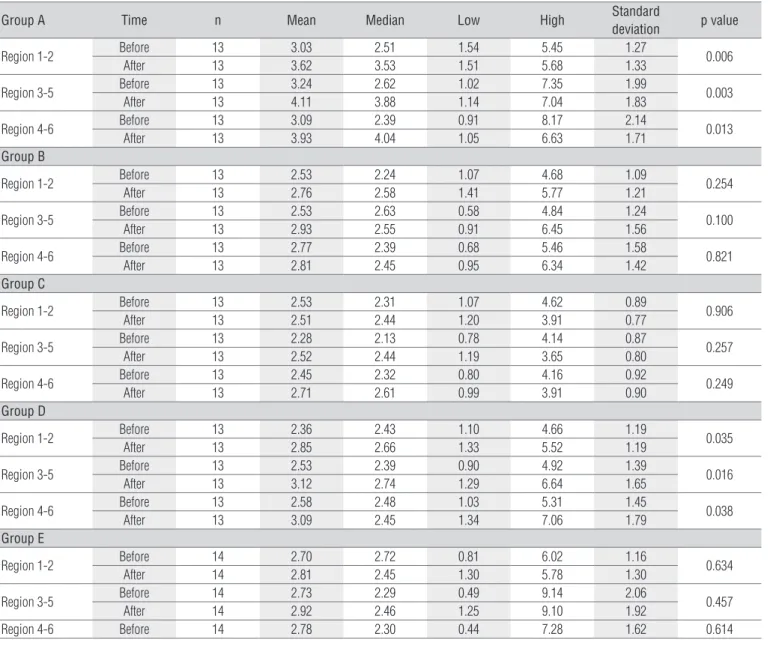
There were no differences between the groups regarding the variables of degree of pain (according to the VAS) and heart rate, and all groups presented analgesic improvement. However, when comparing pressure algometry findings for the same individual before and after the treatment, within the same group, we found analgesic advantages in using 2500Hz (p=0.006 for the base of the occipital region, p=0.003 for the right trapezius and p=0.013 for the left trapezius), followed by 100Hz (p=0.035, p=0.016 and p=0.038 for the same regions, respectively). Conclusion: We preferentially recommend 2500Hz and 100Hz applications of electroacupuncture for analgesia of neck pain due to muscular tension.
Article registered in the Australian New Zealand Clinical Trials Registry (ANZCTR) under the number 083456.
Key words: electroacupuncture; electrical stimulation; analgesia; neck pain.
Resumo
Objetivo: Avaliar a influência da frequência estimulatória envolvida na analgesia induzida por eletroacupuntura em cervicalgia.
Métodos: Comparou-se o desempenho da analgesia produzida em 2Hz, 100Hz, 1000Hz, 2500Hz e um grupo só com acupuntura, sem estímulo elétrico, avaliado por meio de algometria de pressão, Escala Visual Analógica (EVA) e frequência cardíaca. Utilizou-se um estimulador elétrico microprocessado, com forma de pulso em padrão pulsado, monofásico, retangular, balanceado assimétrico, com fase secundária em exponencial decrescente, com período de estimulação de 4 segundos e repouso de 3 segundos. A amostra contou com 66 voluntários com cervicalgia tensional, idade média de 33,67±9,97 anos, 89,5% do gênero feminino e 10,5% do masculino.
Resultados: Não houve diferenças entre os grupos para as variáveis nota atribuída à dor pela EVA e frequência cardíaca, sendo que em todos os grupos houve melhoras analgésicas. No entanto, quando comparado o comportamento antes-depois, por meio da algometria de pressão, para um mesmo indivíduo, dentro de seu próprio grupo, houve vantagens analgésicas para o uso de 2500Hz (p=0,006 para a base da região occiptal; p=0,003 para o trapézio direito; e p=0,013 para o trapézio esquerdo), seguido de 100Hz
(p=0,035, p=0,016 e p=0,038, para as mesmas regiões, respectivamente). Conclusão: Recomenda-se preferencialmente a aplicação
de 2500Hz e 100Hz em eletroacupuntura para analgesia em cervicalgia tensional.
Artigo registrado no Australian New Zealand Clinical Trials Registry (ANZCTR) sob o número 083456.
Palavras-chave: eletroacupuntura; estimulação elétrica; analgesia; cervicalgia.
Received: 30/05/2008 – Revised: 29/08/2008 – Accepted: 21/11/2008
1 Graduate Program in Health Technology, Pontifícia Universidade Católica do Paraná (PUC-PR), Curitiba (PR), Brazil 2 Instituto Brasileiro de Therapias e Ensino (IBRATE), Curitiba (PR), Brazil
Introduction
Acupuncture has incorporated electrical stimulation as an action mechanism to create electroacupuncture, which is con-sidered an indispensable technological resource for the success of this technique1. Due to the great interest and to the propa-gation of electroacupuncture in Brazil, the Ministry of Health has added acupuncture to the Public Health System (SUS)2. Eight health-related councils also consider this technique a specialty, including the Federal Council of Physical herapy and Occupational herapy3. As a physical therapy resource, electrotherapy appears to be more organized with regard to the standardization of procedures and instruments4. In contrast, electroacupuncture has yet to establish safe procedures and deine efective physical parameters that can be applied during therapeutic practice5.
he therapeutic principles of electrotherapy are based on physiological interactions at cellular, tissue and systemic level. he electric current low, through a biological conduc-tive medium, unleashes physiological efects involving elec-trochemical, electrophysical and electrothermal phenomena4. One of the most relevant and studied physical parameters in electroacupuncture is stimulation frequency, especially its relationship with endogenous opioid release in analgesic and anti-inlammatory processes6.
In the irst generation of electroacupuncture research, stud-ies were conducted on rats with induced pain in rats to relate stimulation frequencies to biochemically released substances such as: cholecystokinin 8 (CCK 8) at 100Hz7; endorphin at 2Hz8; encephalin and dynorphin at 2 and 100Hz6; endomorphin at 2Hz9, and substance P at 10Hz10. he studies on humans, as well as those involving higher frequencies, are scarce and use diferent methodologies, such as analgesia in back pain with the application of 50Hz1 and 80Hz11, postoperative analgesia at 100Hz12-14, neck pain at 120Hz and 250Hz15 and indications for acute pain at frequencies of 800 to 1000Hz12. he scarcity of scientiic studies on humans in this area can be explained by the diiculties which surround the assessment of human pain, as well as methodological errors, which have already been criti-cized by other authors16,17. herefore, it is important to evaluate the analgesic efects of therapeutic procedures to determine whether they should continue to be used.
he symptom of neck pain due to muscular tension was chosen because it is part of the population proile since it af-fects a great number of individuals. According to Côté18, neck pain afects 30% of men and 43% of women at some point in their lives, and it is a complaint that keeps a large number of workers away from their professional activities. Neck pain can have several sources, such as postural changes, mechanical traumas, spine rectiications, joint compressions and others19.
It is known that neck pain due to muscular tension is not a pathology in itself, but a symptom or a manifestation of muscle pain syndromes. Another relevant aspect in choosing this symptomatology was the fact that acupuncture has already shown good therapeutic results in neck pain15,20 and, as conse-quence, there is extensive literature on this symptom and the acupuncture application points20-25.
he present article describes a clinical-experimental study on humans. Its aim was to compare the performance of anal-gesia induced by acupuncture alone (without electrical stimu-lation) with the performance induced by electroacupuncture. he employed stimulation frequencies were 2Hz, 100Hz, 1000Hz, and 2500Hz.
Methods
For the experimental protocol, we used stainless steel dis-posable acupuncture needles (0.25 diameter x 40mm length); 70% alcohol solution; absorbent cotton; a chronometer; a Wag-ner digital algometer; and a sharps disposal box. We also used a class I, BF type electrostimulator (NKL, model EL608, ANVISA 80191680002) with microprocessed stimulus generation and control and 8 isolated outputs through pulse transformers. he output current can reach a maximum value of 10mA per pulse or mean intensity of 6mA.
he pulsed shape generated by the stimulator was con-igured as monophasic, rectangular, asymmetrical, with sec-ondary phase in decreasing exponential obeying, a pulsed pattern with 4-second stimulation periods and 3-second resting periods, according to Knihs5. he equipment was calibrated at the Rehabilitation Engineering Laboratory of PUC/PR, following the technical norms NBR IEC 60601-1 and NBR IEC 60601-226,27.
he subjects were recruited at the outpatient clinics of Instituto Brasileiro de herapias e Ensino of Curitiba. Initially, following the inclusion criteria, a population sample of 88 sub-jects was selected. However, at the time of intervention, a few subjects showed inadequacies such as drop in blood pressure, fear, intolerance to the electrical stimulation, use of analgesic drugs, among others. hese subjects received treatment but were not considered as part of the sample. he sample con-sisted of 66 individuals, aged 18 to 53 years with a mean age of 33.67±9.97 years, 89.5% female and 10.5% male. A subject screening instrument was prepared and validated using the technical reports of 10 orthopedics specialists. he objective of this instrument was to characterize the volunteers as neck pain suferers due to muscular tension to outline the sample proile to guarantee group homogeneity. Based on the deined inclusion criteria, we selected: normotensive individuals, with
neck pain due to muscular tension in the trapezius and neck muscle region, at least in the last 4 weeks before the selection. he exclusion criteria were: smokers, because tobacco was pointed out by Piovesan et al.28 as a factor in the decrease in nociceptive sensibility in algometry evaluation; pacemaker carriers and pregnant women, because the use of electroacu-puncture is contraindicated for those individuals29; individu-als who had received physical therapy treatment, massage or acupuncture in the last two weeks before the intervention, or who had taken anesthetic drugs, painkillers, muscle relaxants, psychotropic drugs or anti-inlammatories in the last two days before the intervention. his project was approved by the Re-search Ethics Committee of PUC-PR, protocol CEP 1035/2006. All the volunteers signed a consent form. With the intention of partially blinding the study, a physical therapist was invited to evaluate the subjects, who were systematically distributed between the groups. he measurement instruments were also evaluated before and after the therapeutic intervention.
Initially, the subjects were asked to score the pain on the visual analog scale (VAS) where zero was deined as “no pain”, and ten as “the worst pain”. he subject’s heart rate was then measured. he evaluation through pressure algometry be-gan with an explanation about the test and how the subject should verbalize the tolerance to the pressure. An example was given before the real test for clarity. he example con-sisted of a mechanical stimulus applied to the right elbow crease until the subject expressed discomfort to the pressure by immediately saying “stop”. At that moment, the compres-sion was instantly blocked, and the reading was checked on the algometer. For the pressure measurement, the algometer (with calibration certiicate) was set at the C function (self-calibration in kgf/cm2). he tolerance was standardized as the expression of the onset of discomfort caused by the pres-sure of the algometer’s rubber tip on the skin. he VAS, heart rate and pressure algometry procedures were performed at
least 10 minutes before the intervention, taking advantage of the interview time when the subject remained seated and at rest. he procedures were repeated 10 minutes after the acupuncture needles were removed.
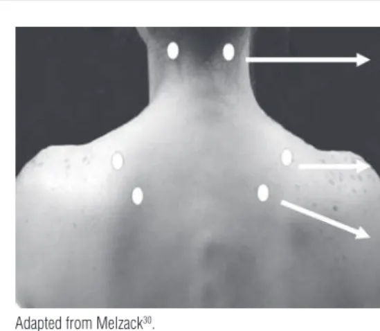
For the pressure readings, we selected three bilateral and symmetrical combinations of points on the neck and trape-zius muscle with a total of six reading areas: 1 and 2 (occipital insertion of the right and left trapezius, respectively); 3 and 4 (midpoint of the upper border of the right and left trapezius, respectively); 5 and 6 (supraspinatus muscle above the medial border of the right and left spine of the scapula, respectively), as demonstrated in Figure 1. hese points were chosen based on the literature because they are painful points in myofascial pain syndromes30.
he subject remained seated during all of the procedures. A sequence of algometry readings was standardized in such a way that, when the irst reading of the six points was com-pleted, a new “round” of readings in the same sequence began. Overall, three readings were performed on each point, before and after the intervention. he values were grouped for mean calculation, considering measure 1 with measure 2, 3 with 5, and 4 with 6. After the pre-intervention evaluations were completed, the acupuncture needles were applied bilaterally. he acupuncture points were selected based on bibliographi-cal indications for neck pain as follows: B10 (tianzhu), VB21
(jianjing), TA15 (tianliao), IG4 (hegu) and ID3 (houxi)23-25. he
needles used on points TA15 and VB21 (trapezius muscle, bi-laterally) were selected to receive electrical stimulus, acting as needle-electrodes. hese points were chosen due to the ana-tomical proximity to the painful region, to the muscle relax-ation function attributed to these points, and the fact that the needles can be easily and more comfortably applied to them. he needle’s depth of insertion was approximately 1.27cm (0.8 in), except in ID3 (on the hand), where the depth was about
Reading 1. Insertion of the right occipital muscle Reading 2. Insertion of left occipital muscle
Reading 3. Midpoint of the top border of the right trapezius
Reading 4. Midpoint of the top border of the left trapezius
Reading 5. Supraspinatus muscle above the medial border of the right spine of the scapula Reading 6. Supraspinatus muscle above the medial border of the left spine of the scapula
Adapted from Melzack30.
0.4cm (0.3 in). he needles were inserted and removed in the same sequence for all the subjects.
he groups were coded by draw with letters A (2500Hz), B (2Hz), C (1000Hz), D (100Hz) and E (without electrical stimula-tion). he subject and the researcher had no knowledge of the frequencies that corresponded to each letter. he stimulation frequency was the variable modiied during the experiments because it was the physical parameter under evaluation. he adjustment of the current intensity respected the stimulus tol-erance of each subject, therefore individualized, based on the electroacupuncture technique5,29.
he subjects were divided into groups A, B, C, D and E by systematic distribution conducted by the invited examiner. he amount of time the needles were left in place, including the time of electrostimulation, was 20 minutes. At the end of this interval, the electrostimulator cables and the needles were removed. Care was taken to avoid pressure close to the reading locations. A rest period of 10 minutes was standardized until the VAS, heart rate and algometry evaluations were repeated, which constituted the post-intervention data collection. he present study included 66 volunteers divided into ive groups: A (2500Hz, n=13), B (2Hz, n=13), C (1000Hz, n=13), D (100Hz, n=13), E (without electrical stimulation, n=14).
Results
In order to accomplish the statistical treatment, the analy-sis of covariance (ANCOVA) was applied to the algometry data. he non-parametric Kruskal-Wallis test was used for the VAS, and the Wilcoxon and t tests were used for within-group evaluations.
Pain score (VAS)
here was statistical signiicance to the reduction in the percentage variation of the mean pain scores, which shows improvements in the analgesic efect noticed by the subjects in all groups. he values were: A (2500Hz) reduction of 52.12% and p=0.003; B (2Hz) reduction of 32.93% and p=0.028; C (1000Hz) reduction of 52.41% and p=0.002; D (100Hz) reduction of 41.92% with p=0.013; and E (without electrical stimulation) reduction of 65.95% and p=0.002.
Tolerance to pressure
An evaluation per anatomical region (Figure 1) found statis-tical signiicance between pre- and post-intervention pressure tolerance. his form of evaluation, from a statistical point of view, reduces individual variability among subjects because it
compares each individual to himself (paired sample). Table 1 shows that there was statistical signiicance for groups A (2500Hz) and D (100Hz) in all evaluated anatomical regions, which demonstrates the efectiveness of the therapeutic inter-vention. he other groups did not show statistical relevance.
Heart rate
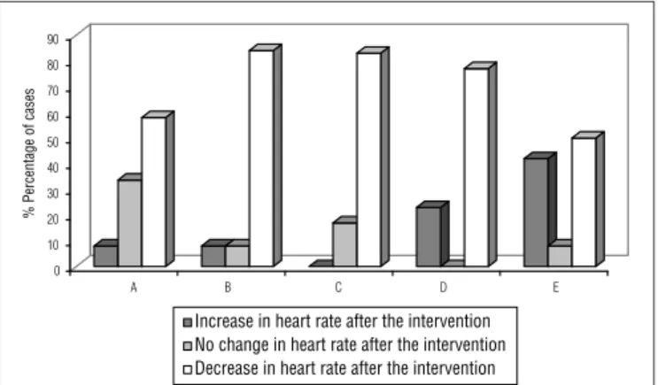
he percentage variation between pre- and post-interven-tion heart rate had no signiicant diference between groups (p=0.716). In all groups, there were subjects with no change in heart rate; however, some had an increase and others had a re-duction, as demonstrated in Figure 2. Group E (without electri-cal stimulation) had the highest number of cases of increased heart rate (43%).
Discussion
he results and statistical analyses show that there was statistical signiicance in all groups between the pre- and post-intervention pain score (VAS) and heart rate, which indicates therapeutic improvement, but without prominence of a spe-ciic group. However, the evaluation of within-group therapeu-tic performance for pressure tolerance showed better results for 2500Hz, followed by the 100Hz frequency. his result was conirmed in all the regions evaluated by pressure algometry.
hese results disagree with some authors such as Han6 and Filshe and White29, who point out the advantages of using low-stimulation frequencies (2Hz) for analgesic efects based on biochemical and immunohistological studies on rats and mice. Research in animals are important because it is based on the analgesic efects of neurotransmitter release. In contrast, it does
0 10 20 30 40 50 60 70 80 90
% Percentage of cases
A B C D E
Increase in heart rate after the intervention No change in heart rate after the intervention Decrease in heart rate after the intervention
Figure 2. Comparison between the evaluated groups with regard to the
incidence of cases increased, changed or decreased heart rate after the intervention. Groups: A (2500Hz), B (2Hz), C (1000Hz), D (100Hz) and E (without electrical stimulation).
not take into account emotional, cultural and biomechanical variables experienced in human pain.
Filshe and White29 conducted a survey of controlled ex-periments on humans which had very few indings, but veriied that lower electroacupuncture frequencies had better analgesic results than the higher frequencies. he authors also reported that the therapeutic efects last longer in chronic painful con-ditions. Unfortunately, electroacupuncture studies in humans are still scarce, particularly the ones which intend to compare parameters.
Tienyou31, Yin22 and Cui32 defend that electroacupuncture has analgesic advantages over acupuncture. he results of the present study partially conirm this statement by showing that there was statistical signiicance for pressure algometry in all evaluated regions in two out of four groups treated with
electroacupuncture (2500Hz and 100Hz), and that there was no diference in the group treated only with acupuncture. However, the results of the VAS evaluation show that group E, which received only acupuncture, demonstrated the highest mean reduction in the pain score (65.95%), although there was no statistical diference in comparison to the other groups. A possible explanation for this result is based on the fact that the mere possibility of an electrical current passing through the body causes anxiety in the subject and consequent negative psychological efect. It is worth noting that the VAS score has a subjective and emotional component, according to Ferreira19.
In pressure algometry, however, the reference is more quantitative and it is associated with nociceptive sensibility based on a concrete mechanical stimulus, which is the rubber tip of the algometer. In addition, the algometry reading points
Group A Time n Mean Median Low High Standard
deviation p value
Region 1-2 Before 13 3.03 2.51 1.54 5.45 1.27 0.006
After 13 3.62 3.53 1.51 5.68 1.33
Region 3-5 Before 13 3.24 2.62 1.02 7.35 1.99 0.003
After 13 4.11 3.88 1.14 7.04 1.83
Region 4-6 Before 13 3.09 2.39 0.91 8.17 2.14 0.013
After 13 3.93 4.04 1.05 6.63 1.71
Group B
Region 1-2 Before 13 2.53 2.24 1.07 4.68 1.09 0.254
After 13 2.76 2.58 1.41 5.77 1.21
Region 3-5 Before 13 2.53 2.63 0.58 4.84 1.24 0.100
After 13 2.93 2.55 0.91 6.45 1.56
Region 4-6 Before 13 2.77 2.39 0.68 5.46 1.58 0.821
After 13 2.81 2.45 0.95 6.34 1.42
Group C
Region 1-2 Before 13 2.53 2.31 1.07 4.62 0.89 0.906
After 13 2.51 2.44 1.20 3.91 0.77
Region 3-5 Before 13 2.28 2.13 0.78 4.14 0.87 0.257
After 13 2.52 2.44 1.19 3.65 0.80
Region 4-6 Before 13 2.45 2.32 0.80 4.16 0.92 0.249
After 13 2.71 2.61 0.99 3.91 0.90
Group D
Region 1-2 Before 13 2.36 2.43 1.10 4.66 1.19 0.035
After 13 2.85 2.66 1.33 5.52 1.19
Region 3-5 Before 13 2.53 2.39 0.90 4.92 1.39 0.016
After 13 3.12 2.74 1.29 6.64 1.65
Region 4-6 Before 13 2.58 2.48 1.03 5.31 1.45 0.038
After 13 3.09 2.45 1.34 7.06 1.79
Group E
Region 1-2 Before 14 2.70 2.72 0.81 6.02 1.16 0.634
After 14 2.81 2.45 1.30 5.78 1.30
Region 3-5 Before 14 2.73 2.29 0.49 9.14 2.06 0.457
After 14 2.92 2.46 1.25 9.10 1.92
Region 4-6 Before 14 2.78 2.30 0.44 7.28 1.62 0.614
chosen for the present study were close to the insertion loca-tion, and the stimulus caused by the electrical current in the groups with electroacupuncture also had an enhanced local efect, unlike the stimulus of acupuncture needles alone. With regard to heart rate variations, before and after the thera-peutic intervention, there were no diferences between the researched groups.
here are no studies in the literature that associate heart rate with analgesic efects of acupuncture or electroacupunc-ture. Although there was no statistical diference between the evaluated groups, one result is worth noting: most of the sub-jects in the groups submitted to electroacupuncture demon-strated a reduction in heart rate after the intervention (Figure 2). he same fact did not occur in the group which received only acupuncture (without electrical stimulation), in which 43% of the subjects had an increase in heart rate after the in-tervention, 50% had reduction and 7% showed no change.
Melzack33 and Guyton34 discussed the inluence of stress and external stimuli on heart rate modulation, as well as the
anatomical and physiological pathways of that inluence. Pomeranz17 found a relationship between low-frequency electroacupuncture and analgesic and sedative efects, which suggests possible indirect efects on heart rate. he studies by Yang et al.35 conirm that electroacupuncture reduces heart rate, blood pressure and catecholamine release, re-ducing stress. Based on these references, the results of the present study indicate that electroacupuncture has a greater efect on the autonomous and hypothalamic tonic regula-tion than acupuncture, which explains the higher propor-tion of subjects with heart rate reducpropor-tion in the groups with electrostimulation.
Although no significant statistical differences were found between groups with regard to pain score and heart rate, the present study recommends electroacupuncture application at a frequency of 2500Hz and 100Hz for anal-gesia of neck pain due to muscular tension because these frequencies demonstrated the highest individual efficiency in the algometry evaluation.
1. Valdés FB, Rabí MCM, Hernández MA, García JCJ. Acupuntura y eletroacupuntura en alivio del dolor de la osteoartrosis de la region lombar. Revista Cubana de Medicina General e Integral. 2001;17(2):143-8.
2. Ministério da Saúde. Portaria nº 971, de 4 de maio de 2006: aprova a política nacional de práticas integrativas e complementares (PNPIC) no Sistema Único de Saúde. Diário Oficial da União. 2006 Mai 4; Seç 1:84.
3. Conselho Federal de Fisioterapia e Terapia Ocupacional. Dispõe sobre a regulamentação da acupuntura. Resoluções n. 60/85 e 219/2000. Disponível em: www.coffito.org.br. Acesso em 07 de maio 2007.
4. Cameron MH. Physical Agents in Rehabilitation: from research to practice. 2ª ed. St. Louis: Saunders-Elsevier; 2003.
5. Knihs FC. Eletroacupuntura: uma proposta de equipamento. [Dissertação]. Florianópolis: Centro Tecnológico da Universidade Federal de Santa Catarina; 2003.
6. Han JS. Acupuncture: neuron peptide release produced by electrical stimulation of different frequencies. Trends Neurosci. 2003;26(1):17-22.
7. Liu SX, Luo F, Shen S, Yu YX, Han JS. Relationship between the analgesic effect of electroacupuncture and CCK-8 content in spinal purfusat in rats. Chinese Science Bulletin. 1999;44:240-3.
8. Han Z, Jiang YH, Wan Y, Wang Y, Chang JK, Han JS. Endomorphin-1 mediates 2 Hz but not 100 Hz eletroacupuncture analgesia in the rat. Neurosci Lett. 1999;274(2):75-8.
9. Han JS. Acupuncture and endorphins. Neurosci Lett. 2004;361(1-3):258-61.
10. Zhang RX, Wang L, Wang X, Ren K, Berman BM, Lao L. Electrocupuncture combined with MK-801 prolongs anti-hyperalgesia in rats with peripheral inflammation. Pharmacol Biochem Behav. 2005;81(1):146-51.
11. Thomas M, Lunberg T. Importance of modes of acupuncture in there treatment of chronic nociceptive low back pain. Acta Anaesthesiol Scand. 1994;38(1):63-9.
12. Amestoy RDF. Eletroterapia e eletroacupuntura. Florianópolis: Bristot; 1998.
13. Wang B, Tang J, White PF, Naruse R, Sloninsky A, Kariger R, et al. Effect of the intensity of transcutaneous acupoint electrical stimulation on the postoperative analgesic requeriment. Anesth Analg. 1997;85(2):406-13.
14. Lin JG, Lo MW, Wen YR, Hsieh CL, Tsai SK, Sun WZ. The effect of high and low frequency electroacupuncture in pain after lower abdominal surgery. Pain. 2002;99(3):509-14.
15. Qing Y, Zhang H, Jin R. Study on the somesthetic evoked potential in eletroacunpucture treatment of cervical spondylopathy. World Journal of Acupuncture and Moxibustion. 2000;10(2):7-10.
16. Ezzo J, Berman B, Hadhazy VA, Jadad AR, Lao L, Singh BB. Is acupuncture effective for the treatment of chronic pain? A systematic review. Pain. 2000;86(3):217-25.
17. Pomeranz B. Analgesia por acupuntura: pesquisas básicas. In: Stux G, Hammerschlap. Acupuntura clínica. São Paulo: Manole; 2005. p.01-31.
18. Côté P, Cassidy JD, Carroll LJ, Kristman V. The annual incidence and course of neck pain the general population-based cohort study. Pain. 2004;112(3):267-73.
157
19. Ferreira PEMS. Dor crônica, avaliação e tratamento oncológico. In: Andrade Filho ACC. Dor, diagnóstico e tratamento. São Paulo: Roca; 2001.
20. Vas J, Pereá-Milla E, Méndez C, Sánchez-Navarro C, Leon-Rubio JM, Brioso M, et al. Efficacy and safety of acupuncture for chronic uncomplicated neck pain: a randomised controlled study. Pain. 2006;126(1-3):245-55.
21. Maciocia G. A prática da medicina chinesa. 2ªed. São Paulo: Roca; 2007.
22. Yin GL, Liu ZH. Advanced modern chinese acupuncture therapy. Beijing: New World Press; 2000.
23. Stux G, Pomeranz B. Bases de acupuntura. 4ªed. São Paulo: Premier; 2004.
24. Yamamura Y. Acupuntura tradicional: a arte de inserir. 2ª ed. São Paulo: Roca; 2004.
25. Lian YL, Hammes MC, Chen Y, Kolster B. Atlas gráfico de acupuntura: um manual ilustrado dos pontos de acupuntura. Slovenia; H.F. Ullmann; 2007.
26. Associação Brasileira de Normas Técnicas. NBR IEC 60601-1: equipamento eletro-médico: prescrições gerais para segurança. Rio de Janeiro: ABNT; 1977.
27. Associação Brasileira de Normas Técnicas. NBR IEC 60601-2: equipamento eletro-médico: prescrições particulares para segurança de equipamento para estimulação neuromuscular. Rio de Janeiro: ABNT, 1997.
28. Piovesan EJ, Tatsui CE, Kowacs PA, Lange MC, Pacheco C, Werneck LC. Utilização da algometria de pressão na determinação dos limiares de percepção dolorosa em voluntários sadios, um novo protocolo de estudos. Arq Neuro Psiquitr. 2001;59(1):92-6.
29. Filshe J, White A. Acupuntura médica: um enfoque científico do ponto de vista ocidental. São Paulo: Roca; 2002.
30. Melzack R, Stillwell DM, Fox EJ. Trigger points of pain: correlations and implications. Pain. 1977;3(1):3-23.
31. Tienyou H. The principle of acupuncture’s pain management. World Journal Acupuncture and Moxibustion. 2000;10(3):47-51.
32. Cui H, Hong X, Chan LH. Estudio clínico de 30 casos de depresión mental tratados mediante electroacupuntura. J Tradit Chin Med. 2004;24(3):172-6.
33. Melzack R. The McGill pain questionnaire: major properties and scoring methods. Pain. 1975;1(3):277-99.
34. Guyton AC, Hall JE. Fisiologia humana e mecanismos das doenças. 6ª ed. Rio de Janeiro: Guanabara Koogan; 1998.