The Role of Nitric Oxide and Reactive Oxygen
Species in the Killing of
Leishmania
braziliensis
by Monocytes from Patients with
Cutaneous Leishmaniasis
Pedro Paulo Carneiro1, Jacilara Conceição1, Michael Macedo1, Viviane Magalhães1, Edgar
M. Carvalho1,2, Olivia Bacellar1,2*
1Serviço de Imunologia, Hospital Universitário Professor Edgard Santos, Universidade Federal da Bahia, Salvador, Bahia, Brazil,2Instituto Nacional de Ciência e Tecnologia de Doenças Tropicais - INCT-DT
(CNPq/MCT), Salvador, BA, Brazil
*olivinhaufba@gmail.com
Abstract
Human cutaneous leishmaniasis (CL) caused byLeishmania braziliensis, presents an
exag-gerated Th1 response that is associated with ulcer development. Macrophages are the pri-mary cells infected byLeishmaniaparasites and both reactive oxygen species (ROS) and
nitric oxide (NO) are important in the control ofLeishmaniaby these cells. The mechanism
involved in the killing ofL.braziliensisis not well established. In this study, we evaluate the
role of ROS and NO in the control ofL.braziliensisinfection by monocytes from CL patients.
Afterin vitroinfection withL.braziliensis, the oxidative burst by monocytes from CL patients
was higher when compared to monocytes from healthy subjects (HS). Inhibition of the ROS pathway caused a significant decrease in the oxidative burst inL.braziliensisinfected
monocytes from both groups. In addition, we evaluated the intracellular expression of ROS and NO inL.braziliensis-infected monocytes. Monocytes from CL patients presented high
expression of ROS after infection withL.braziliensis. The expression of NO was higher in
monocytes from CL patients as compared to expression in monocytes from HS. A strong positive correlation between NO production and lesion size of CL patients was observed. The inhibition of ROS production in leishmania-infected monocytes from CL patients allowed the growth of viable promastigotes in culture supernatants. Thus, we demonstrate that while production of ROS is involved inL.braziliensiskilling, NO alone is not sufficient to
control infection and may contribute to the tissue damage observed in human CL.
Introduction
Leishmaniasis is caused by protozoan parasites of the genusLeishmaniatransmitted by sandfly vectors. The promastigote form of the parasite invades host monocytes/macrophages and transforms into intracellular amastigotes. Host cells are able to control the infection if activated
OPEN ACCESS
Citation:Carneiro PP, Conceição J, Macedo M, Magalhães V, Carvalho EM, Bacellar O (2016) The Role of Nitric Oxide and Reactive Oxygen Species in the Killing ofLeishmania braziliensisby Monocytes from Patients with Cutaneous Leishmaniasis. PLoS ONE 11(2): e0148084. doi:10.1371/journal. pone.0148084
Editor:Simona Stäger, INRS - Institut Armand Frappier, CANADA
Received:May 15, 2015
Accepted:January 12, 2016
Published:February 3, 2016
Copyright:© 2016 Carneiro et al. This is an open access article distributed under the terms of the Creative Commons Attribution License, which permits unrestricted use, distribution, and reproduction in any medium, provided the original author and source are credited.
Data Availability Statement:The restrictions prohibiting the authors from making the minimal data set publicly available are due to maintaining patient privacy. However, the data are available upon request upon approval from the Institutional Ethical
Committee:cep.hupes@gmail.com.
by IFN-γ. [1]. Human cutaneous leishmaniasis (CL) caused byLeishmania braziliensisis
char-acterized by a strong cellular immune response and scarce numbers of parasites within lesions [2]. The presence of activating cytokines, such as IFN-γand TNF, are important for control of
parasite replication, but elimination of leishmania does not occur. The exaggerated Th1 immune response observed duringL.braziliensisinfection has been associated with severe inflammation and pathology [3–5].
During its life cycle,Leishmaniaencounters and readily adapts to various hostile conditions such as oxidative stress due to heme digestion in the blood meal, proteases in the sandfly mid-gut, complement-mediated lysis in the blood upon transmission, and reactive oxygen and nitrogen species (ROS and RNS) generated during phagocytosis by host macrophages [6–8]. Two important molecules that are critical in controllingLeishmaniainfection are superoxide anion (O2-) and nitric oxide (NO). During the initial phase of infection byLeishmania,
super-oxide is produced as part of the oxidative burst of macrophages in response to phagocytosis [6,
8]. The second oxidant produced by macrophages is nitric oxide, which, in contrast to superox-ide, is generated after activation of macrophages by IFN-y and TNF [9], [10]. Nitric oxide is derived from the oxidation of the terminal guanidine nitrogen of L-arginine by an NADPH-dependent enzyme, NO synthase. In murine systems, IFN-γhas been shown to synergize with
TNF, to activate inducible nitric oxide synthase (iNOS or NOS2) to produce nitric oxide (NO) resulting in eradication of intracellular parasites [11,12].
In humans, several studies have shown iNOS is present in lesions of patients with cutaneous leishmaniasis caused byLeishmania braziliensisandLeishmania tropica[5,13,14], [15,16]. Additionally, both iNOS mRNA and protein are expressed at high levels in bone marrow of patients with visceral leishmaniasis. iNOS protein was also found to be increased uponin vitro
infection of human macrophages withLeishmania chagasi[9].
In monocytes from humans unexposed toL.braziliensis, we have shown that both promasti-gote and amastipromasti-gote forms ofL.braziliensisinduce an oxidative burst but control of parasite replication is dependent on ROS [17]. We have previously shown that while macrophages from CL patients produce high amounts of pro-inflammatory cytokines, leishmania killing is impaired [17,18]. Actually, patients with CL due toL.braziliensisproduce high amounts of IFN-γand TNF, but despite an environment adverse to leishmania proliferation, the infection
progresses to disease. One possible explanation of this outcome includes impairment in the ability of monocytes in CL patients to develop an oxidative burst. Alternatively, it is possible that the oxidative burst produced by monocytes is insufficient to control leishmania. In this study, we showed that the oxidative burst is greater in CL monocytes than in healthy subjects (HS) monocytes. The increased oxidative burst is predominantly due to an increase in ROS rather than NO. However, while ROS production participates in leishmania killing, it is insuffi-cient to prevent parasite multiplication. NO production, rather than control parasite growth, is associated with pathology.
Material and Methods
Patients
This study included 28 CL patients and 10 healthy subjects. All patients were examined at the Corte de Pedra clinic, in the state of Bahia, Brazil, a well-known region forL.braziliensis trans-mission. The criteria for diagnosis were a clinical picture characteristic of CL in conjunction with parasite isolation in culture or parasite identification in histopathologic analysis or anL.
braziliensis-positive polymerase chain reaction result. Immunological studies were performed prior to therapy and 6 months to 1 year after cure (Table 1). All patients were treated with i.v. pentavalent antimonial (Sbv) (meglumine antimony; Sanofi-Aventis, Paris-France), 20 mg/kg
Competing Interests:The authors have declared
that no competing interests exist.
Abbreviations:CL, Cutaneous Leishmaniasis; HS,
Healthy Subjects; ROS, Reactive Oxygen Species; NO, Nitric Oxide; TLR, Toll Like Receptor; PMA, Phorbol 12-myristate 13-acetate; DHR,
body weight daily for 20 days. Criteria for cure were complete involution of lesions and/or total scarring of ulcers 90 days after initiation of treatment. Ten healthy subjects (HS) living in a place where there is noL.braziliensistransmission were enrolled in the study as controls. This research was conducted with approval of the Ethical Committee of the Professor Edgard Santos University Hospital, and informed consent was obtained from each participant.
Parasite culture
AnL.braziliensis(MHOM/BR/2003/LTCP11245) isolate obtained from a skin lesion of a CL patient was initially cultivated in biphasic medium (NNN). Following isolation, the parasites were cryopreserved in frozen nitrogen. The parasites selected for this study had not been previ-ously passaged in liquid culture medium. After selection, the parasites were expanded in com-plete Schneider’s medium. The isolate was identified asL.braziliensisby multilocus enzyme electrophoresis [19]. All reagents and Schneider’s medium were endotoxin free as determined by Endotoxin Testing (LAL) (BioReliance, SIGMA-ALDRICH).
Isolation of human peripheral blood cells and infection with
L
.
braziliensis
Peripheral blood mononuclear cells (PBMC) were isolated from heparinized venous blood lay-ered over a Ficoll-Hypaque gradient (GE Healthcare), washed, and resuspended in RPMI 1640 medium (supplemented with 5% of fetal calf serum, 100 U penicillin/mL, 100ug streptomycin/ mL) (GIBCO BRL., Grand Island, NY USA). PBMC (1x10⁶cells/tube) from CL patients and healthy subjects were infected with autologous serum opsonizedL.braziliensisat a ratio of 5:1 cells at 37°C in 5% CO2. After 2 hours, extracellular parasites were removed after
centrifuga-tion. The cells were placed in complete RPMI 1640 medium and incubated for additional 24, 48 and 72 hours. In some experiments, monocytes were preincubated with either 10mM DPI (a specific inhibitor for flavoprotein, a constituent of the NADPH oxidase complex) or 1mM of NG-methyl-L-arginine acetate salt (L-NMMA), an inhibitor of nitric oxide synthase (iNOS) for 10 minutes and were infected withL.braziliensispromastigotes at a 5:1 ratio for 72 hours. After these periods of time, the number of infected cells and the number of intracellular para-sites were determined by microscopic evaluation of 100 monocytes, after May-Grunwald-Giemsa staining from cytocentrifuge preparations.
Evaluation of the oxidative burst
Isolated PBMC (1x10⁶) were stimulated with dihidrohodamine 123 at 10 ng/mL (Cayman Chemical Company) for 10 minutes at 37°C in a 5% of CO2. After that, cells were exposed to opsonizedL.braziliensispromastigotes (ratio 5:1 cells) for 25 minutes at 37°C in a CO2 incuba-tor. PMA (Phorbol 12-myristate 13-acetate) at 1 ug/mL was used as positive control for oxida-tive burst production. Monocytes were stained for surface markers with anti-CD14
(PerCP-Cy5.5 clone HCD14) and anti-CD16 (PE clone 3G8) and the fluorescence intensity of
Table 1. Clinical and epidemiological characteristics of cutaneous leishmaniasis patients (CL) and Healthy Subjects (HS).
CL (n = 28) HS (n = 10)
Age (years) 30.5 (53–17) 30.3 (53–24)
Gender 19M; 6 F 2 M; 8 F
Lesion Size (mm) 14.8x13. 3
-DTH reaction to leishmania antigen (mm) 14.3x14. 8 Negative
the cells assessed by flow cytometry and analyzed using FlowJo software. Monocytes were defined by nonspecific fluorescence with forward scatter (FSC) and side scatter (SSC) as parameters of cell size and granularity, respectively. The cells were then gated based on expres-sion of CD14, CD16 and based on the oxidation of DHR 123 (S1 Fig).
The surface expression of TLR2 and TLR4 on monocytes from peripheral blood of CL patients and HS was analyzed bothex vivoandin vitroafter infection withL.braziliensis. After incubation for 4 hours, analysis was performed by flow cytometry. The following antibodies were used: anti-CD14 conjugated with PerCP-Cy5.5 (clone 61D3); anti-TLR2 conjugated to PE (clone TL2.1) and anti-TLR4 PE-conjugated (clone HTA125) (IMGENEX, San Diego, CA, USA).
Evaluation of oxidative burst after inhibition of enzymes NADPH oxidase
and iNOS in monocytes after infection with
L
.
braziliensis
To evaluate the effects of inhibition of ROS pathway or the NO pathway on the production of oxidative burst, cells were treated for 10 minutes with 10 mM DPI, a specific inhibitor for flavo-protein, a constituent of the NADPH oxidase complex. The inhibition of nitric oxide synthase (iNOS) was performed by treating monocyte cultures with 1mM of NG-methyl-L-arginine ace-tate salt (L-NMMA) for 10 minutes. Cells were then infected withL.braziliensis(5:1 cells) for 25 minutes at 37°C in 5% CO2. The oxidation of DHR-123 was assessed by flow cytometry.
Intracellular production of NO and ROS by monocytes after infection with
L
.
braziliensis
To detect specific intracellular NO production by monocytes, a fluorescent assay using the cell-permeable stain DAF-FM diacetate (4-amino-5-methylamino-2', 7'-difluorofluorescein diace-tate) at 10 mM (Molecular Probe, Life Technologies) was used for 10 minutes before the infec-tion withL.braziliensispromastigotes (5:1 cells). Intracellular ROS production by monocytes was measured by using an intracellular fluorescent probe, CM-H2DCFDA (carboxymethyl-H2-dichlorofluorescein diacetate) at 1μM (Molecular Probe, Life Technologies) for 10 minutes followed by infection for 25 minutes withL.braziliensispromastigotes (5:1 cells) at 37°C in a CO2incubator. To evaluate the effects of the inhibition of ROS and NO pathway, DPI (10 mM, Sigma) and L-NMMA (1 mM) were added to cultures 10 minutes followed by infection for 25 minutes withL.braziliensispromastigotes (5:1 cells). Monocytes were stained for surface markers with anti-CD14 and anti-CD16 antibodies and the fluorescence intensity of the cells was assessed by flow cytometry and analyzed using Flowjo software.
Viability of Parasites
After 72 hours of infection withL.braziliensis, monocytes were washed and the medium was replaced by 0.5 ml of Schneider’s medium (Sigma-Aldrich) supplemented with 10% fetal calf serum, to quantify the number of viable parasites. The tubes were cultured at 26°C for an addi-tional 5 days. Viable number ofL.braziliensiswas estimated by proliferation of extracellular motile promastigotes in the Schneider's medium [20,21].
Evaluation of oxidative burst in patients after therapy and cure of disease
Statistical Analyses
The data are presented as the mean ± SD of the mean. The significance of the results was calcu-lated using nonparametric statistical tests: Mann-Whitney (two-sidedttest), for comparisons between two groups or Kruskal-Wallis, followed by Dunn’s multiple comparison test, for com-parisons between three groups. Correlation analysis was performed using Pearson´s correla-tion. All the analyses was conducted using Prism (GraphPad software) and ap<0.05 was
considered significant.
Results
Induction of the oxidative burst in monocytes from CL patients and HS
after infection for
L
.
braziliensis
Monocytes (1x10⁶) from CL and healthy subjects (HS) were incubated with DHR-123 for 10 minutes before infection. The conversion of DHR-123 to rhodamine following oxidation was assayed by flow cytometry in monocytes cultures without stimulus, stimulated with PMA and after infection withL.braziliensispromastigotes (5:1 cells) for 25 minutes. The samples without stimulus and PMA treated were used to set the negative and positive regions for the fluores-cence of the rhodamine, respectively. A fluoresfluores-cence intensity (FL-1) higher than 103was inter-preted as positive for intracellular oxidative burst (Fig 1a).
We first assessed the oxidative burst in monocytes from CL patients after infection withL.
braziliensiscompared with the production by monocytes from HS. After infection withL. bra-ziliensis, the oxidative burst by monocytes from CL patients represented by the mean fluores-cence intensity (MFI), 2821 ± 1040 was higher than the MFI in monocytes from HS 1091 ± 268 (p<0.01) as shown inFig 1b. There was no difference in the oxidative burst in unstimulated
cells from CL and HS (243 ± 102versus172 ± 74) and in cells stimulated with PMA (4240 ± 1635versus5741 ± 2294).
As Toll-like Receptor (TLR) expression has been associated with oxidative burst [22,23] and can be induced upon leishmania infection, we evaluated theex vivoexpression of TLR2 and TLR4 (Fig 1c). Both TLR2 and TLR4 were more highly expressedex vivoin monocytes from CL (28 ± 9 and 55 ± 18) than HS (10 ± 5 and 7 ± 3), respectively (p<0.001). Afterex vivo
infection withL.braziliensis, the expression of TLR2 and TLR4 increased significantly in mono-cytes isolated from CL patients (32 ± 26versus90 ± 64, and 14 ± 7versus55 ± 32, respectively), p<0.001.
Expression of oxidative burst in
L
.
braziliensis
infected monocytes after
inhibition of NADPH oxidase and iNOS enzymes
To determine the importance of ROS and NO in the generation of oxidative burst in mono-cytes during CL, infection assays were conducted in the presence of inhibitors of nitric oxide synthase (L-NMMA) and of NADPH oxidase (DPI).
Preincubation with DPI (10 mM for 10 minutes) caused a significant decrease in the oxida-tive burst inL.braziliensisinfected monocytes (5:1 cells for 25 minutes) from CL patients (2821 ± 1040versus1225 ± 724), (P<0.001). L-NMMA pretreatment (1 mM for 10 minutes)
did not alter the oxidative burst inL.braziliensisinfected monocytes from CL patients (2948 ± 879versus2821 ± 1040) (Fig 2a). In cells from HS, the addition of DPI also decreased the oxidative burst (1239 ± 494versus514 ± 175), (p<0.05) while L-NMMA had no effect.
Monocytes stimulated with PMA (1ug/mL for 25 minutes) in the presence of DPI or
was 34 ± 32 and 46 ± 45 respectively. The addition of DPI and LNMMA to uninfected or unsti-mulated cells did not change these data.
These results suggest that oxidation of rhodamine in monocytes from patients with CL and HS after infection byL.braziliensisor stimulation with PMA reflects an increased production of reactive oxygen species during the oxidative response.
Intracellular production of ROS and NO by monocytes from CL patients
and HS after infection of
L
.
braziliensis
Our data provides evidence that ROS is more induced than NO during the oxidative response by monocytes after infection withL.braziliensis. To further elucidate these data, we measured intracellular production of ROS and NO after infection with leishmania, using CM-H2DCFDA (1μM for 10 minutes), an indicator of intracellular ROS production [24] and DAF-FM diace-tate (10 mM for 10 minutes), an indicator of NO production [25]. The frequency of monocytes from CL patients expressing ROS after infection withL.braziliensispromastigotes (5:1 cells for
Fig 1.L.braziliensisinduces high levels of oxidative burst in monocytes from CL patients.Monocytes from CL patients (n = 13) and HS individuals (n = 5) were treated with DHR (10ng/mL-10 min) and infected withL.braziliensispromastigotes for 25 minutes at a ratio of 5:1cells. PMA (1ug/ml) was used as positive control. Cells were stained with anti-CD14. Data were collected using flow cytometry and analyzed using FLOWJO software. (A) Representative gating strategy on CD14+and DHR expression in monocytes from one CL patient (B) The data represent the median of mean intensity of fluorescence (MIF) of oxidative burst production by CL and HS monocytes. (C) Theex vivoexpression of TLR2 and TLR4 was evaluated on CD14+cells. Data were collected using flow cytometry and analyzed using FLOWJO software. Statistical analysis was performed using the Manny Whitney test and results were considered significant with a**p<0.01,***p<0.001.
25 minutes) was higher (47 ± 35%) than those observed in cells from HS (37±11%), although without statistical significance. However, the production of NO by cells from CL patients after infection was higher (p<0.05) than in HS (12 ± 10%versus3.9 ± 1.2%), (Fig 3a and 3b).
The ability of monocytes from CL patients and HS in control
L
.
braziliensis
infection
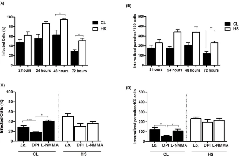
We infected monocytes from CL patients and HS withL.braziliensisfor different periods of time. After cytocentrifugation, the number of infected cells and the parasite load were evalu-ated using light microscopy. The percentage of infected cells and the number of intracellular amastigotes/100 monocytes were similar in the groups 2h after infection. The percentage of cells infected withL.braziliensisand the number of parasites/100 monocytes was lower in monocytes from CL patients as compared to monocytes from HS. The percentage of infected cells was also lower at 24, 48 and 72 hours in monocytes from CL patients than in HS mono-cytes (55 ± 22versus87 ± 8; 62 ± 22versus94 ± 6; and 29 ± 7versus50 ± 11) (p<0.05), as
shown inFig 4a. After 48 and 72 hours, the parasite load was also lower in monocytes from CL
Fig 2. Inhibition of NADPH oxidase decreases the oxidative burst.Monocytes from CL patients (n = 15) and HS individuals (n = 7) were preincubated with either DPI (10mM), an inhibitor of the NADPH oxidase, or L-NMMA (1mM), an iNOS inhibitor, for 10 minutes. The monocytes were pre-incubated with DHR (10 minutes) and infected withL.braziliensispromastigotes (5:1cells) or stimulated with PMA (1 ug/mL) for 25 minutes. Cells were stained with anti-CD14. Data were collected using flow cytometry and analyzed using FLOWJO software. (A) Representative contour plots. (B) The data represent the median of mean intensity of fluorescence (MIF) of DHR expression by monocytes from CL patients and HS individuals (C). Statistical analysis was performing using ANOVA with Bonferoni´s pos-test and Manny Whitney test. The results were considered significant with a p<0.05 (**p<0.01;***p<0.001).
patients as compared to HS (176 ± 40versus341 ± 119; and 119 ± 61versus232±38), p<0.05,
p<0.01 (Fig 4b).
At the time of 2, 24 and 48 hours there was no difference in the number of infected cells or number of internalized parasites in the presence of DPI or LNMMA in monocytes from untreated CL or HS cells (S2 Fig). However, at 72 hours, in monocytes from CL patients, the number of infected cells (Fig 4c) and the number of internalized parasites (Fig 4d) was lower in the presence of DPI (29 ± 7versus17 ± 4; and 119 ± 61versus53 ± 23), p<0.001 and p<0.05.
A possible explanation for this observation is that the inhibition of ROS production allows the parasite survival. Together these data suggest that the production of ROS by monocytes from CL patients may increase the ability of these cells to kill leishmania.
Fig 3. Monocytes from CL patients produced high levels of reactive oxygen species after infection withL.braziliensis.Monocytes from CL patients (n = 13) and HS individuals (n = 7) were stained with DAF-FM diacetate (NO probe, 10mM) and CMH-2DCFDA (ROS probe, 1μM) for 10 minutes, infected withL.braziliensispromastigotes for 25 minutes at a ratio of 5:1cells, and stained with anti-CD14. PMA was used as positive control. Data were collected using flow cytometry and analyzed using FLOWJO software (A). Representative histograms of ROS production, (B) Frequency ofL.braziliensis-infected monocytes expressing ROS, (C) Representative histograms of NO production, (D) Frequency ofL.braziliensis-infected monocytes expressing ROS. Statistical analysis was performing using ANOVA with Bonferoni´s pos-test and Manny Whitney test. The results were considered significant with a p<0.05 (*p<0.05).
Evaluation of the viability of
L
.
braziliensis
promastigotes after inhibition
of NO and ROS pathways
To assess whether the decrease in the parasite load was related to leishmania killing, we evalu-ated the viability of promastigotes in cultures of infected monocytes from CL patients in the presence or absence of oxidant inhibitors. The number of viable promastigotes, estimated by proliferation of extracellular motile parasites in Schneider´s medium, was higher in cultures of monocytes from CL patients (23 ± 5.6) as compared to monocyte from HS (15 ± 8.7), p<0.05
(Fig 5). In the presence of ROS inhibitor, the number of viableL.braziliensiswas higher (66 ± 14) as compared with infected monocytes alone (21 ± 8) or in the presence of NO inhibi-tor (18 ± 7), p<0.001 (Fig 5). However, in cultures of infected monocytes from HS, we did not
observe any difference in number of viable parasites in the presence of inhibitors. The reduced number of internalized parasites at 72 hours shown inFig 4is compatible with the higher num-ber of viable promastigotes observed in supernatants of culture. What may be happening is that intense intracellular proliferation of parasites causes disruption of the cell, allowing the release of live parasites into the supernatant.
Fig 4. Phagocytosis and the killing ofL.braziliensisby monocytes from CL patients.Monocytes from CL patients (n = 9) and HS individuals (n = 6) were infected withL.braziliensispromastigotes at a 5:1 ratio for 2, 24, 48 and 72 hours. The number of infected cells (A) and the number of intracellular parasites (B) were determined by microscopic evaluation after May-Grunwald-Giemsa staining from cytocentrifuge preparations. Monocytes were preincubated with either DPI (10mM) or L-NMMA (1mM), for 10 minutes and were infected withL.braziliensispromastigotes at a 5:1 ratio for 72 hours. (C) The number of infected cells. (D) The number of intracellular parasites. Statistical analysis was performed using the Kruskal-Wallis test (*p<0.05,**
p<0.01).
These results indicate that in CL patients, production of ROS participates in parasite killing, but despite its production a high percentage of monocytes remain infected and a large amount of amastigotes are found in such cells.
Correlation between oxidant production by
L
.
braziliensis
infected
monocytes and lesion size in CL patients
The production of NO has been implicated in the pathogenesis of several inflammatory diseases, such as tuberculoid leprosy and psoriasis [26–28]. In cutaneous leishmaniasis caused by Leish-mania mexicana, iNOS expression was correlated with increased number of skin lesions [29]. We extend our observations showing that inL.braziliensisinfection, there is a positive correla-tion between NO produccorrela-tion by monocytes and lesion size in CL patients (Fig 6a). On the other hand, no correlation between production of ROS and lesion size was observed (Fig 6b).
The production of NO and ROS by monocytes of CL patients after
therapy of American Tegumentary Leishmaniasis
All patients were treated with i.v. pentavalent antimonial, 20 mg/kg body weight, daily for 20 days. Criteria for cure were complete involution of lesions and/or total scarring of ulcers 90 days after initiation of treatment. After treatment and cure of patients with cutaneous leishmaniasis, the production of NO and ROS was evaluated in monocytes afterL.braziliensisinfection (Fig 7). There was a significant decrease in production of oxidative burst by these cells after therapy 3603 ±160versus1179±157, p<0.05 (Fig 7a). A decrease in NO and ROS levels was also observed, 5.8
±1.3versus0.705 ± 0.259 and 40.3±3.5versus8.4 ±1.6, respectively, p<0.05 (Fig 7b and 7c). The
Fig 5. The role of NO and NO in the control ofL.braziliensisinfection by monocytes from CL patients.Monocytes from CL patients (n = 9) and HS individuals (n = 6) were treated with inhibitor of the NADPH oxidase (DPI-10mM) or with an iNOS inhibitor (L-NMMA-1mM) for 10 minutes and infected withL. braziliensisat a 5:1. After 72 hours, the medium of monocytes culture was replaced by Schneider’s medium and after 5 days the number of viable
promastigotes was estimated. Statistical analysis was performed using the Manny Whitney test. (***p<0.001).
Fig 6. Correlation between NO and ROS production by monocytes and lesion size of CL patients.Monocytes from CL patients (n = 8) were treated with DAF-FM diacetate (NO probe, 10mM) or CMH-2DCFDA (ROS probe, 1μM) for 10 minutes and infected withL.braziliensispromastigotes for 25 minutes at a ratio of 5:1cells as described in materials and methods. Production of NO and ROS was evaluated by flow cytometry. (A) Correlation between NO production (%) and lesion size (mm). (B) Correlation between ROS production (%) and lesion size (mm). Statistical analysis was performed using the Pearson correlation.
doi:10.1371/journal.pone.0148084.g006
Fig 7. Oxidative burst production before and after therapy and cure of CL patients.Production of burst oxidative, NO and ROS by monocytes from CL patients (n = 6) after infection withL.braziliensispromastigotes or upon PMA stimulus, were determined before and after therapy (i.v. pentavalent antimonial, 20mg/kg body weight daily for 20 days) and cure of cutaneous leishmaniasis. The data represent the median of mean intensity of fluorescence (MIF) of oxidative burst production (A), frequency of NO production (B) and frequency of ROS production (C). Statistical analysis was performed using Wilcoxon test and results were considered significant (p<0.05).
cure is associated with the decreased or eradication of the parasite and also associated with a decreased inflammatory response. Alternatively, it has been reported that antimony treatment can induce ROS and NO generation to kill leishmania [30]. Thus, we believe that the successful therapy contributes to decreased oxidative burst. This observation indicates that the high inflam-matory response observed in CL regulates ROS and NO production.
Discussion
Monocytes play a critical role in leishmania infection, not only are they a primary leishmania host cell but also the main cell with the ability to eradicate parasites. Human CL is character-ized by an exaggerated Th1 immune response, but despite the production of high levels of
IFN-γand TNF, the parasites survive. Compared with cells from subclinicalL.braziliensisinfected
subjects (SC), CL monocytes/macrophages are more permissive to parasite growth and elimi-nate fewer parasites than cells from SC subjects [18,31]. We evaluated whether or notL. brazi-liensisinfection was able to induce the oxidative burst in monocytes from CL patients, and the production and role of ROS and NO in parasite killing. Our data indicate thatL.braziliensis
induce a greater oxidative burst in CL than in HS monocytes. This observation could be due to the inflammatory environment of patient’s cells and the greater expression of TLR2 and TLR4 on monocytes from CL patients. This increase in oxidative burst by monocytes is mainly related to ROS, a molecule involved in parasite eradication. Moreover, NO was also enhanced afterL.braziliensisinfection, but while NO production was not associated with parasite killing, there was a direct correlation between NO production and the size of the CL ulcer. In post kala-azar dermal leishmaniasis (PKDL), caused byLeishmania donovani, production of NO in cultured cells stimulated with leishmania antigen was elevated compared with post treatment samples, suggesting that this molecule may be associated with the pathogenesis of PKDL [32].
The oxidative burst is the main mechanism used by macrophages for leishmania eradication and O2-and NO are the main molecules utilized by the oxidative burst [9,33]. As expected,
after infection withL.braziliensis, unstimulated monocytes and PMA stimulated cells showed a similar increase in oxidative burst in HS and CL monocytes. These results suggest that the parasite may induce a stronger oxidative burst in monocytes from CL patients.
Factors involved in ROS production include phagocytosis and TLR expression [22,34]. Therefore, increased oxidative burst in CL monocytes could be due to increased leishmania uptake by CL monocytes or increased expression of TLRs. Penetration and uptake ofL. brazi-liensiswas similar between CL and HS monocytes. However, uninfected monocytes from CL had increased TLR-2 and TLR-4 expression compared with HS monocytes. Also, infection withL.braziliensissignificantly increased the TLR-2 and TLR4 expression on monocytes in CL patients. Several studies have shown a role for TLRs in the generation of oxidative burst during leishmania infection [23,35]. For instance, Srivastava et al. demonstrated that higher expres-sion of TLR2 was associated with a higher oxidative response and increased iNOS expresexpres-sion in macrophages infected withL.major[36].
The role of NO in the defense mechanisms of human monocytes/macrophages is controver-sial and may be specific to the leishmania species. We have previously shown that the percent-age of infected monocytes, as well the number of amastigotes ofL.braziliensis, were similar in monocytes stimulated with IFN-γand treated with or without L-NMMA, a nitric oxide
synthe-sis inhibitor. However, treatment of IFN-γstimulated cells with N-acetyl cysteine (NAC), an
in monocytes from CL patients and HS (data not shown), as previously demonstrated [20]. Concurrently, we found a strong correlation between production of NO and lesion size of CL patients. These results suggest that, while NO production alone does not participate in the con-trol of infection, it may contribute to the tissue damage observed in human CL.
To evaluate the ability of monocytes from CL patients to killL.braziliensisand whether or not killing could be associated with the oxidative burst, we determined the frequency of infected monocytes at different times, the numbers of amastigotes inside the cells and parasite viability in supernatants ofL.braziliensisinfected monocytes. Despite CL monocytes display-ing greater oxygen burst than HS cells, there was not a dramatic decrease in the percentage of infected cells, although a few amastigotes were detected through 24 to 72 hours inside of CL monocytes. Moreover, the parasite viability was similar in the supernatants of CL and HS monocytes infected withL.braziliensis, but the parasite viability was increased in supernatants of CL monocytes treated with DIP, a ROS inhibitor, indicating the role of this molecule in para-site killing. However, the addition of DIP did not affect the parapara-site viability in supernatants of HS monocytes indicating that ROS did not affect parasite survival in some infected monocytes. The reason why, despite greater respiratory burst, the parasites survive is not clear. One possi-bility could be the use of evasion mechanisms as production of superoxide dismutase [37,38]. Alternatively, it is possible that some monocytes produce lower amounts of ROS. The mono-cytes are a heterogeneous population and based on the expression of CD14 and CD16, it can be classified as classical, intermediate or inflammatory and non-classical monocytes [39]. While inL.braziliensis, infection there is an increase in the frequency of intermediate monocytes and production of pro-inflammatory cytokines [40],L.braziliensiskilling is mediated by classical monocytes [17]. We have not determined the ROS production in monocyte subsets but it can-not be ruled out that some monocyte subsets may have different expressions of oxygen burst than others. The persistence of parasites on monocytes and macrophages stimulates and con-tributes to maintenance of the inflammatory reaction that leads to ulcer development.
Due toL.braziliensis, CL has an inflammatory environment with high production of Th1 cytokines, IL-17 and pro-inflammatory chemokines [18,41–43]. This inflammatory environ-ment contributes to the increase in respiratory burst inL.braziliensisinfected cells, as success-ful therapy was followed by a significant reduction in the oxidative burst. The participation of the inflammatory response in the pathogenesis of CL ulcers has been well documented. Pro-gression from infection to disease occurs despite IFN-γand TNF production and there is a
direct correlation between the expression of TLR9 and lesion size and a positive correlation between the frequency of cell expressing IFN-γcell, TNF as well as T cell activation markers,
and the lesion size [4,44,45]. Moreover, molecules that down modulate the immune response as GM-CSF and pentoxyfilline are more effective than antimony alone in curing CL ulcers, reducing the healing time and in curing patients that are refractory to antimony therapy [46,
47]. In conclusion, monocytes from CL display greater expression of the oxidative burst, and despite the role of ROS in parasite control by these cells, it is not sufficient to killL.braziliensis
from infected cells. Alternatively, while production of NO does not participate in leishmania eradication, it may contribute to the tissue damage observed in human CL.
Supporting Information
S1 Fig. Representative plots used for the analysis of monocyte expressing CD14 and oxidative burst production.Peripheral blood mononuclear cells (PBMC) were obtained and stimulated with dihidrohodamine 123 (DHR) for 10 minutes. Monocytes were infected withL.braziliensis
S2 Fig. Phagocytosis and killing ofL.braziliensisby monocytes from CL patients after ROS and NO inhibition.Monocytes from CL patients (n = 9) and HS individuals (n = 6) were infected withL.braziliensispromastigotes at a 5:1 ratio for 2, 24, 48 and 72 hours. Monocytes were prein-cubated with either DPI (10mM) or L-NMMA (1mM), for 10 minutes and were infected withL.
braziliensispromastigotes at a 5:1 ratio for 72 hours. The number of infected cells (A and C) and the number of intracellular parasites (B and D) were determined by optical microscopy. Statistical analysis was performed using the Kruskal-Wallis test (p<0.05,p<0.01).
(TIF)
Acknowledgments
We would like to thank Ednaldo Lago for field assistance and Aline Muniz for help with figures.
Author Contributions
Conceived and designed the experiments: OB EMC PPC. Performed the experiments: PPC JC MM VM. Analyzed the data: PPC OB EMC. Contributed reagents/materials/analysis tools: VM MM JC. Wrote the paper: OB EMC PPC. Parasite identification by RT-PCR: VM.
References
1. Kaye P, Scott P. Leishmaniasis: complexity at the host-pathogen interface. Nat Rev Microbiol. 2011; 9 (8):604–15. Epub 2011/07/13. doi:10.1038/nrmicro2608PMID:21747391
2. Bittencourt AL, Barral A. Evaluation of the histopathological classifications of American cutaneous and mucocutaneous leishmaniasis. Mem Inst Oswaldo Cruz. 1991; 86(1):51–6. Epub 1991/01/01. PMID: 1842401.
3. Bacellar O, Lessa H, Schriefer A, Machado P, Ribeiro de Jesus A, Dutra WO, et al. Up-regulation of Th1-type responses in mucosal leishmaniasis patients. Infect Immun. 2002; 70(12):6734–40. Epub 2002/11/20. PMID:12438348; PubMed Central PMCID: PMC132996.
4. Antonelli LR, Dutra WO, Almeida RP, Bacellar O, Carvalho EM, Gollob KJ. Activated inflammatory T cells correlate with lesion size in human cutaneous leishmaniasis. Immunol Lett. 2005; 101(2):226–30. Epub 2005/08/09. doi:10.1016/j.imlet.2005.06.004PMID:16083969.
5. Faria DR, Gollob KJ, Barbosa J Jr., Schriefer A, Machado PR, Lessa H, et al. Decreased in situ expres-sion of interleukin-10 receptor is correlated with the exacerbated inflammatory and cytotoxic responses observed in mucosal leishmaniasis. Infection and immunity. 2005; 73(12):7853–9. PMID:16299275.
6. Channon JY, Roberts MB, Blackwell JM. A study of the differential respiratory burst activity elicited by promastigotes and amastigotes of Leishmania donovani in murine resident peritoneal macrophages. Immunology. 1984; 53(2):345–55. Epub 1984/10/01. PMID:6490087; PubMed Central PMCID: PMC1454813.
7. Almeida TF, Palma LC, Mendez LC, Noronha-Dutra AA, Veras PS. Leishmania amazonensis fails to induce the release of reactive oxygen intermediates by CBA macrophages. Parasite Immunol. 2012; 34(10):492–8. Epub 2012/07/24. doi:10.1111/j.1365-3024.2012.01384.xPMID:22817661; PubMed Central PMCID: PMC3532614.
8. Miao L, St Clair DK. Regulation of superoxide dismutase genes: implications in disease. Free Radic Biol Med. 2009; 47(4):344–56. Epub 2009/05/30. doi:10.1016/j.freeradbiomed.2009.05.018PMID: 19477268; PubMed Central PMCID: PMC2731574.
9. Gantt KR, Goldman TL, McCormick ML, Miller MA, Jeronimo SM, Nascimento ET, et al. Oxidative responses of human and murine macrophages during phagocytosis of Leishmania chagasi. J Immunol. 2001; 167(2):893–901. Epub 2001/07/07. PMID:11441096.
10. Smith MT, Evans CG. Inhibitory effect of superoxide-generating quinones on superoxide dismutase. Biochem Pharmacol. 1984; 33(19):3109–10. Epub 1984/10/01. PMID:6091670.
11. Liew FY, Li Y, Millott S. Tumor necrosis factor-alpha synergizes with IFN-gamma in mediating killing of Leishmania major through the induction of nitric oxide. J Immunol. 1990; 145(12):4306–10. Epub 1990/ 12/15. PMID:2175327.
13. Morgado FN, Schubach A, Vasconcellos E, Azeredo-Coutinho RB, Valete-Rosalino CM, Quintella LP, et al. Signs of an in situ inflammatory reaction in scars of human American tegumentary leishmaniasis. Parasite Immunol. 2010; 32(4):285–95. Epub 2010/04/20. doi:10.1111/j.1365-3024.2009.01188.x PIM1188 [pii]. PMID:20398229.
14. Vieira MG, Oliveira F, Arruda S, Bittencourt AL, Barbosa AA Jr., Barral-Netto M, et al. B-cell infiltration and frequency of cytokine producing cells differ between localized and disseminated human cutaneous leishmaniases. Mem Inst Oswaldo Cruz. 2002; 97(7):979–83. Epub 2002/12/10.
S0074-02762002000700009 [pii]. PMID:12471424.
15. Kumar R, Bumb RA, Salotra P. Evaluation of localized and systemic immune responses in cutaneous leishmaniasis caused by Leishmania tropica: interleukin-8, monocyte chemotactic protein-1 and nitric oxide are major regulatory factors. Immunology. 2010; 130(2):193–201. Epub 2010/01/28. doi:10.1111/ j.1365-2567.2009.03223.xIMM3223 [pii]. PMID:20102417; PubMed Central PMCID: PMC2878464.
16. Serarslan G, Atik E. Expression of inducible nitric oxide synthase in human cutaneous leishmaniasis. Mol Cell Biochem. 2005; 280(1–2):147–9. Epub 2005/11/29. doi:10.1007/s11010-005-8542-3PMID: 16311916.
17. Novais FO, Nguyen BT, Beiting DP, Carvalho LP, Glennie ND, Passos S, et al. Human classical mono-cytes control the intracellular stage of Leishmania braziliensis by reactive oxygen species. J Infect Dis. 2014; 209(8):1288–96. Epub 2014/01/10. doi:10.1093/infdis/jiu013PMID:24403561; PubMed Central PMCID: PMC3969552.
18. Giudice A, Vendrame C, Bezerra C, Carvalho LP, Delavechia T, Carvalho EM, et al. Macrophages par-ticipate in host protection and the disease pathology associated with Leishmania braziliensis infection. BMC Infect Dis. 2012; 12:75. Epub 2012/03/31. doi:10.1186/1471-2334-12-75PMID:22458474; PubMed Central PMCID: PMC3373377.
19. Cupolillo E, Grimaldi G Jr., Momen H. A general classification of New World Leishmania using numeri-cal zymotaxonomy. Am J Trop Med Hyg. 1994; 50(3):296–311. Epub 1994/03/01. PMID:8147488.
20. Novais FO, Santiago RC, Bafica A, Khouri R, Afonso L, Borges VM, et al. Neutrophils and macro-phages cooperate in host resistance against Leishmania braziliensis infection. J Immunol. 2009; 183 (12):8088–98. Epub 2009/11/20. doi:10.4049/jimmunol.0803720PMID:19923470.
21. Santos DM, Carneiro MW, de Moura TR, Soto M, Luz NF, Prates DB, et al. PLGA nanoparticles loaded with KMP-11 stimulate innate immunity and induce the killing of Leishmania. Nanomedicine. 2013; 9 (7):985–95. Epub 2013/04/23. doi:10.1016/j.nano.2013.04.003PMID:23603355.
22. Gill R, Tsung A, Billiar T. Linking oxidative stress to inflammation: Toll-like receptors. Free Radic Biol Med. 2010; 48(9):1121–32. Epub 2010/01/20. doi:10.1016/j.freeradbiomed.2010.01.006PMID: 20083193; PubMed Central PMCID: PMC3423196.
23. Roy S, Mukhopadhyay D, Mukherjee S, Ghosh S, Kumar S, Sarkar K, et al. A Defective Oxidative Burst and Impaired Antigen Presentation are Hallmarks of Human Visceral Leishmaniasis. J Clin Immunol. 2014. Epub 2014/12/07. doi:10.1007/s10875-014-0115-3PMID:25479930.
24. Nogueira NP, de Souza CF, Saraiva FM, Sultano PE, Dalmau SR, Bruno RE, et al. Heme-induced ROS in Trypanosoma cruzi activates CaMKII-like that triggers epimastigote proliferation. One helpful effect of ROS. PLOS One. 2011; 6(10):e25935. Epub 2011/10/25. doi:10.1371/journal.pone.0025935 PMID:22022475; PubMed Central PMCID: PMC3191175.
25. Olekhnovitch R, Ryffel B, Muller AJ, Bousso P. Collective nitric oxide production provides tissue-wide immunity during Leishmania infection. J Clin Invest. 2014; 124(4):1711–22. Epub 2014/03/13. doi:10. 1172/JCI72058PMID:24614106; PubMed Central PMCID: PMC3973105.
26. Kroncke KD, Fehsel K, Kolb-Bachofen V. Inducible nitric oxide synthase in human diseases. Clin Exp Immunol. 1998; 113(2):147–56. Epub 1998/08/26. PMID:9717962; PubMed Central PMCID: PMC1905037.
27. Little JW, Doyle T, Salvemini D. Reactive nitroxidative species and nociceptive processing: determining the roles for nitric oxide, superoxide, and peroxynitrite in pain. Amino Acids. 2012; 42(1):75–94. Epub 2010/06/17. doi:10.1007/s00726-010-0633-0PMID:20552384.
28. Matoshvili M, Katsitadze A, Sanikidze T, Tophuria D, Richetta A, D'Epiro S. The role of nitric oxide in the pathogenesis and severity of psoriasis. Georgian Med News. 2014;(234: ):61–4. Epub 2014/10/24. PMID:25341240.
29. Qadoumi M, Becker I, Donhauser N, Rollinghoff M, Bogdan C. Expression of inducible nitric oxide synthase in skin lesions of patients with american cutaneous leishmaniasis. Infect Immun. 2002; 70 (8):4638–42. Epub 2002/07/16. PMID:12117977; PubMed Central PMCID: PMC128200.
Antimicrob Agents Chemother. 2006; 50(5):1788–97. Epub 2006/04/28. 50/5/1788 [pii] doi:10.1128/ AAC.50.5.1788-1797.2006PMID:16641451; PubMed Central PMCID: PMC1472228.
31. Cardoso TM, Machado A, Costa DL, Carvalho LP, Queiroz A, Machado P, et al. Protective and patholog-ical functions of CD8+ T cells in Leishmania braziliensis infection. Infect Immun. 2015; 83(3):898–906. Epub 2014/12/24. doi:10.1128/IAI.02404-14PMID:25534940; PubMed Central PMCID: PMC4333467.
32. Katara GK, Ansari NA, Singh A, Ramesh V, Salotra P. Evidence for involvement of Th17 type responses in post kala azar dermal leishmaniasis (PKDL). PLOS Negl Trop Dis. 2012; 6(6):e1703. Epub 2012/06/23. [pii]. PMID:22724038; PubMed Central PMCID: PMC3378621.
33. Chang HK, Thalhofer C, Duerkop BA, Mehling JS, Verma S, Gollob KJ, et al. Oxidant generation by sin-gle infected monocytes after short-term fluorescence labeling of a protozoan parasite. Infect Immun. 2007; 75(2):1017–24. Epub 2006/11/23. doi:10.1128/IAI.00914-06PMID:17118986; PubMed Central PMCID: PMC1828521.
34. Sasada M, Pabst MJ, Johnston RB Jr. Activation of mouse peritoneal macrophages by lipopolysaccha-ride alters the kinetic parameters of the superoxide-producing NADPH oxidase. J Biol Chem. 1983; 258 (16):9631–5. Epub 1983/08/25. PMID:6309777.
35. Srivastava A, Singh N, Mishra M, Kumar V, Gour JK, Bajpai S, et al. Identification of TLR inducing Th1-responsive Leishmania donovani amastigote-specific antigens. Mol Cell Biochem. 2012; 359(1–
2):359–68. Epub 2011/08/23. doi:10.1007/s11010-011-1029-5PMID:21858498.
36. Srivastava S, Pandey SP, Jha MK, Chandel HS, Saha B. Leishmania expressed lipophosphoglycan interacts with Toll-like receptor (TLR)-2 to decrease TLR-9 expression and reduce anti-leishmanial responses. Clin Exp Immunol. 2013; 172(3):403–9. Epub 2013/04/23. doi:10.1111/cei.12074PMID: 23600828; PubMed Central PMCID: PMC3646439.
37. Khouri R, Bafica A, Silva Mda P, Noronha A, Kolb JP, Wietzerbin J, et al. IFN-beta impairs superoxide-dependent parasite killing in human macrophages: evidence for a deleterious role of SOD1 in cutane-ous leishmaniasis. J Immunol. 2009; 182(4):2525–31. Epub 2009/02/10. doi:10.4049/jimmunol. 0802860PMID:19201909.
38. Khouri R, Santos GS, Soares G, Costa JM, Barral A, Barral-Netto M, et al. SOD1 plasma level as a bio-marker for therapeutic failure in cutaneous leishmaniasis. J Infect Dis. 2014; 210(2):306–10. Epub 2014/02/11. doi:10.1093/infdis/jiu087PMID:24511100; PubMed Central PMCID: PMC4073785.
39. Ziegler-Heitbrock L, Hofer TP. Toward a refined definition of monocyte subsets. Front Immunol. 2013; 4:23. Epub 2013/02/06. doi:10.3389/fimmu.2013.00023PMID:23382732; PubMed Central PMCID: PMC3562996.
40. Passos S, Carvalho LP, Costa RS, Campos TM, Novais FO, Magalhaes A, et al. Intermediate mono-cytes contribute to pathologic immune response in Leishmania braziliensis infections. J Infect Dis. 2015; 211(2):274–82. Epub 2014/08/21. doi:10.1093/infdis/jiu439PMID:25139016; PubMed Central PMCID: PMC4334833.
41. Ribeiro-de-Jesus A, Almeida RP, Lessa H, Bacellar O, Carvalho EM. Cytokine profile and pathology in human leishmaniasis. Braz J Med Biol Res. 1998; 31(1):143–8. Epub 1998/08/01. PMID:9686192.
42. Bacellar O, Faria D, Nascimento M, Cardoso TM, Gollob KJ, Dutra WO, et al. Interleukin 17 production among patients with American cutaneous leishmaniasis. J Infect Dis. 2009; 200(1):75–8. Epub 2009/ 05/30. doi:10.1086/599380PMID:19476435; PubMed Central PMCID: PMC2732405.
43. Gonzalez-Lombana C, Gimblet C, Bacellar O, Oliveira WW, Passos S, Carvalho LP, et al. IL-17 medi-ates immunopathology in the absence of IL-10 following Leishmania major infection. PLOS Pathog. 2013; 9(3):e1003243. Epub 2013/04/05. doi:10.1371/journal.ppat.1003243PMID:23555256; PubMed Central PMCID: PMC3605236.
44. Vieira EL, Keesen TS, Machado PR, Guimaraes LH, Carvalho EM, Dutra WO, et al. Immunoregulatory profile of monocytes from cutaneous leishmaniasis patients and association with lesion size. Parasite Immunol. 2013; 35(2):65–72. Epub 2012/10/12. doi:10.1111/pim.12012PMID:23050581; PubMed Central PMCID: PMC3575026.
45. Weinkopff T, Mariotto A, Simon G, Hauyon-La Torre Y, Auderset F, Schuster S, et al. Role of Toll-like receptor 9 signaling in experimental Leishmania braziliensis infection. Infect Immun. 2013; 81(5):1575–
84. Epub 2013/02/27. doi:10.1128/IAI.01401-12PMID:23439309; PubMed Central PMCID: PMC3648021.
46. Machado PR, Lessa H, Lessa M, Guimaraes LH, Bang H, Ho JL, et al. Oral pentoxifylline combined with pentavalent antimony: a randomized trial for mucosal leishmaniasis. Clin Infect Dis. 2007; 44 (6):788–93. PMID:17304449.