UDC 577.15 + 543.6 + 543.9 + 543.55 + 544.725
Biosensors. A quarter of a century of R&D experience
A. P. Soldatkin, S. V. Dzyadevych, Y. I. Korpan, T. A. Sergeyeva, V. N. Arkhypova,
O. A. Biloivan, O. O. Soldatkin, L. V. Shkotova, O. A. Zinchenko, V. M. Peshkova,
O. Y. Saiapina, S. V. Marchenko, A. V. El’skaya
Institute of Molecular Biology and Genetics, NAS of Ukraine 150, Akademika Zabolotnogo Str., Kyiv, Ukraine, 03680 [email protected]
The paper is a review of the researches of Biomolecular Electronics Laboratory concerning the development of biosensors based on electrochemical transducers (amperometric and conductometric electrodes, potentiometric pH-sensitive field effect transistors) and different biorecognition molecules (enzymes, cells, antibodies), biomi-mics (molecularly imprinted polymers), as sensitive elements for direct analysis of substrates or inhibitory analy-sis of toxicants. Highly specific, sensitive, simple, fast and cheap detection of different substances renders them as promising tools for needs of health care, environmental control, biotechnology, agriculture and food industries. Diverse biosensor formats for direct determination of different analytes and inhibitory enzyme analysis of a num-ber of toxins have been designed and developed. Improvement of their analytical characteristics may be achie-ved by using differential mode of measurement, negatively or positively charged additional semipermeable memb-ranes, nanomaterials of different origin, genetically modified enzymes. These approaches have been aimed at in-creasing the sensitivity, selectivity and stability of the biosensors and extending their dynamic ranges. During the last 25 years more than 50 laboratory prototypes of biosensor systems based on mono- and multibiosensors for direct determination of a variety of metabolites and inhibitory analysis of different toxic substances were crea-ted. Some of them were tested in real samples analysis. The advantages and disadvantages of the biosensors de-veloped are discussed. The possibility of their practical application is considered.
Keywords: electrochemical biosensor, immobilized enzyme, substrate, inhibitor, multibiosensor.
Introduction. The last decades have shown unprece-dented interest in the development of analytical devices for the detection, quantification, and monitoring of dif-ferent biological and chemical compounds. The dynamic field of biosensors is covered by the extensive number of reviews [1–5].
As compared with existing standard methods of sub-strate determination, electrochemical biosensor methods of analysis have a number of advantages: they provide easy, fast, accurate, highly sensitive, spesific, and cheap procedure of measurement. Besides, real-time measure-ments are possible, while only minimal probe pretreat-ment is necessary. A biosensor is a self-contained device consisting of two functional parts. A bioselective element
is in direct contact with a physical transducer, which transforms the information from biorecognition domain into an electrical or optical signal. The amplitude of such signal depends on the concentration of the analysed compound (analyte) in the sample. Biologically active materials used for the construction of biosensor sys-tems can be divided into two main groups: catalytic (en-zymes, cells, tissues, biomimics) and noncatalytic, or af-finity (antibodies, receptors, nucleic acids, biomimics). Electrochemical (amperometric, potentiometric, con-ductometric or impedimetric), optical, calorimetric, and acoustic transducers are currently used in measuring systems. The main endevours in the biosensor develop-ment are focused on the exploration of various combi-nations of biological components (or their synthetic mi-mics) with different transducers.
In this paper our achievements in the development of electrochemical mono- and multibiosensors are re-viewed. The laboratory prototypes of such biosensors were created and used in analysis of real samples for future application in medicine, environmental monito-ring and food quality control.
Biosensors based on enzyme field effect transis-tors. The theory, technology and instrumentation for ion-selective field-effect transistors (ISFETs) were thorough-ly reviewed in [5]. In pH-sensitive field-effect transis-tors (pH-FETs) an electrical signal depends on surface potential of transistor gate dielectric depending on pH changes near the transistor’s gate.
A differential pair of ISFETs (one – covered with an enzyme-containing membrane, another – a control one with a reference membrane) is usually employed to compensate common interferences such as bulk pH, ion strength, photo- and temperature changes. Enzyme field effect transistors (ENFETs) were obtained by deposi-tion of a thin layer of enzyme-loaded composite over a pH-selective layer of ISFETs. The ENFETs
function-ing is based on the measurement of pH changes, locally occurring inside the bioselective membrane as a result of the enzymatic reaction involving the substrate to be assayed.
ENFETs based on direct analysis. S u b s t r a t e d
i-r e c t d e t e c t i o n. We developed ENFETs foi-r dii-rect determination of glucose, urea, acetyl- and butyrylcholi-ne, penicillin, formaldehyde, 4-chlorophenol, creatini-ne, etc. The pH changes are a result of enzymatically ca-talysed conversion of different substrates:
Immobilized enzyme
Substrate (analyte) ® product + proton or hydroxyl ion.
The corresponding enzymes are glucose oxidase (GOD), urease, acetylcholine esterase (AcChE), buty-rylcholine esterase (BuChE), penicillinase, alcohol oxi-dase (AOX), polyphenoloxioxi-dase (PPox), creatinine de-iminase (CD),etc.
The main analytical characteristics of the develo-ped biosensors are presented in Table 1.
Substrate (enzyme) Dynamic range, M Operational mode Time of analysis, min
Stability
References Operational, h Storage, day
Glucose ( Glucose Oxidase) 10–4
–3×10⁻³ Kinetic 0.1–0.2
> 20 90 [2, 6, 7] Steady-state 1–2
Urea (Urease) 10–4
–5×10⁻³ Kinetic 0.1–0.2
> 20 90 [2, 8] Steady-state 1–3
Acetylcholine chloride (Acetylcholine
Esterase) 10
–5 –5×10⁻³
Kinetic 0.1–0.2
> 30 90 [9, 10] Steady-state 1–3
Butyrylcholine chloride (Butyrylcholine
Esterase) 10
–4
–10–2 Kinetic 0.1–0.2
> 30 90 [9, 10] Steady-state 1–3
Penicillin (Penicillinase) 10–4
–1.5×10⁻²
Steady-state 1–2 > 20 90 [11]
Formaldehyde (Alcohol Oxidase) 5×10⁻³ –2×10⁻¹
Steady-state 1–3 > 20 30 [12]
Creatinine (Creatinine Deiminase) 1×10⁻⁵ –5×10⁻³
Steady-state 1–2 > 20 180 [13, 14] 4-Chlorophenol (Tyrosinase) 2×10⁻⁴–8×10⁻³ Steady-state 2–4 > 150 15 [15, 16] Protein substrates (Trypsin, Pronase E,
Trypsin with Carboxypeptidase B) 1–60 µg/ml Steady-state 5–7 > 10 20 [17, 18] Multi detection: specific proteinase activity
determination Trypsin–Chemotrypsin (proteinase-a2M molecule complexes)
0.1–20 U/ml Kinetic 5–7 > 15 30 [19]
Table 1
A comprehensive analysis showed that the develo-ped biosensors demonstrated reproducible, stable and fast (1–4 min) responses to the substrates to be measured [6–19]. Unfortunately, application of ENFETs can be re-stricted because of the dramatic decrease in the sensor response at increasing buffer capacity and ionic strength, pH-dependence of the enzyme kinetics, co-substrate li-mitation of enzymatic reaction rate (the glucose sensor).
I m p r o v e m e n t o f ENFETs a n a l y t i c a l c h a-r a c t e a-r i s t i c s. The investigation of glucose and ua-rea biosensors showed that main analytical characteristics of ENFETs can be improved by using charged permse-lective additional membranes [20–24].
A narrow dynamic range of urea biosensors is a dis-advantage which does not allow their appplication for measurements in whole human blood (where urea normal concentration is 6–8 mM but much higher in patholo-gy) and, thus, previous sample dilution is required [23]. The dynamic range can be shifted toward higher concen-trations by adding sodium tetraborate, a competitive in-hibitor of urease [25]. Another way to overcome the problem is modulation of urease affinity as a biorecog-nition element. Noteworthy is a trend of the last few years to use genetically modified enzymes for the imp-rovement of biosensor characteristics. Thus, we used a new enzyme preparation – recombinant urease from
Es-cherichia coliwith an active site, genetically modified
so that urease affinity toward the specific substrate de-creased. According to the information of the enzyme preparation producer, it has high, about 200 mM,Km.
The improved biosensor had quite broad, 1–80 mM,
dy-namic range [26], covering urea concentrations in blood and dialysate samples for both normal patients and tho-se with renal dysfunction. Moreover this biotho-sensor de-monstrated high reproducibility and storage stability. So, the developed biosensor has working characteri-stics fitting the medical requirements as for direct urea measurement in blood and dialysis samples.
D e t e r m i n a t i o n o f c r e a t i n i n e a n d u r e a c o n c e n t r a t i o n s. Determination of creatinine and urea, the main compounds of human nitrogen metabo-lism, is important for diagnostics of pathological states of kidneys and assessment of the hemodialysis effecti-veness. For this purpose we developed new enzyme po-tentiometric monobiosensors and designed their labo-ratory prototypes. As bioselective elements served im-mobilized enzymes – creatynindeiminase of microbial origin, urease from soybeans and recombinant urease
fromE. coli[26].
The biosensors were characterized by short time of analysis (1–2 min), high selectivity, reproducibility (standard deviation did not exceed 5 %) and storage sta-bility (30–40 days). They were successfully tested in samples of blood serum and dialysate of patients with re-nal failure [27, 28]. The measured values of creatinine and urea concentrations in dialysate were compared with the results of traditional methods, a high level of data correlation (R= 0.98) being observed in both cases (Fig. 1).
ENFETs based on inhibitory analysis. For
determina-tion of different toxic compounds we developed ENFET biosensors based on enzyme inhibition [2, 9, 10, 30–
0,0 0,1 0,2 0,3 0,4 0,5 0,6
0,00 0,05 0,10 0,15 0,20 0,25 0,30
y = 0,56x-0,02 R = 0,98
C
reat
in
in
e,
m
M
(J
af
fe
m
et
hod)
Creatinine, mM (biosensor method)
0,0 0,1 0,2 0,3 0,4 0,5 0,6
0,00 0,05 0,10 0,15 0,20 0,25 0,30
y = 0,56x-0,02 R = 0,98
C
reat
in
in
e,
m
M
(J
af
fe
m
et
hod)
Creatinine, mM (biosensor method)
A B
35]. Their main analytical characteristics are presented in Table 2.
I r r e v e r s i b l e i n h i b i t i o n o f u r e a s e. The immobilised urease can be inactivated by heavy metal ions through their direct interaction with sulfhydryl groups of the enzyme active site [2, 29]. The assay proto-col included measurement of the biosensor response to a fixed urea concentration before and after the biosensor incubation in a solution of heavy metal ions. The detec-tion limits were: 1.0×10⁻⁶
M for Hg2+
, 2.0×10⁻⁶ M for Cu2+
, 5.0×10⁻⁶
M for Cd2+
, 1.0×10⁻⁵
M for Co2+
, 2.0×10⁻⁵
M for Pb2+and 1.0×10⁻⁴M for Sr2+. Thus, the row of heavy metal ions regarding their toxicity toward urease is: Hg2+
> Cu2+
> Cd2+
> Co2+
> Pb2+
> Sr2+
. This inhibition is reversible in the presence of a strong chelating agent EDTA [46] which was used as a scavenging agent for re-activation of the enzyme activity and biosensor multi-use. I r r e v e r s i b l e i n h i b i t i o n o f c h o l i n e s t e-r a s e s. Detee-rmination of oe-rganophosphoe-rous and cae-r- car-bamate compounds is based on their ability to inhibit cho-linesterases irreversibly via interaction with the serine
–OH group in the enzyme active site. A decrease in cho-linesterase activity after its interaction with pesticides can be effectively monitored by ENFET biosensors, which allows the toxicity assessment (Table 2).
The conditions of practical application of ENFET biosensors based on inhibitory analysis were investiga-ted and critically evaluainvestiga-ted for optimisation. The enzyme reactivation after inhibition by organophosphorous pes-ticides was demonstrated when using pyridine-2-aldo-xime methyliodide (PAM-2) [9, 30]. At the enzyme in-hibition by hypochlorite species, PAM-2 treatment did not give a restoration effect. This fact can be used as a selectivity test for discrimination of inhibition by orga-nophosphorous pesticides or hypochlorite species.
R e v e r s i b l e i n h i b i t i o n o f c h o l i n e s t e r a-s e a-s. Steroidal glycoalkaloida-s (e. g.,a-solanine,a -cha-conine) are natural neurotoxins, the main source of which are the plants of familySolanaceae, including potato, widely used in food by millions of people. Consump-tion of potatoes and potato products can be potentially dangerous because high concentrations of steroidal
gly-Toxic components Enzyme Dynamic range, M analysis, minTime of
Stability
References Operational, h Storage, day
Heavy metal ions Urease 10–6–5×10⁻³ 20 > 10 30 [2, 29]
Diisopropyl fluorophosphate AcChE 3×10–11 –5×10⁻⁷
15 > 10 30 [2, 9, 30]
Diisopropyl fluorophosphate BuChE 5×10–11 –10–7
15 > 10 30 [2, 9, 30]
Trichlorfon AcChE 2×10⁻⁷
–10–5
15 > 10 30 [2, 9, 30]
Trichlorfon BuChE 1×10⁻⁶
–10–5
15 > 10 30 [2, 9, 30]
Paraoxon-ethyl AcChE 10–6–5×10⁻⁵ 15 > 10 30 [2, 9, 30]
Paraoxon-ethyl BuChE 5×10⁻⁷
–5×10⁻⁵
15 > 10 30 [2, 9, 30]
Paraoxon-methyl AcChE 10–6
–5×10⁻⁵
15 > 10 30 [2, 9, 30]
Paraoxon-methyl BuChE 5×10⁻⁶
–5×10⁻⁵
15 > 10 30 [2, 9, 30]
Parathion-methyl AcChE 5×10⁻⁶
–10–4
15 > 10 30 [30, 31]
Carbofuran AcChE 2×10⁻⁶–10–4 15 > 10 30 [30, 31]
Hypochlorite AcChE 10–5–3×10⁻⁴ 15 Disposable 30 [31]
a-Solanine BuChE 5×10⁻⁷
–10–4
5 > 30 30 [30, 32–34]
a-Chaconine BuChE 2×10⁻⁷
–10–4
5 > 30 30 [30, 32–34]
Tomatine BuChE 2×10⁻⁷
–10–4
5 > 30 30 [35]
Table 2
coalkaloids lead to poisoning, sometimes with fatal out-come, for both humans and animals [37]. Glycoalkaloids cause double toxic effects, firstly, as detergents, destroy-ing cell membranes, and second, inhibitdestroy-ing cholineste-rases, which play a key role in the transmission of nerve impulses. Thus, continuous monitoring of the glycoal-kaloids level in food is considered to be of extreme im-portance.
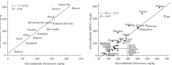
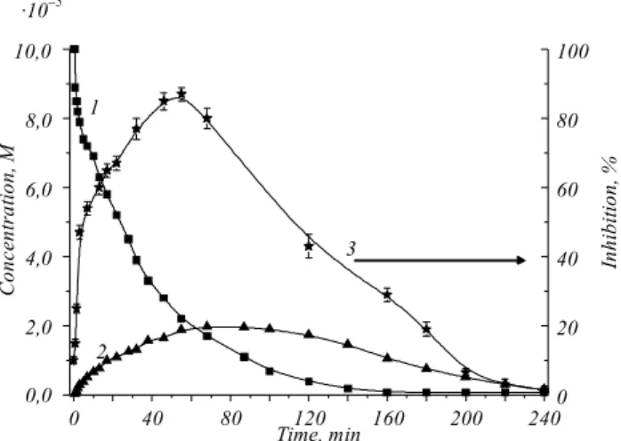
The determination of alkaloids concentrations by biosensors is based on the alkaloids ability to inhibit BuChE reversibly. The potentiometric biosensor with ISFETs and immobilized BuChE was developed for the express analysis of the glycoalkaloid total content which ranges within 0.2–100mM depending on the type of alkaloid, low detection limits of 0.2mM fora -cha-conine, 0.5mM fora-solanine, and 0.5mM for tomatine [32–35]. Glycoalkaloids were measured in potato and tomato juices by two methods, namely standard addi-tions and using a calibration curve [36, 37]. Compari-son of the results of biosensor determination with the data of thin film chromatography and HPLC analysis (Fig. 2) showed a high correlation. The biosensor deve-loped is sensitive and highly reproducible, it provides simple and quick direct screening of glycoalkaloids in fresh potato and tomato juices without any sample pre-treatment. Thus, it can be considered as a basis for the development of a commercial device for the express analysis of total glicoalcaloids in potato tubers of exis-ting varieties and those obtained by selection or genetic modification, as well as in foodstuffs.
M u l t i b i o s e n s o r f o r e c o l o g i c a l m o n i-t o r i n g o f i-t o x i c c o m p o u n d s. We were i-the firsi-t to propose the conception of toxins determination by a multibiosensor based on the enzyme inhibition analysis [10]. The multibiosensor with a matrix of ISFETs and several bioselective elements (AcChE, BuChE, urease, glucose oxidase, and three-enzyme system – invertase, mutarotase, glucose oxidase) was developed and its main charateristics were examined [40–43]. The results of in-hibition of the enzyme system by different concentra-tions of individual toxicants and their mixtures were analyzed by the methods of mathematical statistics to develop the approach to quantitative or semi-quantita-tive determination of toxicants in real water samples of the environment [42].
On the basis of this multibiosensor, we created a portable laboratory device to analyze real water samples from a number of Kyiv water reservoirs, and complex multicomponent water assays from the landfill waste for the presence of toxic substances [43]. A comparison of the sensor data with traditional methods shows a high degree of correlation (Table 3) [43].
The developed multibiosensor can serve as a basis for designing and industrial manufacture of measuring apparatus for express integral and selective determina-tion of water pollutants.
Conductometric biosensors. The biosensors based on conductometric principle seem to be very advanta-geous in several aspects: i) thin-film electrodes are suitable for miniaturisation and large scale production
0 50 100 150 200 250
0 50 100 150 200 Slov'yanka Zabava Yavir Serpanok Obriy Promin' Kupava Vodogray
Borodyans'ka rojeva Svitanok Kiivs'kiy Povin' Zarevo Racurs Lugovs'ka G lycoal ka lo id s (T L C ), m g/ kg
Glycoalkaloids (biosensor), mg/kg y = 7,1+0,92x
R = 0,96
0 100 200 300 400 500 600 700
0 100 200 300 400 201 308 Albane 306 Virginia 302 Redlaure Hinga Lucie 303 309 Elkana 209 Roseval Pompadour Starter Pollux Venus 91 F220.9 Phoebus Voyager 310 Charlotte Epona 207 Kaptah V Monalisa 204 Daifla y = 0,6 x - 27,9 R = 0,93
G ly co a lk a lo id s (H P L C ), m g /k g
Glycoalkaloids (biosensor), mg/kg
by inexpensive technology; noble metals can be chan-ged for cheaper ones,e. g. Ni; ii) there is no need in re-ference electrode, no light sensitivity, small driving vol-tage decreases power consumption; iii) large spectrum of analytes of different nature can be determined on the basis of various reactions and mechanisms.
The method of solution conductance monitoring was originally developed for determining chemical reaction rates and only recently it has been applied to the reactions catalysed by enzymes, namely, hydrolases, proteases, oxidases, amidases, peptidases,etc. [44].
Taking into consideration the mentioned attractive features of conductometric principle and simplicity of corresponding devices, we directed our efforts on the development of conductometric biosensors with imp-roved working characteristics.
Enzyme-based biosensors. The enzyme-based
con-ductometric biosensors can be used for both direct and
inhibitory analysis. In the last years we developed a num-ber of enzyme conductometric biosensors for determi-nation of main natural metabolites and toxins, created their laboratory prototypes, and optimized them for real samples. The main analytical characteristics are presen-ted in Table 4.
In these biosensors we applied conductometric trans-ducers consisting of two pairs of planar interdigitated electrodes, produced by vacuum deposition of gold or platinum on a ceramic support. Immobilized enzymes, either individual ones or their cascade, served as biose-lective elements. Immobilization of enzymes with BSA on the transducer surface was performed by glutaralde-hyde crosslinking. The bioselective membranes compo-sition and the conditions of enzyme immobilization we-re optimized.
We developed conductometric biosensors based on direct analysis for determination of glucose, urea,
ace-Place of sampling
Traditional methods, mg/l
Multibiosensor method
AAAHg2+ AAS TLC
Vyrlytsia (Poznyaki, Kyiv) – – – –
Vyrlytsia + 400 nM Hg2+
0.079 – – +
Dnieper river (Osokorki, Kyiv) – – – –
Dnieper river + 10 µM trichlorfon – – 2.57 +
Solnechnoe (Osokorki, Kyiv) – – – –
Solnechnoe + 5 µM Cu2+
– 0.321 (Cu2+
) – +
Ministerske (Obolon, Kyiv) – – – –
Opechen (Obolon, Kyiv) – – – –
Opechen lower (Obolon, Kyiv) – – – –
Verbne (Obolon, Kyiv) – – – –
Dnieper gulf (Obolon, Kyiv) – – – –
Dnieper (Obolon, Kyiv) – – – –
Landfill waste N 5, Pidgirtsi village,
Obukhivski, Kyiv region –
0.317 (Cu2 + )
– +++
0.034 (Co2+) 1.471 (Zn2+) 0.988 (Cr2+
) N o t e s. AAAHg2+
– atomic absorption mercury analyzer; AAS – atomic absorption spectroscopy; TLC – thin layer chromatography; «+» – exceeding MPC; «++» – exceeding MPC by several orders of magnitude.
Table 3
tylcholine, butyrylcholine, penicillin, formaldehyde, ar-ginine,etc. (Table 4). The changes of conductivity du-ring enzymatically catalysed conversion of the substra-te are proportional to its concentration:
Appropriate
Substrate ® Chargedproduct(s) Enzyme
and can be registered by the conductometric sensor system.
The conductometric enzyme biosensors, using inhi-bitory analysis, were created for determination of orga-nophosphorous pesticides and heavy metal ions. The scheme of pesticides detection is based on their ability to inhibit the AcChE and BuChE activity by
phos-Substrate/inhibitor (enzyme) Dynamic range, M Operational mode Time of analysis, min
Stability
References Operational, h Storage, day
Substrate analysis
Glucose (Glucose Oxidase) 10–4
–3×10⁻³ Kinetic 0.1–0.2
> 20 90 [45]
Steady-state 1–2 Urea (Urease) 10–4–5×10⁻³
Kinetic 0.1–0.2
> 20 30 [45]
Steady-state 1–3 Acetylcholine chloride (Acetylcholine
Esterase) 10
–4
–1.2×10⁻² Kinetic 0.1–0.2
> 10 30 [46]
Steady-state 1–2 Butyrylcholine chloride (Butyrylcholine
Esterase) 10
–4 –10–2
Kinetic 0.1–0.2
> 10 30 [46]
Steady-state 1–2 Penicillin (Penicillinase) 10–4
–1.5×10⁻²
Steady-state 1 > 20 90 [11]
Formaldehyde (Alcohol Oxidase) 0.1–5 mg/ml Steady-state 1–2 > 2 30 [47, 48] Arginine (Arginase + Urease) 0.1–5 mg/ml Steady-state 1–2 > 2 30 [49]
Inhibitory analysis
Hg2+
(urease) 10–6
–5×10⁻⁵
Kinetic 20** Disposable 30 [50, 51] Cu2+
(urease) 2×10⁻⁶
–10–4
Kinetic 20** Disposable 30 [50, 51] Cd2+
(urease) 5×10⁻⁶
–2×10⁻⁴
Kinetic 20** Disposable 30 [50, 51]
Co2+(urease) 10–5–5×10⁻⁴ Kinetic 20** Disposable 30 [50, 51]
Pb2+(urease) 2×10⁻⁵–5×10⁻³ Kinetic 20** Disposable 30 [50, 51] Sr2+
(urease) 10–4
–5×10⁻³
Kinetic 20** Disposable 30 [50, 51]
DFP (AChE) 5×10–11
–10–7
Steady-state 15** > 2 30 [50, 51] Trichlorfon (AChE) 5×10⁻⁷
–10–5
Steady-state 15** > 2 30 [50, 51] Paraoxon-ethyl (AChE) 10–8–10–4 Steady-state 15** > 2 30 [46, 51] Paraoxon-methyl (AChE) 5×10⁻⁷–10–4 Steady-state 15** > 2 30 [46, 51]
Diuron (tyrosinase) 1–2000 ppb Steady-state 15** Disposable 30 [52]
Atrazine (tyrosinase) 1–2000 ppb Steady-state 15** Disposable 30 [52] N o t e. *Without additional charged membranes; **measurement time includes preincubation with inhibitor.
Table 4
phorylating serine OH–
groups in the enzyme active si-tes. In the case of heavy metal determination, immobili-sed urease can be inactivated by heavy metal ions via their direct interaction with the thiol group of the en-zyme active site.
The assay protocol included measurement of the bio-sensor response to a fixed concentration of the specific substrate before and after the biosensor incubation in a solution containing the toxic compound.
The biosensors demonstrated reproducible and stab-le responses to substrates and inhibitors with a measure-ment time of 0.5–2 min for direct and 15–20 min for inhibitory analysis. Influence of pH, buffer capacity and ionic strength was studied [45–49]. Responses of the conductometric enzyme sensors presented in Table 4 were shown to be strongly dependent on buffer capaci-ty and ionic strength. We tested different additional membranes, which may control diffusion of substrates and products of the biochemical reaction for optimizing sensor operation to satisfy practical requirements [53]. It is noteworthy that the urease- and cholinesterase-based conductometric biosensors may also serve as a re-liable tool to evaluate the overall toxicity of liquid samp-les [46, 50, 51]. The conductometric AcChE-based bio-sensor was applied for control of organophosphorous pes-ticides photodegradation. Fig. 3 shows a correlation bet-ween HPLC measurement of parathionmethyl photode-gradation and the toxicity of sample solution obtained by the biosensor [54, 55]. As the curve 4 shows, the inhi-bition effect registered by the biosensor increases dra-matically as soon as photodegradation begins. In addi-tion, the toxicity curve does not exactly follow the curve of appearance of paraoxon-methyl, which is more toxic toward AcChE than the precursor pesticide.
The maximal sample toxicity is obtained about 40 min after UV irradiation. It is important that even after almost complete degradation of parathion-methyl (t> 160 min) the mixture still exhibited a relatively high toxi-city, mainly due to paraoxon-methyl. Such information can not be obtained by traditional methods of analysis.
Biosensors based on enzyme cascades.
Determina-tion of mono- and disaccharides (carbohydrates) at dif-ferent stages of technological processes is necessary in food and beverages industry (sugar, dairy, wine produc-tion), breweries, agriculture, pharmaceutical manufac-ture,etc.
In recent years, we first developed new conducto-metric biosensors based on enzyme cascades to identi-fy major natural saccharides – sucrose, maltose, lactose with three-enzyme system (mutarotase, glucose oxida-se, and related glycosidases (invertaoxida-se,a-glucosidase,
b-galactosidase, respectively), and glucose – with only one enzyme – glucose oxidase [78–80]. The laboratory prototypes were created and optimized for real samp-les, the analytical procedure was worked out. The con-tent of sucrose and glucose in samples of sugary drinks and juices was analyzed by the sucrose and glucose bio-sensors, a good correlation of the results with the HPLC data was shown (R= 0.996). The same biosensors were used to determine the content of sucrose and glucose in samples of sugar beet homogenate. The results were in good correlation with those of polarimetric method com-monly used in sugar production to measure sucrose in sugar beet (R= 0.914).
Thus, the developed biosensor can be employed as a basis for industrial production of analytical tools for selective determination of maltose, sucrose, lactose and glucose. Application of such biosensors in food, phar-maceutical, biotechnical industries, agriculture, etc., can significantly simplify and improve the analysis of carbohydrates.
Cell-based biosensors. The yeast cell-based
con-ductometric biosensor was developed for the ethanol quantification in alcoholic beverages [59]. When etha-nol is added to the tested solution, the alcohol molecu-les penetrating through a cell membrane are oxidised
Co
n
ce
n
tr
a
tio
n
,
M
Time, min
0 40 80 120 160 200 240
0,0 2,0 4,0 6,0 8,0 10,0
3
2 1
0 20 40 60 80 100
In
h
ib
itio
n
,
%
·10-5
by methylotrophic yeastCandida boidinii706 (wild ty-pe) in two steps. First, alcohol dehydrogenase (type II) catalyses formation of acetaldehyde in oxidative utilisa-tion of ethanol. Then, acetaldehyde is converted to acetic acid by acetaldehyde dehydrogenase. The resulting con-ductivity changes due to specific acidic metabolite se-cretion out of the membrane are registered by the sen-sor system.
The results of ethanol determination in different di-luted alcoholic beverages by the cell-based biosensor are presented in Fig. 4 in comparison with those obtained by gas chromatography. Good correlation is shown (R =
= 0.9988).
A bi-enzymatic conductometric biosensor with im-mobilisedChlorella vulgarismicroalgae as bioreceptors was described in [60, 61]. The use of micro-organisms for multi-detection can be a good alternative, since each living cell contains a large number of enzymes. Local conductivity variations caused by the algae alkaline phos-phatase and AcChE activities were detected. These two enzymes are known to be inhibited by distinct families of toxic compounds: alkaline phosphatase – by heavy me-tals, AcChE – by carbamates and organophosphorous pesticides.
It was shown that these biosensors are quite sen-sitive to Cd2+
and Zn2+
(the detection limit is 10ppb for a 30-min exposure). For pesticides, the experiments show-ed that paraoxon-methyl inhibits AChE ofC. vulgaris
contrary to parathion-methyl and carbofuran. The biosen-sors were then exposed to different mixtures (Cd2+
/Zn2+
, Cd2+
/paraoxon-methyl) but neither synergistic nor anta-gonistic effect could be observed. A good repeatability
was obtained – relative standard deviation did not ex-ceed 8 %, the response time was 5–7 min.
Capacitance biosensors and numerical evalua-tion of their analytical parameters. One of the most crucial problems arising in practical application of poten-tiometric and conductometric biosensors is a dramatic de-crease in the sensor response when the buffer concentra-tion of the measured sample increases. The same pheno-menon has been observed for the formaldehyde-specific sensor based on recombinant formaldehyde dehydroge-nase when measuring capacitance versus voltage [62]. The numerical simulation of the obtained experimental results using the classical site-binding model (Eq. 1) has shown that the resulted theoretical curve does not completely fit the experimental observations.
P X X Zq
KT
Z Z
[ ] log[ ]
.
+ = - + = y0 +
23
+ æ
-è ç ç
ö
ø ÷ ÷
-log qNs ,
Ceqy0 1 pK (1)
wherey0is the potential of the functionalized insulator/
electrolyte interface,Nsis the surface density of sites,
pKis the complexation constant,Ceqis the double layer
capacitance, andZis the charge of the ionic species. We suggested that classical model should be impro-ved, since it does not take into account the phenomenon of specific adsorption of counter ions and, consequent-ly the bio-recognition membrane potential fluctuations. To describe the membrane fractality effect we introdu-ced a scale law term:
S(C) =aC–b, (2)
whereSis the sensor output signal,Cis the buffer con-centration in bulk solution,a andbare the parameters fitting.
The addition of the above mentioned scale effect term, which is attributed to the fractal nature of membrane/electrolyte interface in the Eq. 1, allowed us to get the modified equation for site-binding model:
P X X Zq
KT
Z Z
[ ] log[ ]
.
+ = - + = y0 +
23
+ æ
-è ç ç
ö
ø ÷
÷ - +
-log qNs
Ceqy pK aC
b
0
1 . (3)
0 20 40 60 80 100
0 20 40 60 80 100
Cethanol, % vol,-Gas chromatography
Cet
han
ol
,
%
vol
,
Sens
or
The numerical simulation of the obtained data using modified equation has resulted in theoretical curves fit-ting well the experimental data.
The response of the formaldehyde-sensitive biosen-sor has also been examined in Borate and Tris buffers, pH 8.4. It has been shown that in Tris buffer theC(V) curves reveal a weak shift of the initialC(V) (without formaldehyde injection) and consequently a variation of the flat-band potential (DVFB) to positive bias at in-creasing formaldehyde concentration, while the oppo-site behavior has been monitored in Borate buffer [62]. We have suggested a change in the mechanism of the signal generation due to the enzymatic transformation of formaldehyde in Tris buffer. Moreover, the biosen-sor sensitivity in Tris buffer decreased dramatically and became only 2.4 mV per decade (more than ten times lower than that obtained in Borate buffer). The first phe-nomenon was explained by the different nature of the used buffers: for Boric buffer – weak acid and for Tris one – weak base are responsible for pH-buffering, so different charge of pH-forming ions (anionic – for Bo-rate, and cationic – for Tris) and their different interac-tion with bio-funcinterac-tionalized surface could result in op-posite direction of the curves’ shift. The drastic drop in sensitivity of the bio-membrane response to formalde-hyde in Tris buffer was explained by chemical interac-tion of the Tris-base with formaldehyde [12], masking the target analyte from enzymatic conversion to formic acid. The numerical simulation of the obtained results using modified site-binding model has resulted in theo-retical calibration curves, which are in full agreement with the experimental data.
The calculated number of sites (Ns) available for binding the charged molecules released due to the enzy-matic FA transformation is 5×10¹⁷
sites/cm2
in Borate buffer and 7×10¹⁵
sites/cm2
in Tris buffer. These values are the additional proofs of the described above obser-vations and explanations. The key characteristics and fitting parameters of the capacitive formaldehyde
bio-sensor with Borate and Tris buffers are summarized in Table 5. We envisage that our adjusted site-binding mo-del can also be applied further for the analysis of analy-tical parameters of potentiometric and conductometric sensors owing to the similarity in sensor signal genera-tion mechanisms (charged ion species producgenera-tion/con- production/con-sumption).
Amperometric enzyme biosensors. The functioning of amperometric biosensors is based on enzymatic re-actions where the concentrations of electroactive sub-strates/products change. These variations can be measu-red directly by an amperometric transducer. The enzy-mes generally catalyzing reactions of this kind are va-rious oxidoreductases. The scheme of such reactions is:
S+EFAD Û EFADS Þ EFADH2+P;
EFADH2+ O2 Þ EFAD+ H2O2.
Most of amperometric sensors of this kind operate at the potential of +0.65–+0.9 V.
Biosensors for determination of main components
of wine. Ethanol, glycerol, glucose and lactate are
im-portant components of wine, their concentration is an indicator of quality, naturalness, taste and stability of drinks. Traditionally, wine components are analyzed by chromatographic, enzymatic, spectrophotometric, re-fractometric, densitometric methods, capillary electro-phoresis, etc. Biosensors providing express, highly sen-sitive, selective and cheap analysis can be a promising modern alternative.
We developed enzyme amperometric biosensors for determination of lactate, glucose, glycerol and etha-nol, designed their laboratory prototypes and specified the main analytical characteristics (Table 6) [63–67].
Along with the development of monobiosensors, there is a need for simultaneous determination of seve-ral analytes. The creation of multibiosensor combining a series of sensitive elements on a single chip is an actual
Transducer Bio-recognitionmolecule Buffer type Sensitivity,mV/decade pK Ns, sites/cm2 a b
Si/SiO2/Si3N4 rFDH Borate 37 5.8 5×10
17
2.1×10⁻⁴
0.9
Tris 2.5 2.1 7×10¹⁵ 25×10–4 0.9
Table 5
challenge. Therefore, we developed an amperometric enzyme multibiosensor and designed its portable labo-ratory prototype for wine quality control. It had the li-near ranges of 0.005–0.8 mM lactate, 0.01–6.4 mM ethanol, 0.005–1 mM glucose, and sufficient storage stability. The developed system was tested in the analy-sis of real samples of wine products. The procedure of standardization of mono- and multisensor devices for measurements in vegetable and fruit juices and wines has been launched.
Amperometric biosensors for biomedical purpose.
One more field of scientific interest of our laboratory was the research aimed at the development of micro-biosensors of biomedical application for analysis of neu-rotransmitters and metabolites. A number of ampero-metric microbiosensors based on various enzyme systems were developed for determination of acetylcholine, cho-line, glutamate, D-serine, glucose, lactate, and ATP (Table 7) [68–72]. In these biosensors we used the am-perometric transducers produced in our laboratory on the basis of monocarbon fiber and platinum wire. To increase sensitivity, the carbon-based transducers were modified by electrochemically deposited ruthenium. The biosensor selectivity was increased by electrodeposi-tion of an addielectrodeposi-tional polyphenylenediamine membrane on the transducer surface. The main analytical characte-ristics of the developed microbiosensors are given in Table 7. Some of the developed microbiosensors were successfully used to monitor the secretion of neuro-transmitters (glutamate, D-serine) by the cell cultures of
astrocytes, andin vivo– to determine the level of neu-rotransmitters and metabolites (glucose, lactate, gluta-mate) in the brain of rats.
The microbiosensors designed for ATP determina-tion can be also used in the development of drugs based on inhibitors of certain target enzymes such as kinases, synthetases and other enzymes for which ATP is a sub-strate. The electrochemical biosensor will enable rapid analysis of ATP level without using radiolabeled pro-ducts that will significantly improve the working con-ditions.
Application of nanomaterials for enhancement of analytical characteristics of biosensors. Immobiliza-tion is the key-step in biosensor construcImmobiliza-tion. However, in general the conventional methods of biomolecule immobilization (physical adsorption, covalent binding, cross-linkings and entrapment in gels or membranes) have some disadvantages – low reproducibility and sta-bility of bioselective elements, poorly controlled spatial deposition. In this context, the use of nanomaterials for the construction of biosensing devices is one of the most exciting approaches. The extremely promising prospects of these devices accrue from the unique properties of na-nomaterials.
Application of zeolites. Zeolites are potentially
help-ful for the biosensor development due to their specific properties – low toxicity, chemical, mechanical and ther-mostability, tolerance to microorganisms [73]. More-over, a wide range of modifications allows obtaining zeolites with improved properties.
Substrate (enzyme) Dynamic range, mM Operational mode analysis, minTime of
Stability
References Operational, h Storage, day
Glucose (Glucose Oxidase) 0.03–8 Steady-state 1–3 > 20 90 [63, 64]
Lactate (Lactate Oxidase) 0.004–0.5 Steady-state 1–3 > 20 25 [63, 64] Ethanol (Alcohol Oxidase) 0.08–1 Steady-state 1–3 > 120 33 [63, 64, 66] Glycerol (Glycerol Oxidase) 0.001–25 Steady-state 1–3 > 30 40 [67] Multisensor detection:
Steady-state 1–5 > 20 32 [63]
Lactate (Lactate Oxidase) 0.0005–0.64 Ethanol (Alcohol Oxidase) 0.1–6.4 Glucose (Glucose Oxidase) 0.0005–1.6
Table 6
Zeolites can be embedded into the bioselective ele-ments to improve analytical characteristics of biosensors, i. e. their sensitivity to the substrate, linear detection range, signal reproducibility. We applied zeolites of va-rious kinds as alternative carriers for enzyme immo-bilisation [74–76]. It was shown, that use of zeolites al-lowed optimization of sensitivity, selectivity and stabi-lity of glucose amperometric biosensors and urea po-tentiometric and conductometric biosensors [74–76].
Promising results were obtained when using natu-ral zeolites clinoptilolite and silicalite [77–80]. The de-veloped clinoptilolite-based biosensors [77–79] demon-strated high operational stability (the coefficient of va-riation was within the range 0.8–7 %) and storage stabi-lity for about five months. In silicalite-based biosensors, urease was physically absorbed on zeolite by simple and fast procedure without toxic and auxiliary compounds. These biosensors were characterized by improved intra-(RSD 9 %) and inter- intra-(RSD 4 %) reproducibility and operational stability (less than 10 % loss of activity after 10 days).
Application of carbon nanomaterials. The
applica-tion of carbon nanomaterials in the elaboraapplica-tion of sensors is among novel trends in modern analytical bio-technologies. Ultra-low sizes of carbon nanotubes and nanodiamonds, large specific surface area, the possibi-lity of their modification by active groups, good bio-compatibility, low toxicity and good electrical conduc-tivity provide for active application of these materials in targeted management of the key analytical
characteris-tics of biosensors, in particular, for the enhancement of their stability, sensitivity and selectivity. In recent years the laboratory has elaborated a number of highly sensiti-ve and stable amperometric sensors based on nanocom-posite biomembranes using carboxylated and aminated carbon nanotubes and detonation nanodiamonds [18– 20]. In particular, the biosensor, based on gold printed amperometric electrodes («DropSens», «Llanera» («As-turias»), Spain) and immobilized glucose oxidase using multi-walled carbon nanotubes (MWCNT) was deve-loped for the purpose of detecting glucose in wine. It was demonstrated that the introduction of MWCNT to the amperometric biosensor membrane promotes the increase in the signal value, expands the linear range of glucose concentration evaluation and allows evalua-ting the substrate in a wide range of working potential (0.3–0.8 V). Several amperometric sensors were elabo-rated on the basis of nanocomposite sensitive elements, based on immobilized choline oxidase for quantitative estimation of choline. The sensitivity of the elaborated biosensor is 500.0mA M–1
cm–2
, the minimal reporting concentration of choline is 0.3–0.4 µM, the linear range of choline concentration evaluation is in the range of 0.3 up to 500 µM of analyte. It was demonstrated that the introduction of MWCNT into the enzyme-con-taining membrane structure contributes to the signifi-cant enhancement of its storability (after storing for five months in 100 mM phosphate buffer, pH 7.9 at 4 °C the residual activity of the enzyme is 70 %). It is also no-teworthy that the application of nanocomposite
memb-Enzymatic system Transducer Specificity Linear dynamic
range, mM References
GOD Carbon fiber electrode + ruthenium layer + poly-m-PD Glucose 0.1–4 [68] LOD Carbon fiber electrode + ruthenium layer + poly-m-PD Lactate 0.1–2.0 [68] ChOD Carbon fiber electrode + Os-based Red-Ox hydrogel electrode Choline 0.01–0.5 [69] ChOD + AcChE Carbon fiber electrode + Os-based Red-Ox hydrogel electrode Choline +
Acetylcholine 0.01–0.5 [69] GlOD Carbon fiber electrode + ruthenium layer + poly-m-PD + Nafion and
polyurethane L-glutamate 0.001 – 0.1 [68, 70] Hexokinase + GOD Platinum electrode + poly-m-PD D-serine 0.0001–0.05 [71]
Oxidase of D-AA Platinum electrode + poly-m-PD ATP +
glucose 0.0025–2.5 [72]
Table 7
ranes, based on carbon nanomaterials and conductive polymers allows creating bioselective membranes with complex architecture. We have optimized the method of obtaining a multilayer membrane, based on Nafion film (1 %) with nanodiamonds (1 %) on the surface of car-bon electrodes, modified by cobalt phthalocyanine, and choline oxidase, immobilized into protein gel, for quan-titative determination of choline. It was demonstrated that the modification of carbon electrodes with a film, based on nanodiamonds, provides for the three-fold in-crease in the sensitivity of substrate detection and the de-crease in the minimal reporting choline concentration. The above mentioned results of investigations lead us to the following generalizing conclusion: the application of carbon nanomaterials and nanocomposite bioselec-tive membranes on their basis is a promising trend both for the elaboration of new amperometric sensors and the improvement and targeted modification of their analy-tical characteristics. Another promising possibility is deemed to be the application of carbon nanomaterials in combination with other types of nanoparticles for the elaboration of highly organized nanocomposite memb-ranes, which would facilitate specific targeted immobili-zation of bio- and chemoselective molecules of diffe-rent origin practically on any surface of electrochemi-cal, optic, acoustic and calorimetric transformers.
Biosensors based on synthetic mimics of natural antibodies and enzymes obtained by the technique of molecular imprinting. Due to high selectivity of biomolecules, that are usually used as recognition ele-ments of biosensors, combined with high sensitivity,
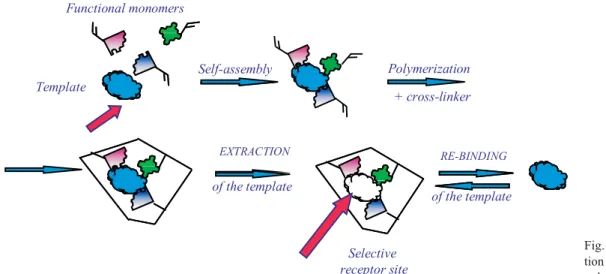
fast response and low cost of physical transducers, bio-sensor-based analytical methods are recognized to be the most effective methods of modern analytical bio-technology. However, despite the fact that many labo-ratory prototypes of biosensors were developed up to now, the number of commercially-available biosen-sors is relatively small. Apparently, that is mainly asso-ciated with low stability of all biomolecules in extreme environments. Moreover, the methods of isolation and purification of enzymes, antibodies, and receptors are quite sophisticated and time-consuming, which determi-nes their high cost. Therefore, development of highly-se-lective, effective and inexpensive analytical systems without unstable biological material is of great impor-tance. Recently artificial receptors and enzymes (poly-mers-biomimics) attracted significant attention of ana-lysts working in the biosensor technology field. These materials mimic binding sites of natural antibodies and receptors as well as enzyme active centers and, at the sa-me tisa-me, combine their high selectivity with desirable stability in aggressive media. One of the most effective approaches towards synthesis of polymers-biomimics is a technique of molecular imprinting, first reported by Wulff [83]. The method assumes synthesis of highly-cross-linked polymers around so-called template mole-cules (the analytes of interest). Extraction of the temp-late molecules from the fully-formed polymer results in formation of cavities in the polymeric network, which in size, shape and spatial arrangements of functional groups are complementary to the templates used in syn-thesis (Fig. 5). The polymers synthesized according to
Template
Functional monomers
Polymerization Self-assembly
+ cross-linker
of the template
EXTRACTION
of the template
RE-BINDING
Selective receptor site
this principle are capable of subsequent recognition of the template molecules, which occurs via combination of reversible binding and shape complementarity.
Molecularly imprinted polymers (MIPs) are being traditionally used for the analytical purposes in a form of MIP particles prepared by grinding and sieving of synthesized polymer blocks or particles prepared by suspension polymerization. However, use of MIP par-ticles in sensor technology is not effective since the ma-jority of synthetic binding sites are loosen in course of the polymer preparation. Moreover, a number of techno-logical problems arise during integration of polymeric particles with physical transducers. Therefore, a general approach towards synthesis of MIPs in a form of free-standing polymeric membranes and thin films on the surface of physical transducers was developed in our lab [84–88].
Using the novel approaches for MIP synthesis, con-ductometric, amperometric, and capacitive sensor devi-ces were developed for detection of environmental pol-lutants,i. e. triazine herbicides atrazine [84, 85] and des-metryn [89], cholesterol [90] and o-hydroxyphenols [91]. The main working characteristics of the MIP-ba-sed sensors are summarized in Table 8.
The principle of operation of all MIP-based sensors is similar to that of immunosensor devices. Biomime-tics function as selective elements of the sensors, which are responsible for recognition of the analyte. MIP is immobilized on the surface of a physical transducer, which transforms a signal arising after analyte binding in the electrochemical, thermal or optical signal. The lat-ter is proportional to the analyte concentration in the
ana-lyzed sample. The interaction of MIPs with the corres-ponding analyte (similarly to immunosensor devices) is not accompanied with the formation of electroactive products (protons or electrons). Therefore, the sensors for direct detection of a MIP-analyte binding event are based on a change of properties of the immobilized MIP (its electrical conductivity [84, 85] or capacity [89]) after its interaction with the analyte.
Application of the new approaches towards MIP synthesis in a form of membranes and thin films allow-ed us to detect corresponding analytes in nanomolar ran-ge (Table 7). The detection limit for desmetryn, provi-ded by the capacitive sensor based on thin MIP films, comprised 100 nM, while the linear dynamic range was 100–300 nM. The detection limit for atrazine, provided by the conductometric sensor based on free-standing MIP membranes synthesized by the method ofin situ po-lymerization comprised 15 nM. The linear dynamic ran-ge of the conductometric sensor comprised 15–50 nM. Time of the sensor responses did not exceed 10–12 min.
The most promising approach towards development of MIP-based sensors is synthesis of polymers-biomi-mics that are capable of not only highly-selective re-cognition of corresponding analytes, but also of genera-tion of a sensor response, which can be easily registered. From this point of view, synthesis of polymers-biomi-mics with catalytic properties, capable of highly-selecti-ve cleavage of the analytes of interest, is of great impor-tance. This would provide a possibility of their effecti-ve electrochemical detection. For instance, synthetic mi-mics of a natural enzyme tyrosinase (EC 1.14.18.1) were synthesized by our group according to the principle of
Analyte Dynamic range, M Operational mode Time of analysis, min Storage stability,
month References
Atrazine (15–50)×10–9
Steady-state 6–12 18 [84, 85]
Desmetryn (1–30)×10–8
Steady-state 10 12 [89]
o-Hydroxyphenol (0.078–5)×10–3
Steady-state 10 12 [91]
Phenol (0.0005–1)×10–2 Steady-state 20 18 [93]
Cholesterol (5–50)×10–6 Steady-state 10 1 [90]
Aflatoxin B1 (1–500)×10–9
Steady-state 20 18 [92]
Creatinine (0.25–2.5)×10–3
Steady-state 15 12 [93, 94]
Table 8
molecular imprinting. Taking into account literature data on a structure of tyrosinase active center, we assu-med that a molecule of o-hydroxyphenol, which coordi-nates two Cu(II) ions at a distance of 3,6–4 C (as it is happens in a natural enzyme) can be imprinted using urocanic acid ethyl ester as a functional monomer. This functional monomer in the MIP would mimic histidine residues that are present in an active center of natural tyrosinase. The synthesized polymers-biomimics (in a form of both polymeric particles and polymeric membra-nes) were used as selective elements of electrochemical biosensors for express-detection of o-hydrohyphenols. Application of the polymers with the optimized compo-sition as selective elements of electrochemical sensors allowed us to detecto-hydroxyphenols in aqueous solu-tions with the 0.078 mM detection limit, while the linear dynamic range of the sensor comprised 0.078–5 mM.
Free-standing MIP membranes obtained by either the method ofin situpolymerization or the method of photo-initiated grafting polymerization can be also used as a basis for easy-to-use optical (colorimetric or fluo-rescent) biosensor systems. The principle of their opera-tion is the following. The analytes of interest are selec-tively adsorbed by the synthetic receptor sites in the po-lymer structure after filtration of the analyzed samples through the MIP membranes. The revealing of the ad-sorbed analytes is based either on generation of own fluorescence of the analyte after UV-irradiation of the MIP membranes.
Alternatively, the non-fluorescent analytes can be revealed through the formation of the colored comp-lexes with the certain substances. Intensity of the MIP membranes’ staining or fluorescence is proportional to the concentration of certain analytes in the analyzed
samples. Fluorescent sensor systems for aflatoxin B1 detection were developed using the described principle [92]. The developed sensor systems provide aflatoxin B1 detection in the range 1–500 ng/ml with the detec-tion limit of 1 ng/ml.
Application of the computational modeling method for optimization of the polymer-biomimic composition provided synthesis of highly-selective receptors capable of selective recognition of aflatoxin B1 in mixtures con-taining close structural analogues of this toxin – afla-toxins B2 and G2.
Colorimetric biosensor systems for detection of phe-nol [93] and creatinine [94] for environmental monito-ring and medical diagnostics were developed on the ba-sis of MIP membranes obtained by the methods ofin situand grafting polymerization.
Storage stability of all the described sensors and sensor systems based on polymers-biomimics was very high and comprised 12–18 months (Table 1) for the MIP membranes stored in a dry state at room temperature. All the synthesized biomimics demonstrated extremely high selectivity, which is comparable to that of their na-tural counterparts.
High selectivity and stability of these materials ma-kes them an attractive alternative to natural receptors and enzymes in biosensor technology.
Conclusions. A number of electrochemical mono-and multibiosensors based on different enzymes, living cells and biomimics were developed, their laboratory prototypes were fabricated (Fig. 6) and thoroughly in-vestigated for real conditions of application (blood se-rum, blood dialisate, wine and wine must, natural fruit juices, environmental water samples, etc.). It is note-worthy that electrochemical biosensors are adaptable to
A B C D
Fig. 6. The laboratory prototypes of developed biosensors:A– conductometric enzyme system for determination of carbohydrates and aldehyde;
the technologies of large-scale production of miniaturi-sed devices. Concerning further wide application of the biosensors developed, the obtained results demonstrate the possibility to modulate their main characteristics to comply with the requirements specific for potential prac-tical purposes. Diverse biosensor modifications (with genetically modified enzymes, microorganisms, addi-tional membranes, different nanoscaled materials,etc.) can be elaborated.
It is important, that all the biosensors designed are complementary to traditional analytical techniques. Bio-sensors are an additional system for fast and early war-ning about the presence of various substances. They can provide a way to save time and costs under urgent con-ditions due to a possibility of making rapid decision re-garding local environmental problems or in the medical field where cheap and throw-away gadgets can be a tool for home monitoring of the patient’s state or early disease diagnostics. More accurate but time-consu-ming and expensive classical methods could be used (if necessary) for further validation and additional inves-tigation of the samples previously tested by biosensor arrays.
Acknowledgements. Part of the work was done in collaboration with Institute of Cell Biology, NAS of Ukraine, V. Ye. Lashkaryov Institute of Semiconduc-tor Physics, NAS of Ukraine, Institute of Electrodyna-mics, NAS of Ukraine, Ecole Centrale de Lyon (Fran-ce), C. Bernard University Lyon 1 (Fran(Fran-ce), Middle East Technical University of Ankara (Turkey) and Ins-titut Superieur des Sciences Appliquees et de Techno-logie de Sousse (Tunisia).
Î. Ï. Ñîëäàòê³í, Ñ. Â. Äçÿäåâè÷, ß. ². Êîðïàí, Ò. À. Ñåðãåºâà, Â. Ì. Àðõèïîâà, Î. À. Á³ëî³âàí, Î. Î. Ñîëäàòê³í, Ë. Â. Øêîòîâà, Î. À. dzí÷åíêî, Â. Ì. Ϻøêîâà, Î. ß. Ñàÿï³íà, Ñ. Â. Ìàð÷åíêî, À. Â. ªëüñüêà
Á³îñåíñîðè. ×âåðòü ñòîë³òòÿ äîñâ³äó íàóêîâî-äîñë³äíèõ ðîçðîáîê Ðåçþìå
Ïðåäñòàâëåíî îãëÿä âèêîíàíèõ ó ëàáîðàòî𳿠á³îìîëåêóëÿðíî¿ ýëåêòðîí³êè äîñë³äæåíü â îáëàñò³ ðîçðîáêè á³îñåíñîð³â íà îñíîâ³ åëåêòðîõ³ì³÷íèõ ïåðåòâîðþâà÷³â (àìïåðî- ³ êîíäóêòîìåòðè÷í³ åëåêòðîäè, ïîòåö³îìåòðè÷í³ ðÍ-÷óòëèâ³ ïîëüîâ³ òðàíçèñòîðè) ³ ð³çíèõ á³îðîçï³çíàâàëüíèõ ìîëåêóë (ôåðìåíòè, êë³òèíè, àíòèò³-ëà), á³îì³ì³ê³â àáî ñèíòåòè÷íèõ ìåìáðàí, âêëþ÷àþ÷è ìàòðè÷í³ ïîë³ìåðè, ÿê ÷óòëèâèõ åëåìåíò³â äëÿ ïðÿìîãî àíàë³çó ñóáñòðàò³â àáî ³íã³á³òîðíîãî àíàë³çó òîêñèí³â. Çàâäÿêè âèñîê³é ñïåöèô³÷íî-ñò³ ³ ÷óòëèâîñïåöèô³÷íî-ñò³, ïðîñòîò³ òà íèçüê³é âàðòîñïåöèô³÷íî-ñò³ âèçíà÷åííÿ
ð³ç-íèõ ðå÷îâèí á³îñåíñîðè º ïåðñïåêòèâíèìè ïðèëàäàìè äëÿ ïîòðåá îõîðîíè çäîðîâ’ÿ, êîíòðîëþ äîâê³ëëÿ, á³îòåõíîëî㳿, ñ³ëüñüêîãî ãîñïîäàðñòâà ³ õàð÷îâî¿ ïðîìèñëîâîñò³. Ðîçðîáëåíî é äîñë³äæå-íî á³îñåíñîðè äëÿ ïðÿìîãî âèçíà÷åííÿ íèçêè àíàë³ò³â òà ³íã³á³-òîðíîãî àíàë³çó ð³çíèõ òîêñè÷íèõ ðå÷îâèí. Ïîë³ïøåííÿ ¿õí³õ àíà-ë³òè÷íèõ õàðàêòåðèñòèê ìîæíà äîñÿãòè çà ðàõóíîê çàñòîñóâàí-íÿ äèôåðåíö³éíîãî ðåæèìó âèì³ðþâàíü, íåãàòèâíî àáî ïîçèòèâ-íî çàðÿäæåíèõ äîïîì³æíèõ íàï³âïðîíèêíèõ ìåìáðàí, íàïîçèòèâ-íîìàòå- íàíîìàòå-ð³àë³â ð³çíîãî ïîõîäæåííÿ, ãåíåòè÷íî ìîäèô³êîâàíèõ ôåðìåíò³â òîùî. Âèêîðèñòàííÿ öèõ ï³äõîä³â çðîáèòü ìîæëèâèì ï³äâèùèòè ÷óòëèâ³ñòü, ñåëåêòèâí³ñòü ³ ñòàá³ëüí³ñòü á³îñåíñîð³â, à òàêîæ ðîçøèðèòè äèíàì³÷íèé ä³àïàçîí âèì³ðþâàíü. Óïðîäîâæ îñòàí-í³õ 25 ðîê³â âèãîòîâëåíî á³ëüø ÿê 50 ëàáîðàòîðíèõ ïðîòîòèï³â á³îñåíñîðíèõ ñèñòåì íà îñíîâ³ ìîíî- ³ ìóëüòèá³îñåíñîð³â äëÿ ïðÿ-ìîãî âèçíà÷åííÿ ð³çíîìàí³òíèõ ìåòàáîë³ò³â òà ³íã³á³òîðíîãî àíàë³çó òîêñèêàíò³â. Äåÿê³ ç íèõ âèïðîáóâàíî çà óìîâ àíàë³çó ðå-àëüíèõ çðàçê³â.  îãëÿä³ îáãîâîðåíî ïåðåâàãè ³ íåäîë³êè ðîçðîáëå-íèõ á³îñåíñîð³â òà ðîçãëÿíóòî ìîæëèâîñò³ ¿õíüîãî ïðàêòè÷íîãî çàñòîñóâàííÿ.
Êëþ÷îâ³ ñëîâà: åëåêòðîõ³ì³÷íèé á³îñåíñîð, ³ììîá³ë³çîâàíèé ôåðìåíò, ñóáñòðàò, ³íã³á³òîð, ìóëüòèá³îñåíñîð.
À. Ï. Ñîëäàòêèí, Ñ. Â. Äçÿäåâè÷, ß. È. Êîðïàí, Ò. À. Ñåðãååâà, Â. Í. Àðõèïîâà, Î. À. Áåëîèâàí, À. À. Ñîëäàòêèí, Ë. Â. Øêîòîâà, Å. À. Çèí÷åíêî, Â. Í. Ïåøêîâà, Î. ß. Ñàÿïèíà, Ñ. Â. Ìàð÷åíêî, À. Â. Åëüñêàÿ
Áèîñåíñîðû. ×åòâåðòü âåêà îïûòà íàó÷íî-èññëåäîâàòåëüñêèõ ðàçðàáîòîê
Ðåçþìå
Êëþ÷åâûå ñëîâà: ýëåêòðîõèìè÷åñêèé áèîñåíñîð, èììîáèëè-çîâàííûé ôåðìåíò, ñóáñòðàò, èíãèáèòîð, ìóëüòèáèîñåíñîð.
REFERENCES
1.Turner A. P.Biosensors: sense and sensibility // Chem. Soc. Rev.–2013.–42, N 8.–P. 3184–3196.
2.Dzyadevych S. V., Soldatkin A. P., Korpan Y. I. et al.Biosensors based on enzyme field-effect transistors for determination of so-me substrates and inhibitors // Anal. Bioanal. Chem.–2003.–377, N 3.–P. 496–506.
3.Dzyadevych S. V., Arkhypova V. N., Soldatkin A. P. et al. Con-ductometric enzyme biosensors // Handbook of Biosensors and Biochips / Eds S. Marcs, D. C. Cullen, I. Karube et al.–Chi-chester: J. Willey & Sons Ltd., 2007.–P. 379–393.
4.Dzyadevych S. V., Arkhypova V. N., Soldatkin A. P. et al. Ampe-rometric enzyme bio- sensors: past, present and future // ITBM-RBM.–2008.–29, N 2.– P. 171–180.
5.Dzyadevych S. V., Soldatkin A. P., El’skaya A. V. et al.Enzyme biosensors based on ion-selective field-effect transistors // Anal. Chim. Acta.–2006.–568, N 1–2.–P. 248–258.
6.Shul’ga A. A., Sandrovsky A. K., Strikha V. I. et al.Overall cha-racterization of ISFET-based glucose biosensor // Sens. Actua-tors B: Chem.–1992.–10, N 1.–P. 41–46.
7.Dzyadevich S. V., Korpan Y. I., Arkhipova V. N. et al. Appli-cation of enzyme field-effect transistors for determination of glu-cose concentrations in blood serum // Biosens. Bioelectron.– 1999.–14, N 3.–P. 283–287.
8.Boubryak O. A., Soldatkin A. P., Starodub N. F. et al. Determi-nation of urea in blood serum by a urease biosensor based on an ion-sensitive field-effect transistor // Sens. Actuators B: Chem.– 1995.–27, N 1–3.–P. 429–431.
9.Hendji A. N., Jaffrezic-Renault N., Martelet C. et al.Sensitive de-tection of pestecides using differential ISFET-based scheme and immobilized cholinesterases // Anal. Chim. Acta.–1993.–281, N 1.–P. 3–11.
10.Arkhypova V. N., Dzyadevych S. V., Soldatkin A. P. et al. Multi-biosensor based on enzyme inhibition analysis for determina-tion of different toxic substances // Talanta.–2001.–55, N 5.– P. 919–927.
11.Gorchkov D. V., Soldatkin A. P., Maupas H. et al.Correlation bet-ween the electrical charge propertes of polymeric membranes and the characteristics of ion selective field effect transistors or peni-cillinase based enzymatic field ef- fect transistors // Anal. Chim. Acta.–1996.–331, N 3.–P. 217–223.
12.Korpan Y. I., Gonchar M. V., Sibirny A. A. et al.Development of highly selective and stable potentiometric sensors for formalde-hyde determination // Biosens. Bioelectron.–2000.–15, N 1–2.– P. 77–83.
13.Soldatkin A. P., Montoriol J., Sant W. et al.Creatinine sensitive biosensor based on ISFETs and creatinine deiminase immobili-sed in BSA membrane // Talanta.– 2002.–58, N 2.–P. 351–357. 14.Soldatkin A. P., Montoriol J., Sant W. et al.Development of po-tentiometric creatinine-sensitive biosensor based on ISFET and creatinine deiminase immobilised in PVA/SbQ photopolymeric membrane // Mater. Sci. Eng. C.–2002.–21, N 1–2.–P. 75–79. 15.Anh T. M., Dzyadevych S. V., Soldatkin A. P. et al.Development
of tyrosinase biosensor based on pH-sensitive field-rffect tran-sistor for phenols determination in water solution // Talanta.– 2002.–56, N 4.– P. 627–634.
16.Dzyadevych S. V., Mai Anh T., Soldatkin A. P. et al. Develop-ment of enzyme biosensor based on pH-sensitive field-effect transistors for detection of phenolic compounds // Bioelectroche-mistry.–2002.–55, N 1–2.–P. 79–81.
17.Biloivan O. A., Dzyadevich S. V., El’skaya A. V. et al. Develop-ment of bi-enzyme microbiosensor based on solid-contact ion-selective microelectrodes for protein detection // Sens. Actuators B: Chem.–2007.–123, N 2.–P. 1096–1100.
18.Marrakchi M., Dzyadevych S. V., Biloivan O. A. et al. Develop-ment of trypsine biosensor based on ion-sensitive field-effect tran-sistors for proteins deter mination // Mater. Sci. Eng. C.–2006.–26, N 2–3.–Ð. 369–373.
19.Biloivan O. A., Dzyadevich S. V., Boubriak O. A. et al. Develop-ment of enzyme biosensor based on ISFETs for Quantitative ana-lysis of serine proteinases // Electroanaana-lysis.–2004.–16, N 22.– P. 1883–1889.
20.Soldatkin A. P., El’skaya A. V., Shul’ga A. A. et al.Glucose sen-sitive field-effect transistor with additional Nafion membrane: reduction of influence of buffer capacity on the sensor response and extention of its dynamic range // Anal. Chim. Acta.–1993.–
283, N 3.–P. 695–701.
21.Volotovsky V., Soldatkin A. P., Shulga A. A. et al. Glucose-sen-sitive ion-senGlucose-sen-sitive field-effect transistor-based biosensor with additional positively charged membrane. Dynamic range ex-tension and reduction of buffer concentration influence on the sensor response // Anal. Chim. Acta.–1996.–322, N 1–2.– P. 77–81.
22.Gorchkov D. V., Soldatkin A. P., Poyard S. et al.Application of charged polymeric materials as additional permselective memb-ranes for improvement of the performance characteristics of urea-sensitive enzymatic field effect transistors. 1. Determination of urea in model solutions // Mater. Sci. Eng. C.–1997.–5, N 1.– P. 23–28.
23.Gorchkov D. V., Soldatkin A. P., Poyard S. et al.Application of charged polymeric materials as additional permselective memb-ranes for improvement of the performance characteristics of urea-sensitive enzymatic field effect transistors. 2. Urea determination in blood serum // Mater. Sci. Eng. C.–1997.–5, N 1.–P. 29–34. 24.Soldatkin A. P., Dzyadevych S. V., El’skaya A. V. et al.Pathways
for improving potentiometric and conductometric enzymatic bio-sensors // Encyclopedia of Sensors / Eds C. A. Grimes, E. C. Di-ckey, M. V. Pishko.–Stevenson Ranch: Amer. Sci. Publ., 2006.– Vol. 7.–P. 331–348.
25.de Melo J. V., Soldatkin A. P., Martelet C. et al.Use of compe-titive inhibition for driving sensitivity and dynamic range of urea ENFETs // Biosens. Bioelectron.–2003.–18, N 4.–P. 345–351. 26.Soldatkin A. P., Montoriol J., Sant W. et al.A novel urea
sensiti-ve biosensor with extended dynamic range based on recombi-nant urease and ISFETs // Biosens. Bioelectron.–2003.–19, N 2.– P. 131–135.
27.Zinchenko O. A., Marchenko S. V., Sergeyeva T. A. et al. Appli-cation of creatinine-sensitive biosensor for hemodialysis control // Biosens. Bioelectron.–2012.–35, N 1.–P. 466–469.
28.Marchenko S. V., Soldatkin A. P. Potentiometric biosensor based on recombinant urease for urea detection in real biological samp-les // Sensor Electronics and Microsystem Technologies.– 2012.–3(9), N 4.–P. 40–48.
29.Soldatkin A. P.Urease-based biosensor with improved sensitivi-ty for analysis of heavy metal ions // Biopolym. Cell.–1997.–13, N 5.–P. 377–379.