J of Evolution of Med and Dent Sci/ eISSN- 2278-4802, pISSN- 2278-4748/ Vol. 3/ Issue 49/Oct 02, 2014 Page 11726
FUNCTIONAL OUTCOME FOLLOWING RECONSTRUCTION FOR CHRONIC
ISOLATED DORSAL DISTAL RADIOULNAR JOINT INSTABILITY BY
FULKERSON-WATSON METHOD-A PROSPECTIVE STUDY
Santhamoorthy T1, Arun K2HOW TO CITE THIS ARTICLE:
Santhamoorthy T, Arun K. Functional Outcome Following Reconstruction for Chronic Isolated Dorsal Distal Radioulnar Joint Instability by Fulkerson-Watson Method - A Prospective Study. Journal of Evolution of Medical and Dental Sciences 2014; Vol. 3, Issue 49, October 02; Page: 11726-11735,
DOI: 10.14260/jemds/2014/3540
ABSTRACT: BACKGROUND: Chronic isolated distal radioulnar joint instability is a relatively rare entity. Several methods of reconstruction were available to stabilize the joint and each method has some advantage over others. We proposed to assess the functional outcome following reconstruction of chronic dorsal distal radio ulnar instability using extra articular reconstruction by Fulkerson – Watson method. AIM: To assess the functional outcome following reconstruction for chronic isolated dorsal distal radio ulnar instability using Fulkerson –Watson method. METHODS: We conducted a prospective study in five patients over three years from 2010 to 2013 with chronic isolated dorsal distal radio ulnar instability who were treated by Fulkerson-Watson method of reconstruction. All patients underwent MRI evaluation before surgery to assess ligament pathology and for adequacy of sigmoid notch. Arthroscopy performed in all patients. Functional outcomes were assessed using VAS score, quick-DASH score and Mayo wrist score at every 6 months follow-up. Radiological assessment done using plain x-rays at each follow up. RESULTS: Three patients required Arthroscopic debridement for TFCC. All five patients had achieved stability at distal radio ulnar joint after surgery and remained so till their last follow up. One patient had persistent pain near ulnar styloid. The average loss of motion for pronation was 10 degrees and supination was 3 degrees in reference to the normal side. All except one patient achieved ulnar grip strength of >90 % compared to normal side. The mean pre and postoperative VAS score, quick-DASH score, Mayo wrist score were 76.6 and 17.2, 37.3 and 11.3, 45 and 77 respectively. CONCLUSION: Though extra articular reconstruction for DRUJ by Fulkerson-Watson method is non-anatomical, the procedure is simple than intra articular reconstruction and gives similar functional outcome like intra articular reconstructions as shown by our results.
KEYWORDS: Distal radio ulnar joint, triangular Fibrocartilage complex. Extra Articular reconstruction.
INTRODUCTION: Stability of the distal radio ulnar joint (DRUJ) is provided by bony architecture and by soft tissues such as the triangular fibrocartilage complex (TFCC), the joint capsule, and surrounding muscles. DRUJ is a diarthrodial trochoid synovial joint relatively new in evolution, evolved from the primitive pectoral fin of early fish to the bipedal primate wrist to its current form in human wrist.1 The radio ulnar articulation is formed by the lower end of ulna (seat) and the sigmoid notch (medial articular facet) of the distal radius.
J of Evolution of Med and Dent Sci/ eISSN- 2278-4802, pISSN- 2278-4748/ Vol. 3/ Issue 49/Oct 02, 2014 Page 11727 fibrocartilage (TFC or articular disk), meniscal homologue, ulnocarpal [ulnolunate (UL) and lunotri quetral] ligaments, the dorsal and volar radio ulnar ligaments, ulnar collateral ligament, and the extensor carpi ulnar is (ECU) sub sheath. The ECU sub sheath is reinforced medially by linear fibers referred to as the linea jugata. The radio ulnar ligaments (dorsal and volar) are the primary stabilizers of the DRUJ. The secondary stabilizers include the ECU sub sheath, the UL and ulnotriquetral (UT) ligaments, the lunotriquetral interosseous ligament (LTIOL).
Incidence and mechanism of isolated dorsal dislocation of DRUJ: Subluxations and dislocations without concomitant fracture of the forearm, distal radius, or ulnar styloid are uncommon but have been described.3 By convention, DRUJ dislocation is defined on the basis of the final location of the ulna head. Dorsal dislocations are more common: the mechanism is hyper pronation. The mechanism for dorsal subluxation and dislocation is extreme pronation and extension, with a tightened ECU and ulnar carpal ligaments, which pull the ulnar head out through the dorsal capsule. TFCC avulsion and attenuation of the palmar radio ulnar ligament will allow this dislocation.2
Acute dislocations can be categorized as simple or complex. Simple dislocations can be readily reduced or even undergo spontaneous reduction. Complex dislocations can be characterized by irreducibility or by an inability to maintain reduction.4Typical obstructions to reduction are the ECU and extensor communis, the TFC, and the extensor digiti minimi (EDM), either in concert or alone.
Dorsal dislocations are usually reduced in supination. In chronic isolated DRUJ dislocations more than one components of TFCC would have been injured apart from dorsal and volar radio ulnar ligaments.5 Hence it is important to address these associated injuries to get a satisfactory functional outcome. This involves either repair of peripheral tears or debridement of central tears by arthroscopy.
One of the problem in intra articular reconstruction of DRUJ would be chances of injuring the normal components of TFCC and its related morbidity.6 unlike the knee joint where one can greatly appreciate the anatomy of individual ligaments it was feasible for intra articular anatomical reconstruction and hence satisfactory functional outcome, the same would not be possible with TFCC as it is more complex and less discernable to naked eye.
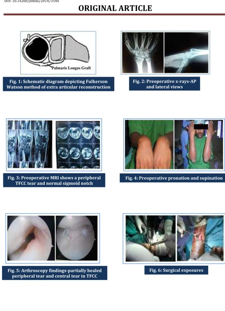
Extra articular reconstruction though non-anatomical theoretically should produce less morbidity.7 Hence we proposed to do a prospective study on functional outcome after reconstruction of chronic isolated dorsal distal radio ulnar instability by Fulkerson-Watson method (Fig-1).
PATIENTS AND METHODS: We prospectively followed up five patients from 2010 to 2013 who were operated by Fulkerson-Watson method of reconstruction for chronic isolated dorsal distal radio ulnar instability. Our inclusion criteria were patients with posttraumatic isolated dorsal DRUJ instability of 2 or more months in duration. Four were males and one was female. The mean age of the patients was 37.
J of Evolution of Med and Dent Sci/ eISSN- 2278-4802, pISSN- 2278-4748/ Vol. 3/ Issue 49/Oct 02, 2014 Page 11728 As there were no bony deformities, corrective osteotomy or sigmoid notchplasty were not performed. Palmaris longus graft harvested from same side. Through the same graft harvested wound on volar aspect distal radius approached and a vertical tunnel made 3mm above and lateral to sigmoid notch. One end of the graft passed through this tunnel and retrieved on the dorsum through a 2.5 cms skin incision lying over the ulna. This end was looped around the neck of ulna and brought through the interosseous membrane to the dorsum. The other free end of the graft also brought through the interosseous membrane to the dorsum.
While maintaining the reduction in full supination the free ends are sutured to the main loop using non-absorbable sutures. Temporary k-wire fixation also performed in all cases. Long arm cast support in neutral position maintained for 2 weeks. K-wire was removed at 6th week. Rehabilitation started from then. Pre and postop VAS score, Quick DASH score and Mayo wrist score were calculated at every 6months. Radiological assessment was done using plain x-ray at each follow up.
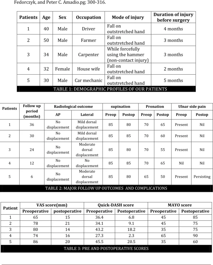
RESULTS: The average duration after injury before surgical intervention was 3.4 months (Table-1). Plain x-ray revealed increased radio ulnar space in all patients in AP view and dorsal subluxation in lateral view (Fig-2). MRI revealed peripheral tear of TFCC in 2 patients, central tear of TFCC in 3 patients and tear of dorsal radio ulnar ligament in all patients (Fig-3).
Arthroscopic findings correlated well with MRI findings (Fig-5). Debridement was done for the central tears and no intervention done for peripheral tears, as they were small and partially healed. All patients underwent reconstruction by Fulkerson-Watson method (Fig-6). The average follow up period was 21.6 months (minimum 6 and maximum 36). All patients achieved clinical stability at their final follow-up.
One patient had persistent ulnar side pain in his last follow-up. Immediate Postoperative x-rays showed satisfactory reduction and stabilization in all patients in both AP and lateral views (Fig-7). Except one patient rest of them showed variable amount of dorsal subluxation in lateral view x-rays, but well maintained reduction in AP view at their last follow-up (Fig-8 and 9).
Four patients returned to their previous occupation and one patient with persistent ulnar side pain changed his profession. The mean preoperative Quick-DASH score was 37.3 (27.3–45.5) and decreased to 11.3 (2.3–20.5) after surgery. The mean VAS score decreased from 76.6 (65-86) to 17.2 (14-21) after surgery. The mean Mayo wrist score improved from 45 (35-65) to 77(60-90) after surgery (Table -3).
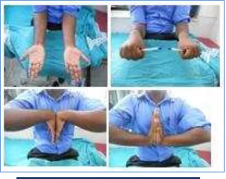
All except one patient achieved ulnar grip strength of >90 % compared to normal side. The average loss of motion for pronation was 10 degrees and supination was 3 degrees in reference to the normal side and there was no restriction of wrist dorsiflexion or palmar flexion (Fig-10).
DISCUSSION: The distal radio ulnar joint is the distal articulation of the biarticulate rotational arrangement of the forearm where the ulna is fixed segment and the radius rotates around it in supination and pronation. The peculiarity of this joint is it allows simultaneous rotation and antero posterior translation.13 Stability of the distal radio ulnar joint is provided by the joint surface morphology, the joint capsule, the dorsal and palmar radio ulnar ligaments, the interosseous membrane, and the musculotendinous units, primarily the extensor carpi ulnaris and pronator quadratus.8,9
J of Evolution of Med and Dent Sci/ eISSN- 2278-4802, pISSN- 2278-4748/ Vol. 3/ Issue 49/Oct 02, 2014 Page 11729 and is a result of soft tissue injury or osseous malunion or a combination of both. Chronic distal radio ulnar joint instability is a painful and disabling condition with reduced hand grip, restricted rotation of forearm and pain on axial loading.
Isolated dislocation of DRUJ is relatively rare entity described only as case studies and case series in literature. In that the dorsal dislocation is more common than volar. The usual mechanism for dorsal subluxation and dislocation is hyper pronation and extension, with a tightened ECU and ulnar carpal ligaments, which pull the ulnar head out through the dorsal capsule. TFCC avulsion and attenuation of the palmar radio ulnar ligament also allow this dislocation.2 In four of our patients the mechanism of injury was fall on outstretched hand with hyper pronation. In one patient the injury occurred as non-contact mechanism while using the hammer (Table-1).
The indications and criteria for ligament reconstruction include unidirectional or bidirectional chronic instability of the distal radio ulnar joint, absence of substantial arthritis, and a competent sigmoid notch rim with no residual axial instability of the forearm.19 Most common symptom of chronic dislocation of DRUJ would be pain in the wrist often associated with symptoms of mechanical instability, including weakness and a joint clunk. The distal ulna may remain tender and appear prominent. In mild instability, pain and weakness often occur only with activities that require power rotation of the forearm, especially while gripping.17
In our study also the predominant symptom was pain associated with loss of grip strength. We could clinically diagnose the dislocation with prominence of ulnar head on the dorsum with variable tenderness on ulnar side and a positive piano key test. The table top test though was described for DRUJ instability, was not easily reproducible in all patients.20 The grind test was used to test for DRUJ arthrosis. Unusually in all our patients full supination was preserved before surgery, but variable pronation restriction was found (Fig-4).
We confirmed our diagnosis with plain radiography and assessed the status of sigmoid notch and associated TFCC injuries using MRI in all our patients. We felt that CT scan evaluation was not essential in our patients as sigmoid notch was well delineated in MRI imaging itself. T2-weighted images in the coronal plane are of the greatest diagnostic value. The TFCC has homogeneous low signal intensity. The synovial fluid of the joint appears as a bright image on T2 and will outline tears in the TFCC. The addition of a gadolinium arthrogram enhances the visualization of TFCC tears.20
Arthroscopy is the gold standard to diagnose TFCC injuries. It is also used to treat various TFCC injuries. Central tears are not amenable to repair due to avascularity of the central zone of the TFCC, which precludes healing and peripheral ulnar sided tears involve the vascular zone of the TFCC and can be repaired either arthroscopically or by open technique.19 In our study arthroscopy revealed central tear of TFCC in 3 patients and peripheral tear in 2 patients. Central tears were effectively debrided. Although the peripheral tears ideally to be repaired in our patients they were small and partially healed due to late presentation. Hence we decided to leave it alones.
In a biomechanical cadaver model, intra articular reconstructions of the radio ulnar ligaments were found to be superior to radio ulnar tethering procedures although the results of capsular repair alone most closely matched the kinematics of an intact distal radio ulnar joint.11 Fulkerson and Watson procedure is a direct radio ulnar tether extrinsic to the joint.10 The other methods of extra articular reconstructions described were by Eliason, Boyes and Bunnel, Hui and Linseid etc.
J of Evolution of Med and Dent Sci/ eISSN- 2278-4802, pISSN- 2278-4748/ Vol. 3/ Issue 49/Oct 02, 2014 Page 11730 provide immediate DRUJ stability in cadaveric tests.14,16 Intra articular reconstructions of distal radio ulnar joint, though anatomical is technically more demanding with a propensity for injuring normal structures and hence not easily reproducible.11,12
Husrev Purisa et al in a retrospective study shown satisfactory short term results following extra articular reconstruction for chronic isolated DRUJ instability in five patients using Fulkerson-Watson procedure.18 Based on their results we proposed to conduct a prospective study using Fulkerson –Watson method in patients with chronic isolated dorsal instability of DRUJ. Five consecutive patients fulfilling the inclusion criteria underwent this surgery.
Three surgical approaches to the DRUJ were described in literature-dorsoulnar, ulnar, and palmar. We used combined palmar and dorsoulnar approach in our patients. We harvested Palmaris longus from same side. A vertical tunnel made in distal radius. Palmaris longus tendon graft passed through this and looped around neck of ulnar with forearm in complete supination. It is secured with nonabsorbable sutures while maintaining the reduction and joint stabilized temporarily with a K -wire. Forearm and wrist joint immobilized in long arm posterior slab for 2 weeks. K-wire removed at 6th week.
In all our patients we used a thin cerclage wire tied to the graft end for easy passage of the graft through the tunnel and for its retrieval to make the loop around ulnar neck. Patient’s functional outcomes were assessed using the preoperative and postoperative Visual Analogue Score (VAS), quick DASH score and MAYO wrist score at 6 months interval till their last follow up. On last follow up it was noted that the VAS score and Quick DASH score have decreased and the Mayo wrist score had significantly increased (Table-2). All except one patient had grip strength of more than 90 %.
Our functional outcome were comparable to previous studies which used similar method of reconstruction as well as to studies in which other methods of reconstruction were used.12,13,15,18,19
The postoperative x rays of all our patients showed well reduced distal radio ulnar joint in AP view, but mild to moderate dorsal subluxation was evident in four of our patients in their last follow-up (Table-2). But, none of them had clinical findings of instability. This would suggest that the important cause for clinical symptoms in these patients may not be due to antero-posterior displacement, rather may be due to rotational and abnormal axial forces as described in various biomechanical studies.7,8
Scheker LR.et al in their study stated that preserving balanced rotation of the forearm after reconstruction around the ulnar head is difficult, and limitation of supination and pronation is a possible sequelae.7 In our study the average loss of motion of pronation was more than supination. We didn’t encounter any specific surgical complications. We understood from our study that the main technical difficulties in this method were retrieval of the graft and securing the sutures in well reduced position.
One patient had persistent pain over the ulnar styloid region till his last follow-up. The probable reason would be an unaddressed peripheral tear of TFCC. We didn’t encounter any indentation by graft on the ulna in postoperative x-rays, as we stayed close to the DRUJ while forming the loop intra operatively. To our knowledge this is the first prospective study done for isolated dorsal dislocation of DRUJ using Fulkerson-Watson method. One of the constraints of our study is small sample size. But considering the rarity of this disease entity we could not avoid it.
J of Evolution of Med and Dent Sci/ eISSN- 2278-4802, pISSN- 2278-4748/ Vol. 3/ Issue 49/Oct 02, 2014 Page 11731 simple and gives satisfactory functional outcome as shown by our results. We like to conclude that future long term studies comparing intra articular and extra articular methods would be strongly recommended.
REFERENCES:
1. Thomas BP, Sreekanth R. Distal radio ulnar joint injuries. Indian J Orthop 2012; 46: 493-504 2- Szabo RM. Distal radio ulnar joint instability. J Bone Joint Surg. Am. 2006; 88 (4): 884-94.
2. Palmer AK, Werner FW. The triangular fibrocartilage complex of the wrist: anatomy and function. J Hand Surg Am. 1981; 6 (2):153-62.
3. Lichtman DM, Joshi A. Acute injuries of the distal radio ulnar joint and triangular fibrocartilage complex. Instr. Course Lect.2003; 52: 175-83.
4. Bruckner, J.D. D.M. Lichtman, and A.H. Alexander. Complex dislocations of the distal radio ulnar joint. Recognition and management. Clin. Ortho. Relat. Res. 1992 (275): 90-103.
5. Ward LD1, Ambrose CG, Masson MV, Levaro F. The role of the distal radio ulnar ligaments, interosseous membrane, and joint capsule in distal radio ulnar joint stability J Hand Surg. Am. 2000 Mar; 25 (2): 341-51.
6. Palmer AK, Werner FW. The triangular fibrocartilage complex of the wrist--anatomy and function. J Hand Surg Am. 1981 Mar; 6 (2): 153-62.
7. Scheker LR, Ozer K. Ligamentous stabilization of the distal radio ulnar joint. Tech Hand Up Extrem Surg 2004; 8: 239-46.
8. Gofton WT, Gordon KD, Dunning CE, Johnson JA, King GJ. Soft-tissue stabilizers of the distal radio ulnar joint: an in vitro kinematic study. J Hand Surg [Am]. 2004; 29: 423-31.
9. Kihara H, Short WH, Werner FW, Fortino MD, Palmer AK. The stabilizing mechanism of the distal radio ulnar joint during pronation and supination. J Hand Surg [Am]. 1995; 20: 930-6. 10.Fulkerson JP, Watson HK. Congenital anterior subluxation of the distal ulna. A case report. Clin
Orthop Relat Res. 1978; 131: 179-82.
11.Gofton WT, Gordon KD, Dunning CE, Johnson JA, King GJ. Comparison of distal radio ulnar joint reconstructions using an active joint motion simulator J Hand Surg [Am]. 2005; 30: 733-42. 12.Adams, B. D., & Berger, R. A. (2002). An anatomic reconstruction of the distal radio ulnar
ligaments for posttraumatic distal radio ulnar joint instability. The Journal of Hand Surgery, 27 (2), 243–251.
13.Henry. Anatomic reconstruction of the radio ulnar ligament. Hand (N Y). 2012; 7 (4): 413–419. 14.Kyu Nam Seo, MD, Min Jong Park, MD, Hong Je Kang, MD. Anatomic Reconstruction of the Distal
Radio ulnar Ligament for Posttraumatic Distal Radio ulnar Joint Instability. Clinics in Orthopedic Surgery. 2009; 1: 138-145.
15.Hui FC, Linscheid RL. Ulnotriquetral augmentation tenodesis: a reconstructive procedure for dorsal subluxation of the distal radio ulnar joint. J Hand Surg [Am] 1982; 7: 230–236.
16.Petersen MS, Adams BD. Biomechanical evaluation of distal radio ulnar reconstructions. J Hand Surg [Am] 1993; 18: 328–334.
17.Hand Surgery 1st Edition 2004. Lippincott Williams & Wilkins pg; 338-353.
J of Evolution of Med and Dent Sci/ eISSN- 2278-4802, pISSN- 2278-4748/ Vol. 3/ Issue 49/Oct 02, 2014 Page 11732 19.Scheker LR, Ozer K. Ligamentous stabilization of the distal radio ulnar joint. Tech Hand Up
Extrem Surg 2004; 8: 239-46.
20.Rehabilitation of the Hand and Upper Extremity. Terri M. Skirven, A. Lee Osterman, Jane M. Fedorczyk, and Peter C. Amadio.pg; 300-316.
Patients Age Sex Occupation Mode of injury Duration of injury before surgery
1 40 Male Driver Fall on
outstretched hand 4 months
2 50 Male Farmer Fall on
outstretched hand 3 months
3 34 Male Carpenter
While forcefully using the hammer (non-contact injury)
3 months
4 32 Female House wife Fall on
outstretched hand 2 months
5 30 Male Car mechanic Fall on
outstretched hand 5 months TABLE 1: DEMOGRAPHIC PROFILES OF OUR PATIENTS
Patients Follow up period (months)
Radiological outcome supination Pronation Ulnar side pain
AP Lateral Preop Postop Preop Postop Preop Postop
1 36 No
displacement
Mild dorsal
displacement 85 80 70 65 Present Nil
2 30 No
displacement
Mild dorsal
displacement 85 85 70 60 Present Nil
3 24 No
displacement
Moderate dorsal displacement
85 80 70 55 Present Nil
4 12 No
displacement
No
displacement 85 85 70 65 Nil Nil
5 6 No
displacement
Moderate dorsal displacement
85 80 65 50 Present Persisting
TABLE 2: MAJOR FOLLOW UP OUTCOMES AND COMPLICATIONS
Patient VAS score(mm) Quick-DASH score MAYO score
Preoperative postoperative Preoperative Postoperative Preoperative Postoperative
1 65 15 36.4 6.8 45 85
2 78 21 34.1 9.1 45 75
3 80 14 43.2 18.2 35 75
4 74 16 27.3 2.3 65 90
5 86 20 45.5 20.5 35 60
J of Evolution of Med and Dent Sci/ eISSN- 2278-4802, pISSN- 2278-4748/ Vol. 3/ Issue 49/Oct 02, 2014 Page 11733
Fig. 1: Schematic diagram depicting Fulkerson Watson method of extra articular reconstruction
Fig. 2: Preoperative x-rays-AP and lateral views
Fig. 3: Preoperative MRI shows a peripheral TFCC tear and normal sigmoid notch
Fig. 4: Preoperative pronation and supination
Fig. 5: Arthroscopy findings-partially healed peripheral tear and central tear in TFCC
J of Evolution of Med and Dent Sci/ eISSN- 2278-4802, pISSN- 2278-4748/ Vol. 3/ Issue 49/Oct 02, 2014 Page 11734 Fig. 7: Post operative x-rays showing well
reduced dislocation and stabilization
Fig. 8: x-rays at 1 year follow-up showing well maintained reduction
Fig. 9: Moderate dorsal displacement of ulna at 1 year follow-up
J of Evolution of Med and Dent Sci/ eISSN- 2278-4802, pISSN- 2278-4748/ Vol. 3/ Issue 49/Oct 02, 2014 Page 11735
AUTHORS:
1. Santhamoorthy T. 2. Arun K.
PARTICULARS OF CONTRIBUTORS:
1. Assistant Professor, Department of Orthopaedics, Sri Venkateswara Medical College Hospital & Research Centre, Ariyur, Puducherry.
2. Senior Resident, Department of
Orthopaedics, Sri Venkateswara Medical College Hospital & Research Centre, Ariyur, Puducherry.
NAME ADDRESS EMAIL ID OF THE CORRESPONDING AUTHOR:
Santhamoorthy T, # 5, Ragavendra Illam, Indhira Gandhi St, Nainarmandapam, Puducherry-605004.
Email: [email protected]