Assessment of upper airways measurements in patients
with mandibular skeletal Class II malocclusion
Nayanna Nadja e Silva1, Rosa Helena Wanderley Lacerda2, Alexandre Wellos Cunha Silva3, Tania Braga Ramos4
How to cite this article: Nadja e Silva N, Lacerda RHW, Silva AWC, Ra-mos TB. Assessment of upper airways measurements in patients with mandibular skeletal Class II malocclusion. Dental Press J Orthod. 2015 Sept-Oct;20(5):86-93. DOI: http://dx.doi.org/10.1590/2177-6709.20.5.086-093.oar
Submitted: January 13, 2015 - Revised and accepted: May 18, 2015
Contact address: Rosa Helena Wanderley Lacerda Associação Brasileira de Ortodontia
Rui Barbosa 38 , João Pessoa, Paraíba - CEP: 58.020-040 - Brazil E-mail: [email protected]
1 Specialist in Orthodontics, Associação Brasileira de Odontologia (ABO-PB),
João Pessoa, Paraíba, Brazil.
2 Coordinator, Postgraduate Program in Orthodontics, Associação Brasileira de
Odontologia (ABO-PB), João Pessoa, Paraíba, Brazil.
3 Professor, Postgraduate Program in Orthodontics, Associação Brasileira de
Odontologia (ABO-PB), João Pessoa, Paraíba, Brazil.
4 Professor of Orthognathic Surgery, Postgraduate Program in Orthodontics,
Associação Brasileira de Odontologia (ABO-PB), João Pessoa, Paraíba, Brazil.
» The authors report no commercial, proprietary or financial interest in the prod-ucts or companies described in this article.
Objective: Mandibular Class II malocclusions seem to interfere in upper airways measurements. The aim of this study was to assess the upper airways measurements of patients with skeletal Class II malocclusion in order to investigate the association between these measurements and the position and length of the mandible as well as mandibular growth trend, comparing the Class II group with a Class I one. Methods: A total of 80 lateral cephalograms from 80 individuals aged between 10 and 17 years old were assessed. Forty radiographs of Class I malocclusion individuals were matched by age with forty radiographs of individuals with mandibular Class II malocclusion. McNamara Jr., Ricketts, Downs and Jarabak’s measurements were used for cephalometric evaluation. Data were submitted to descriptive and inferential statistical analysis by means of SPSS 20.0 statistical package. Student’s t-test, Pearson correlation and intraclass correla-tion coefficient were used. A 95% confidence interval and 5% significance level were adopted to interpret the results. Results: There weredifferences between groups. Oropharynx and nasopharynx sizes as well as mandibular position and length were found to be reduced in Class II individuals. There was a statistically significant positive correlation between the size of the oropharynx and Xi-Pm, Co-Gn and SNB measurements. In addition, the size of the nasopharynx was found to be correlated with Xi-Pm, Co-Gn, facial depth, SNB, facial axis and FMA. Conclusion: Individuals with mandibular Class II malocclusion were shown to have upper airways measurements diminished. There was a correlation between mandibular length and position and the size of oropharynx and nasopharynx.
Keywords:Angle Class II malocclusion. Oropharynx. Nasopharynx. Airway obstruction. DOI: http://dx.doi.org/10.1590/2177-6709.20.5.086-093.oar
Introdução: as más oclusões de Classe II mandibulares parecem interferir nas dimensões das vias aéreas superiores. As-sim, o objetivo do presente estudo foi avaliar as vias aéreas superiores de pacientes com Classe II esquelética, verificando a asso-ciação entre essas dimensões e a posição mandibular, o comprimento mandibular e a tendência de crescimento, comparando-os com um grupo pareado de pacientes com Classe I. Métodos: foram avaliadas 80 telerradiografias de perfil de 80 pacientes com 10 a 17 anos de idade, sendo 40 com má oclusão de Classe I e 40 com Classe II mandibular, pareados por idade. Para a avaliação cefa-lométrica, foram utilizadas medidas de McNamara Jr, Ricketts, Downs e Jarabak. Os dados foram submetidos à análise estatística descritiva e inferencial, por meio do software SPSS 20.0, utilizando-se os testes t de Student, coeficiente de correlação de Pearson e coeficiente de correlação intraclasse. Para interpretação dos resultados, adotou-se um intervalo de confiança de 95% e nível de significância de 5%. Resultados: houve diferença entre os grupos, e as medidas da orofaringe e nasofaringe foram menores no grupo de Classe II, assim como as medidas de comprimento e posição mandibular. Houve correlação positiva estatisticamente significativa entre a orofaringe e as medidas Xi-Pm, Co-Gn e SNB; já a nasofaringe apresentou correlação com as medidas Xi-Pm, Co-Gn, profundidade facial, SNB, eixo facial e FMA. Conclusão: indivíduos portadores de Classe II mandibular apresentaram as medidas das vias aéreas superiores diminuídas. Observou-se uma correlação entre o comprimento mandibular e a posição mandibular e as dimensões da orofaringe e da nasofaringe.
INTRODUCTION
Skeletal Class II malocclusion is a dentofacial defor-mity caused by a growth disorder of the bones frequently associated with mandibular retrusion relative to upper facial structures.1 This deformity is also associated with functional disorders, mainly afecting upper airways and the temporomandibular joint.2,3
Patients with skeletal Class II malocclusion who have this deformity due to deiciency in mandibular growth present with a retrognathic mandible either because of growth vector or by deicient mandibular length.
According to Muto et al,4 craniofacial abnormalities, including mandibular retrognathism, short mandibular body length and backward/downward rotation, can lead to decreased pharyngeal airway. These indings indi-cate that nasopharyngeal obstruction may be related to changes in mandibular morphology.5
The study of upper airways and their relationship with mandibular position and size is extremely im-portant in orthodontic diagnosis because of their asso-ciation with obstructive respiratory disorders, especially sleep apnea. This knowledge is deinitive to the indica-tion of mandibular advancement, whether orthopedic or surgical, for treatment of these disorders.
Several studies have been carried out with a view to mea-suring the pharyngeal airway; however, comparison with Class I individuals and the correlation between the variables involved in Class II malocclusion and airways measurements are still scarce, which encouraged the present study.
MATERIAL AND METHODS
This study was submitted to and approved by the Ethics Committee on Human Research through Plata-forma Brasil, following the norms of the law 466/2012, under approval protocol #835.928.
The sample comprised 80 digital lateral cephalo-grams belonging to 80 patients of both sexes, without associated abnormalities, aged between 10-17 years, with a mean age of 12.3 years, treated by postgradu-ate orthodontic students (ABO/PB, Brazil). Of the 80 images, 40 were from patients with mandibular Class II malocclusion, whose diagnosis was conirmed by Xi-Pm, Co-Gn, Go-Me, facial depth and SNB measurements (at least three of these measures should be reduced so that the image would not be withdrawn from the sample). The other 40 radiographs belonged to Class I individuals. Groups were matched by age.
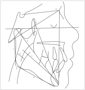
Anatomical tracings of all radiographic images were made on acetate paper, in a dark room, by an examiner us-ing graphite pencil (point 0.3). Each ilm was traced by one investigator and checked by a second one, so as to verify the accuracy of anatomical outline determination and landmark placement. Measurements of mandibular length and spatial position, as well as size of nasopharyngeal and oropharyngeal airways, were taken using the cephalograms (Fig 1, Table 1).
Measurements were taken twice, with a 10-day interval in between, with the aid of a millimeter ruler and a 180° protractor. The irst assessment was carried out with the entire sample while the second one was carried out with 30% of the sample.
Procedures of statistical inference were performed based on parametric statistics. Correlation coeicient and intra-class correlation coeicient (ICC) were used to assess in-traexaminer agreement. The choice for statistical test was based on normal distribution of data, according to Ko-mogorov-Smirnov normality test (p > 0.05). Intergroup comparison was performed by Student’s t-test and Pearson r correlation coeicient. For descriptive procedures, abso-lute and relative data and measurements of central tendency and variability were presented. A 95% conidence interval and 5% signiicance level (p < 0.05) were adopted to inter-pret the results. Data were submitted to SPSS 20.0 statistical package for Windows and analyzed by means of descriptive and inferential statistics.
Figure 1 - Cephalogram and cephalometric measurements used.
9
3
1
8 6
7
2
4
Table 1 - Cephalometric measurements used.
Measure Clinical standard Appropriate age Description
1. Xi-Pm 65 ± 3 mm 9 years (1.6/year) Axis of the mandibular body – a line extending from point Xi to the mental
protuberance.
2. Facial axis
(Ba.NA x Frankfurt) 90 ± 3°
Does not change upon growth
Provides the direction of growth of the chin and the ratio between facial height and depth.
3. Facial depth
(NA-Pog x Frankfurt) 87 ± 3° 9 years (0.33/ year) Indicates the anteroposterior position of the mandible.
4. Co-Gn (Efective mandibular length)
Consists in the geometric relationship between the maxillomandibular length, directly linked either to patient’s age or sex.
5. Oropharynx 10 to 12 mm
Measured by the width of the pharynx at the point where the posterior border of the tongue (in the radiograph) crosses the lower border of the
mandible up to the posterior pharyngeal wall.
6. Nasopharynx Mixed dentition: 12 mm
Permanent dentition: 17.4 mm
It is measured linearly from a midpoint on the posterior wall of the soft palate to the posterior pharyngeal wall where there is the greatest closure
of the airway.
7. Ar-Go 44 mm 11 years (male: 1.01 – 7.2)
(female: 0.71 – 4.2) Height of the mandibular ramus.
8. Go-Me 71 mm 11 years (male: 1.11 -7.11)
(female: 0.73 – 3.12) Length of the mandibular body.
9. SNB 80° Anteroposterior position of the mandible in relation to the
base of the cranium.
RESULTS
In order to assess the reliability of measurements of oropharynx and nasopharynx, mandibular length, man-dibular position and direction of manman-dibular growth, the examiner conducted two assessments which were followed by determination of intraexaminer agreement. This calculation was done using intraclass correlation coeicient (ICC). Results were statistically signiicant and indicated intraclass coeicients ranging from 0.97 (facial depth) and 1.00 (oropharynx), thereby denoting strong intraexaminer agreement (Table 2).
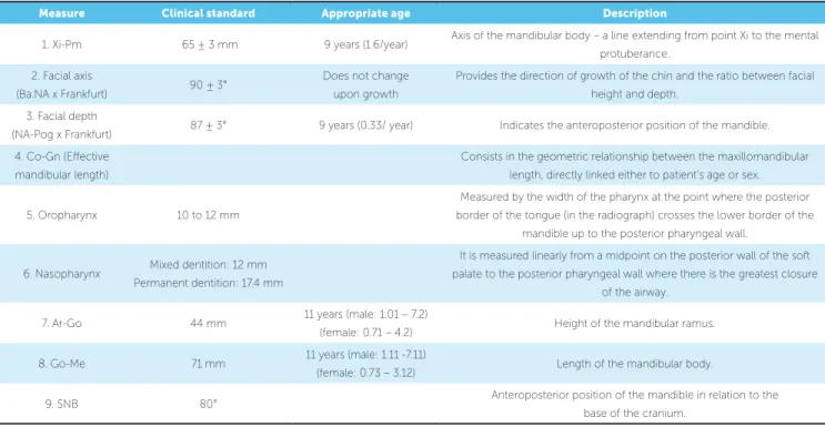
As for upper airways measurements, statistically sig-niicant diferences were found between both groups (p < 0.001). That is, the size of nasopharynx and oro-pharynx is reduced in Class II individuals (Fig 2).
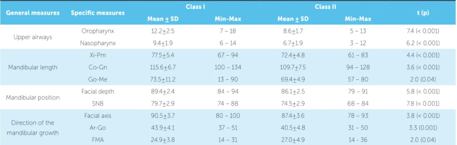
The same results were observed for mandibular length, with signiicant diferences between groups. The following measurements were found to be greater in Class I individuals: Xi-Pm, Co-Gn and Go-Me (Table 3, Fig 3).
As shown in Table 3, measurements of mandibular position also indicated signiicant diferences between
groups, with facial depth and SNB being greater among Class I individuals (Table 3). These results are graphi-cally shown in Figure 4.
Measurements related to the direction of mandibular growth also difered signiicantly between groups. Facial axis and Ar-Go were greater in Class I individuals, while FMA was found to be greater in Class II individuals (Table 3, Fig 5).
In order to assess the correlation between oropharynx/nasopharynx size and mandibular length, position as well as growth, Pearson r correlation coei-cient was performed.
Figure 2 - Assessment of upper airway measurements of Class I and Class II groups.
Figure 3 - Assessment of mandibular length of Class I and Class II groups. Figure 4 - Assessment of mandibular position of Class I and Class II groups.
Table 3 - Assessment of upper airways measurements, mandibular length, mandibular position and direction of mandibular growth of each group.
Table 2 - Assessment of intraexaminer agreement.
Measures ICC p Interpretation
Xi-Pm 0.99 < 0.001 Strong intra-examiner agreement
Co-Gn 0.99 < 0.001 Strong intra-examiner agreement
Go-Me 0.99 < 0.001 Strong intra-examiner agreement
Facial depth 0.97 < 0.001 Strong intra-examiner agreement
SNB 0.99 < 0.001 Strong intra-examiner agreement
Facial axis 0.99 < 0.001 Strong intra-examiner agreement
Ar-Go 0.99 < 0.001 Strong intra-examiner agreement
FMA 0.99 < 0.001 Strong intra-examiner agreement
Oropharynx 1.00 - Perfect agreement
Nasopharynx 0.99 < 0.001 Strong intra-examiner agreement
General measures Specific measures Class I Class II t (p)
Mean ± SD Min-Max Mean ± SD Min-Max
Upper airways Oropharynx 12.2±2.5 7 – 18 8.6±1.7 5 – 13 7.4 (< 0.001)
Nasopharynx 9.4±1.9 6 – 14 6.7±1.9 3 – 12 6.2 (< 0.001)
Mandibular length
Xi-Pm 77.5±5.4 67 – 94 72.4±4.8 61 – 83 4.4 (< 0.001)
Co-Gn 115.6±6.7 100 – 134 109.7±7.5 94 – 128 3.6 (< 0.001)
Go-Me 73.5±11.2 13 – 90 69.4±4.9 57 – 80 2.0 (0.04)
Mandibular position Facial depth 89.4±2.4 84 – 94 86.1±2.5 79 – 91 5.8 (< 0.001)
SNB 79.7±2.9 74 – 88 74.5±2.9 68 – 84 7.8 (< 0.001)
Direction of the mandibular growth
Facial axis 90.5±3.7 80 – 100 87.4±3.6 78 – 93 3.8 (< 0.001)
Ar-Go 43.9±4.1 37 – 51 40.5±4.8 31 – 50 3.3 (0.001)
FMA 24.9±3.8 14 – 31 27.0±4.9 14 - 36 2.0 (0.04)
20
15
10
5
0
Oropharynx Nasopharynx Oropharynx
Class
Class I Class II
Nasopharynx
125
100
75
25 50
0
Class
Class I
Xi-Pm CoGn Go-Me Xi-Pm CoGn Go-Me
Class II
*
95
90
85
80
75
70
65
Class
Class I
Facial depth SNB Facial depth SNB
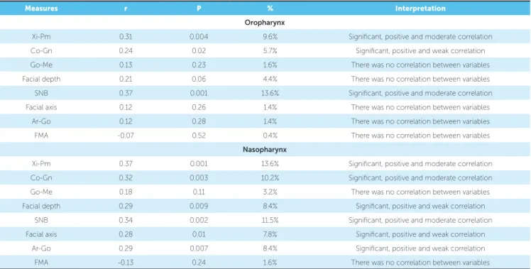
Table 4 - Correlation between upper airways measurements and mandibular length, position as well as direction of mandibular growth in both groups.
Measures r P % Interpretation
Oropharynx
Xi-Pm 0.31 0.004 9.6% Signiicant, positive and moderate correlation
Co-Gn 0.24 0.02 5.7% Signiicant, positive and weak correlation
Go-Me 0.13 0.23 1.6% There was no correlation between variables
Facial depth 0.21 0.06 4.4% There was no correlation between variables
SNB 0.37 0.001 13.6% Signiicant, positive and moderate correlation
Facial axis 0.12 0.26 1.4% There was no correlation between variables
Ar-Go 0.12 0.28 1.4% There was no correlation between variables
FMA -0.07 0.52 0.4% There was no correlation between variables
Nasopharynx
Xi-Pm 0.37 0.001 13.6% Signiicant, positive and moderate correlation
Co-Gn 0.32 0.003 10.2% Signiicant, positive and moderate correlation
Go-Me 0.18 0.11 3.2% There was no correlation between variables
Facial depth 0.29 0.009 8.4% Signiicant, positive and weak correlation
SNB 0.34 0.002 11.5% Signiicant, positive and moderate correlation
Facial axis 0.28 0.01 7.8% Signiicant, positive and weak correlation
Ar-Go 0.29 0.007 8.4% Signiicant, positive and weak correlation
FMA -0.13 0.24 1.6% There was no correlation between variables
Figure 5 - Assessment of mandibular growth of Class I and Class II groups.
DISCUSSION
Although some recent studies have reported a need for tridimensional evaluation by magnetic resonance,6,7,8 its high cost and lack of standardization of patient’s head position still hamper the use of this method for research. According to Muto et al,9 a change of 10o in craniofacial tilt may afect measurement taking in the area of upper airways in approximately 4 mm. Lateral cephalograms have been used in this type of assessment as part of
patients’ basic orthodontic records, with the advantage of having low costs and low radiation dose, being of easy access, and providing standardization of measures with high reproducibility for diagnosis.6,10,11 These advan-tages render this method common in research,7,9,12,13,14 which validates the methodology adopted in the present study and allows comparison of results. The reproduc-ibility of the method was conirmed statistically, with strong intraexaminer agreement.
100
80
60
40
20
0
Class
Class I
Facial axis ArGo FMA Facial axis ArGo FMA
The studied sample comprised patients aged be-tween 10 and 17 years old, with a mean age of 12.3 years, similarly to other studies,5,12,15,16. Because there are minor changes in the nasopharynx as a result of growth,17 the sample was matched by age; thus, avoiding potential bias as regards data interpretation. In terms of sex, groups were similar, although we found three more males than females in the Class II group.
Regarding airways measurements, there were sig-niicant diferences between groups, with Class I pa-tients having oropharynx and nasopharynx greater in size (Table 3, Fig 2). These indings corroborate the majority of studies found in the literature.14,18,19,20 The studies by Freitas et al12 as well as Memon, Fida and Shaikh21 found no interference of malocclusion in oro-pharynx and nasooro-pharynx width when they compared Class I to Class II patients. Diferences in our results may be related to the methods employed, since those studies included a Class II sample based on dental occlusion and may have included subjects with Class II resulting from maxillary prognathism, whereas in our study, mandibu-lar Class II was conirmed cephalometrically.
In order to have a better understanding of which factors inherent to malocclusion could be related to changes in upper airways, we initially diagnosed dif-ferences in skeletal features between groups, as follows: mandibular length (Xi-Pm, Co-Gn and Go-Me), man-dibular position (facial depth and SNB), and direction of growth (facial axis, Ar-Go and FMA).
As regards mandibular length, measurements found in the Class I group were greater than those found in the Class II group (Table 3, Fig 3), thereby conirm-ing mandibular Class II diagnosis. These data validate the assumption that mandibular length can be related to the size of upper airways, which is in agreement with Muto et al4 who pointed out that craniofacial abnor-malities, including mandibular retrognathism, short mandibular body and downward rotation, may cause a decrease in the size of airways, as reported by other studies.9,13,19,22,23 The same behavior was observed in the variables related to spatial position of the mandible. As expected, the mandible in the Class II group was found retropositioned in relation to the cranial base when compared to the Class I group. This information allows us to conclude that both position and length of the mandible, i.e., the efective length of the mandible, must be considered in the diagnosis of patients with
Class II malocclusion. Nevertheless, a greater or less interference of either one of these variables cannot be assumed. In the literature, this comparison is scarce and only cited by a few authors.1,23,24
Our study was carried out considering that sev-eral others have assessed the association between facial growth pattern and upper airways measure-ments.5,12,15,16,19 When comparing Class I and Class II groups, FMA and facial axis indicated an increased ver-tical trend among Class II individuals as well as a shorter mandibular ramus. According to Jarabak,25 this ind-ing refers to mandibular morphology with a clockwise growth pattern. This same feature was reported in the study by Joseph et al15 who used a sample of individuals with Class II malocclusion. This information does not allow us to claim that all mandibular Class II individuals will have a vertical growth trend, although such feature was found in the sample. However, there seem to be an association between vertical pattern and reduced air-ways measurements, which has already been reported by several studies.5,12,14,19
The correlation between oropharynx and naso-pharynx was studied separately from other variables, as shown in Table 4. There was a positive correlation between the size of the oropharynx and mandibular length, represented by Xi-Pm and Co-Gn, and the po-sition of the mandible, represented by SNB. In agree-ment with our indings, studies carried out in the last ive years7,20,23,24 have concluded that mandibular length and position inluence airways measurements.
Although Class II malocclusion patients have mostly presented with a vertical growth pattern in relation to Class I individuals, our results could not support a correlation between vertical pattern and a shorter oropharynx. We did not observe a positive correlation between growth pattern measurements (FMA, Ar-Go and facial axis) and the size of the oropharynx, even though there was an association. This is in agreement with the reports by Castro and Vasconcelos.16 On the other hand, Freitas et al,12 Zhong et al19 as well as Ucar and Uysal5 found a correlation between growth pattern and the size of the oropharynx.
positive correlation between Ar-Go values and the size of the nasopharynx. In addition, they showed a positive correlation between Xi-Pm, Co-Gn, facial depth, SNB and facial axis; thus, concluding that mandibular length and position are related to the size of the nasopharynx.
Mandibular retrusion is one of the factors that may cause obstructive sleep apnea syndrome (OSA), characterized by a collapse site hindering the passage of air located in the pharynx. A reduction in this region can be the etiology of this syndrome both in children and adults. Characterized by respiratory disorders and nocturnal snoring, OSA may cause psychological and social impairment for the individual.11,22,23
As the results of our study suggest that mandibular length and position as well as the direction of growth can inluence measurements of pharyngeal airways, we emphasize the importance of mandibular advancement
in growing children through orthopedics by means of functional appliances; and in adults, with surgical ad-vancement in order to promote enlargement of airways for functional and quality of life improvement, as well as decreased morbidity.8,13,14,26,27
CONCLUSION
» Individuals with mandibular Class II malocclusion were shown to have upper airways measurements reduced when compared to Class I individuals.
» Mandibular length is related to a decrease in upper airways measurements. Similarly, anteroposterior positioning of the mandible exerts inluence on airways measurements.
1. McNamara Jr JA. Components of class II malocclusion in children 8-10 years of age. Angle Orthod. 1981;51(3):177-201.
2. Alcazar NMPV, Freitas MR, Janson G, Henriques JFC, Freitas KMS. Estudo cefalométrico comparativo dos espaços naso e bucofaríngeo nas más oclusões Classe I e Classe II, Divisão 1, sem tratamento ortodôntico, com diferentes padrões de crescimento. Rev Dental Press Ortod Ortop Facial. 2004;9(4):68-76.
3. Joseph AA, Elbowaa J, Cisneros GJ, Eisigsb A. Cephalometric comparative study of the soft tissue airway dimensions in persons with hyperdivergent and normodivergent facial pattern. J Oral Maxillofac surg. 1998;56:135-9. 4. Muto T, Yamazaki A, Takeda S. A cephalometric evaluation of the
pharyngeal airway space in patients with mandibular retrognathia and prognathia, and normal subjects. Int J Oral Maxillofac Surg. 2008;37(3):228-31.
5. Ucar FI, Uysal T. Orofacial dimensions in subjects with Class I malocclusion and diferent growth patterns. Angle Orthod. 2011;81(3):460-8.
6. Pirilä-Parkkinen K, Löppönen H, Nieminen P, Tolonen U, Pääkkö E, Pirttiniemi P. Validity of upper airway assessment in children: a clinical, cephalometric, and MRI study. Angle Orthod. 2011;81(3):433-9. 7. Kim Y-J, Hong J-S, Hwang Y-I, Park Y-H. Three-dimensional analysis of
pharyngeal airway in preadolescent children with diferent anteroposterior skeletal patterns. Am J Orthod Dentofacial Orthop. 2010;137(3):303-11. 8. Schutz TCB, Dominguez GC, Pradella-Hallinan M, Cunha TCA, Tuik S. Class II correction improves nocturnal breathing in adolescents. Angle Orthod. 2011;81(2):222-28.
9. Muto T, Yamazaki A, Takeda S, Kawakami J, Tsuji Y, Shibata T, Mizoguchi I. Relationship between the pharyngeal airway space and craniofacial morphology, taking into account head posture. Int J Oral Maxillofac Surg. 2006;35:132-6.
10. Major MP, Flores-Mir C, Major PW. Assessment of lateral cephalometric diagnosis of adenoid hypertrophy and posterior upper airway obstruction: a systematic review. Am J Orthod Dentofacial Orthop. 2006;130(6):700-8. 11. Bittencourt LRA, Haddad FLM. Diagnóstico e abordagem clínica do
paciente com distúrbio respiratório do sono. In: Fabro CD, Chaves CM Jr, Tuik S. A odontologia na medicina do sono. Maringá: Dental Press; 2012. cap. 6, p. 144-58.
12. Freitas MR, Alcazar NM, Janson G, De Freitas KM, Henriques JFC. Upper and lower pharyngeal airways in subjects with Class I and Class II malocclusions and diferent growth patterns. Am J Orthod Dentofacial Orthop. 2006;130(6):742-45.
REFERENCES
13. Susarla AM, Abramson ZR, Dodson TB, Kaban LB. Cephalometric measurement of upper airway length correlates with the presence and severity of obstructive sleep apnea. J Oral Maxillofac Surg. 2010;68:2846-55. 14. Restrepo C, Santamaria S, Pela EZ, Tapias A. Oropharyngeal airway
dimensions after treatment with functional appliances in class II retrognathic children. J Oral Rehabil. 2011;38(8):588-94.
15. Joseph AA, Elbaum J, Cisneros GJ, Eisig SB. A cephalometric comparative study of the soft tissue airway dimensions in persons with hyperdivergent and normodivergent facial patterns. J Oral Maxillofac Surg.1998;56(2):135-9. 16. Castro AMA, Vasconcelos MHF. Avaliação da inluência do tipo facial nos
tamanhos dos espaços aéreos nasofaríngeo e bucofaríngeo. Rev Dental Press Ortod Ortop Facial. 2008;13(6):43-50.
17. McNamara JA Jr. A method of cephalometric evaluation. Am J Orthod. 1984;86(6):449-69.
18. Mergen DC, Jacobs RM. The size of nasopharynx associated with normal occlusion and Class II malocclusion. Angle Orthod. 1970;40(4):342-6. 19. Zhong Z, Tang Z, Gao X, Zeng XL. A comparison study of upper airway
among diferent skeletal craniofacial patterns in nonsnoring Chinese children. Angle Orthod. 2010;80(2):267-74.
20. El H, Palomo JM. Airway volume for diferent dentofacial skeletal patterns. Am J Orthod Dentofacial Orthop. 2011;139(6):511-21.
21. Memon S, Fida M, Shaikh A. Comparison of diferent craniofacial patterns with pharyngeal widths. J Coll Physicians Surg Pak. 2012;22(5):302-6. 22. Schwab RJ, Goldbert AN. Upper airway assessment: radiographic and other
imagining techniques. Otolaryngol Clin North Am. 1998;31(6):931-68. 23. Guarim JA. Evaluation of the growth mandibular in a buccal respirator after
the treatment with the use of the orthopedical prefabricated apparel. Rev Paul Odontol. 2009;32:15-23.
24. Kim JS, Kim JK, Hong SC, Cho JH. Changes in the upper airway after counterclockwise maxillomandibular advancement in young Korean women with class II malocclusion deformity. J Oral Maxillofac Surg. 2013;71:1603-5. 25. Jarabak JR, Fizzel JA. Technique and treatment with light wire edgewise
appliances. 2nd ed. St. Louis: Mosby; 1972.
26. Lye KW. Efect of orthognathic surgery on the posterior airway space (PAS). Ann Acad Med Singapore. 2008;37(8):677-82.