Stability of smooth and rough mini-implants:
clinical and biomechanical evaluation — an
in vivo
study
Giselle Naback Lemes Vilani1, Antônio Carlos de Oliveira Ruellas2, Carlos Nelson Elias3, Cláudia Trindade Mattos4
How to cite this article: Vilani GNL, Ruellas ACO, Elias CN, Mattos CT. Stability of smooth and rough mini-implants: clinical and biomechanical evalua-tion — an in vivo study. Dental Press J Orthod. 2015 Sept-Oct;20(5):35-42. DOI: http://dx.doi.org/10.1590/2177-6709.20.5.035-042.oar
» The authors report no commercial, proprietary or financial interest in the products or companies described in this article.
Contact address: Giselle Naback Lemes Vilani E-mail: [email protected]
1 PhD in Orthodontics, Universidade Federal do Rio de Janeiro (UFRJ), Rio de
Janeiro, Rio de Janeiro, Brazil.
2 Professor of Orthodontics, Universidade Federal do Rio de Janeiro (UFRJ),
School of Dentistry, Rio de Janeiro, Rio de Janeiro, Brazil.
3 Professor, Instituto Militar de Engenharia, School of Engineering, Department
of Material Sciences, Rio de Janeiro, Rio de Janeiro, Brazil.
4 Professor of Orthodontics, Universidade Federal Fluminense (UFF), School of
Dentistry, Niterói, Rio de Janeiro, Brazil.
Submitted: September 20, 2014 - Revised and accepted: May 20, 2015
Objective: To compare in vivo orthodontic mini-implants (MI) of smooth (machined) and rough (acid etched) surfaces, as-sessing primary and secondary stability. Methods: Thirty-six (36) MI were inserted in the mandibles of six (6) dogs. Each animal received six (6) MI. In the right hemiarch, three (3) MI without surface treatment (smooth) were inserted, whereas in the left hemiarch, another three (3) MI with acid etched surfaces (rough) were inserted. The two distal MI in each hemiarch received an immediate load of 1.0 N for 16 weeks, whereas the MI in the mesial extremity was not subject to loading. Stability was measured by insertion and removal torque, initial and final mobility and by inter mini-implant distance. Results: There was no statistical behavioral difference between smooth and rough MI. High insertion torque and reduced initial mobility were observed in all groups, as well as a reduction in removal torques in comparison with insertion torque. Rough MI presented higher removal torque and lower final mobility in comparison to smooth MI. MI did not remain static, with displacement of rough MI being smaller in comparison with smooth MI, but with no statistical difference. Conclusions:
MI primary stability was greater than stability measured at removal. There was no difference in stability between smooth and rough MI when assessing mobility, displacement and insertion as well as removal torques.
Keywords:Orthodontic anchorage procedures. Osseointegration. Orthodontics. DOI: http://dx.doi.org/10.1590/2177-6709.20.5.035-042.oar
Objetivo: comparar, in vivo, mini-implantes (MI) com superfície lisa (usinada) e porosa (tratada com ácido), avaliando sua estabilidade primária e secundária. Métodos: trinta e seis MI foram inseridos na mandíbula de seis cães, e cada animal rece-beu seis MI. Na hemiarcada direita, foram inseridos três MI sem tratamento da superfície (liso); na esquerda, outros três com a superfície tratada com ácido (poroso). Os dois MI distais de cada hemiarcada receberam carga imediata de 1,0N durante dezesseis semanas, e o MI da extremidade mesial não recebeu carregamento. A estabilidade foi medida pelos torques de in-serção e de remoção, pela mobilidade inicial e final, e pela distância inter-MI. Resultados: não houve diferença estatística do comportamento entre os MI lisos e porosos. No entanto, observou-se torque de inserção elevado e mobilidade inicial reduzida em todos os grupos. Para todos os grupos, houve redução dos torques de remoção, em relação ao de inserção. Os MI porosos apresentaram maior torque de remoção e menor mobilidade final, em relação aos MI lisos. Os MI não permaneceram estáticos, sendo o deslocamento dos MI porosos menor em relação aos MI lisos, mas sem diferença estatística. Conclusões: a estabilidade primária dos MI foi maior do que a estabilidade medida na sua remoção; não houve diferença na estabilidade entre os MI lisos e porosos ao avaliar-se a mobilidade, o deslocamento e os torques de inserção e remoção.
ter results in comparison to other anchorage systems due to being inserted and removed with ease, and particularly due to the reduced size of the devices, which broadens their scope of use.1
With a reduction in mini-implant size, the screws are now made of titanium alloy (Ti6Al4V), which increases fracture strength.2 The disadvantage of
Ti6Al4V alloy is its lower degree of osseointegration and greater susceptibility to corrosion in vivo, both of which may hinder stability.3
Osseointegration stands for direct contact between bone and implant without interposition of soft tissue layers. It is beneficial since it increases stability and raises success rates of MI as tempo-rary anchorage devices, thus expanding their bio-mechanical possibilities.4 Various factors must be
taken into account in order to achieve implant os-seointegration, namely: material biocompatibility, implant surface conditions, patient’s conditions, the surgical technique employed and the load applied on implants after placement.5 Studies have shown
that surface treatment applied to the active parts of mini-implants result in roughness that favors bone-implant contact.6-9 Acid etching is a simple method
that requires little infrastructure and results in im-plant roughness, making imim-plant surface homoge-neous and with a large active surface area that en-ables better bioadhesion.10
At present, there is an increasing trend towards applying immediate loading for orthodontic pur-poses, particularly because studies have shown that mini-implants are able to bear continuous forces im-mediately after placement11 without hindering
an-chorage and success rates.12 Nevertheless, it is
neces-sary to assess the effects of acid etching on stability
MATERIAL AND METHODS
This animal study protocol was approved by the Ethics Committee on Animal Use (CEUA) of the Health Science Center (CCS) of Universidade Federal do Rio de Janeiro, Brazil (protocol: ODONTO 010).
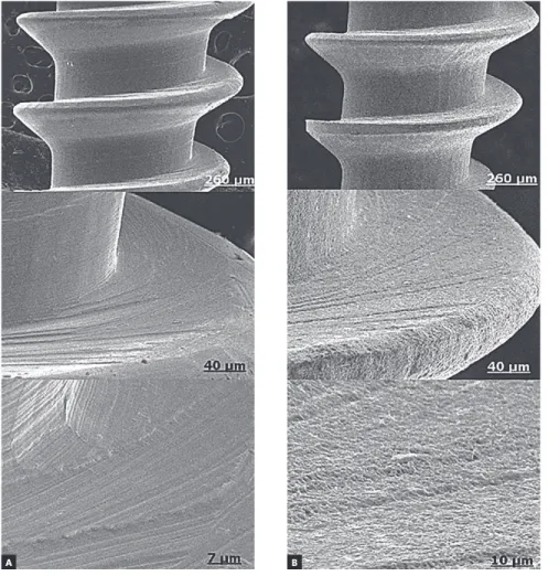
Thirty-six (36) mini-implants made of Ti6Al4V alloy (Conexão Sistemas e Próteses, Arujá, SP, Brazil), measuring 1.5 x 6.0 x 2.0 mm, were used in the present research. Of them, 18 had no sur-face treatment (smooth) while 18 were subject to acid etching specifically carried out for this study. To this end, an aqueous solution made of nitric acid (HNO3), hydrochloric acid (HCl) and sul-furic acid (H2SO4) (rough standard by Conexão) (Fig 1) was used. Six (6) adult male mongrel dogs weighing approximately 18.0 kg were used. Each animal had six (6) mini-implants placed buccally between roots in the alveolar bone of the mandi-ble. On the right side, three smooth mini-implants were inserted, whereas on the left side, three rough implants were inserted. The two distal mini-implants were subject to immediate load while the mesial extremity remained without loading. Mini-implants were divided into four groups: S = smooth without load; SL = smooth with immediate load; R = rough without load; RL = rough with immedi-ate load. Figure 2 discloses a diagram illustrating the position of smooth and rough mini-implants.
Figure 1 - Electromicrographs of smooth (A) and rough (B) mini-implant surfaces.
Figure 2 - Diagram showing the position of smooth and rough mini-implants inserted in the external buccal cortical bone and loaded with NiTi springs. SL = Smooth with immediate load-ing; S = Smooth without load; RL = Rough with immediate loading; R = Rough without load.
Smooth mini-implants Rough mini-implants
SL S R RL
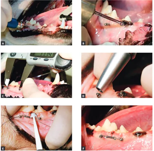
Mini-implant insertion was concluded with the manual key coupled to a portable digital torque me-ter (Instrutherm TQ 680, Korea) used to obtain the maximum insertion torque value (N.cm) (Fig 3B).
The distance between loaded mini-implants was recorded in each quadrant soon ater mini-implants were inserted, before ixation of the spring and ater a period of 16 weeks. The center of the upper portion of the device head was used as reference. Measurements were performed with a digital caliper (Starret Indústria e Comércio Ltda, São Paulo, Brazil) (Fig 3C).
Mini-implant mobility was clinically assessed at two time intervals: at mini-implant placement and ater 16 weeks. Quantitative mobility assessment was performed by Periotest(Medizintechnik Gulden e.K., Modautal, Germany), and consisted of a vibration analysis performed to detect lateral movement of an implant inside the bone. Ater the instrument was cali-brated, it was placed perpendicular to the head of the mini-implant, horizontal towards the ground, with the head of the handpiece placed 2.0 to 3.0 mm from the mini-implant head. Measurements oscillated at a fre-quency of around four times per second. Results were digitally and audibly shown by a descriptive numerical value and ranged from -8 to +5014 (Fig 3D).
Mobil-ity and distance between mini-implants were recorded twice and the mean values obtained.
The two distal mini-implants received load-ing immediately ater insertion. A load of 1.0 N was applied by NiTi springs for 16 weeks. Mesi-al mini-implants were not subject to loading. The force released by the spring was quantiied by a
nixin meglumine (Schering Plough Indústria Quími-ca e FarmacêutiQuími-ca S.A., Rio de Janeiro/RJ, Brazil). The animals were fed with animal food, ground and moisturized with water, suitable for puppies, and were provided with water ad libitum. They also received dental prophylaxis performed with a brush and anti-tartar tooth paste (C.E.T.® Pasta Enzimática, Virbac,
São Paulo, Brazil) once a week during the experiment. Subsequently, the mini-implants were cleaned with 0.12% chlorhexidine gluconate (PerioGard®,
Colgate-Palmolive Indústria Comércio Ltda, São Bernardo do Campo, SP, Brazil). To this end, the dogs were sedated with an intramuscular injection of 0.4 mg/kg xylazine (Bayer S/A, São Paulo, SP, Brazil) and 0.5 mg/kg mor-phine (União Química Farmacêutica Nacional S/A, São Paulo, SP, Brazil).
By the end of the 16-week period, mini-implants were removed and maximum removal torque was recorded.
Statistical analysis
RESULTS
Of 36 mini-implants, six were lost during the ex-periment (one S, three SL, one R and one RL). The success rate of all mini-implants was 83.3%. Rough mini-implants presented a higher success rate (88.8%) when compared to smooth ones (77.7%). The results of the tests performed with two mini-implants were not used due to loss of mini-mini-implants attached to the spring. Assessment was carried out in 28 mini-implants, five from group S, eight from SL, five from R and ten from RL.
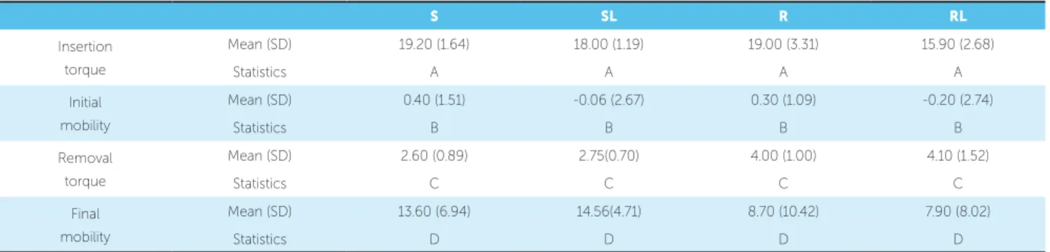
When mini-implants performance was com-pared, no statistically significant difference was found (p > 0.05) between groups for any variables (Table 1). High insertion torque and reduced initial mobility values were observed. Conversely, at the end of the experiment, removal torque was low and final mobility was high; with different values were found between smooth and rough mini-implants.
Rough mini-implants presented higher secondary stability, with higher removal torque and lower final mobility when compared to smooth mini-implants, but without statistical significance.
Smooth mini-implants presented with higher mean displacement (0.94 ± 1.33 mm) when com-pared to rough mini-implants (0.39 ± 0.19 mm) at the end of the experiment; however, this difference was not statistically significant (p = 0.387) (Table 2).
DISCUSSION
In the present study, primary stability was assessed quantitatively by insertion torque (IT) and initial mobility (IMb). Mean IT values were high for groups S (19.20 N.cm), SL (18.00 N.cm), R (19.00 N.cm) and RL (15.90 N.cm), with no statistical differ-ence between them. High IT values may be relat-ed to greater thickness of dogs’ cortical bone,14,15,16
small bone perforation in relation to mini-implants
Figure 3 - Photographs illustrating the steps for mini-implant placement: A) Initial mini-implant placement with manual key; B) Conclusion of placement with torque wrench; C) Measure-ment of inter mini-implant distance; D) Use of Periotest; E) Measurement of force of 1 N;
F) NiTi spring in position.
A
C
E
B
D
Removal torque
Mean (SD) 2.60 (0.89) 2.75(0.70) 4.00 (1.00) 4.10 (1.52)
Statistics C C C C
Final mobility
Mean (SD) 13.60 (6.94) 14.56(4.71) 8.70 (10.42) 7.90 (8.02)
Statistics D D D D
Table 2 - Inter mini-implant distance values for smooth and rough mini-implants with load.
Diference in mean inter MI distance (SD)
Statistical signiicance (p-value) Smooth MI x Rough MI
SL 0.94 (1.33)
0.387
RL 0.39 (0.19)
diameter17 and deeper mini-implants insertion, with
potential compression of the cortical bone by the transmucosal profile.18,19,20 Previous research
con-ducted with dogs have shown similar high IT val-ues in mini-implants subject to surface treatment (15.27 ± 6.65 N.cm) and in smooth mini-implants (19.25 ± 8.34 N.cm) when slightly larger mini-im-plants (1.8 x 8.5 mm) were used.8 Other studies
con-ducted with dogs presented even higher IT values,19,21
with high success rates, which suggests that IT is higher in mini-implants placed in dog’s mandibles, which does not necessarily lead to failure.
In the present study, mini-implants stability was also quantiied by means of Periotestused to detect mobil-ity.22 The index measured by Periotest varies on a scale
ranging from -8 to +50, with values between -8 and +9 indicating that teeth are ixed or implants osseointegrat-ed; between +10 and +19, palpable mobility; between +20 and +29, visible mobility; and between +30 and +50, mobility caused by pressure of the tongue or lip.14 In the
present study, all groups presented with adequate
pri-Secondary stability was assessed by removal torque (RT), final mobility (FMb) and difference in inter mini-implants distance. High RT and re-duced FMb indicate adequate secondary stability. In the present study, RT was much lower than IT, a behavior that may be associated with peri-implant inflammation caused by biofilm accumulation.23
Data available in the literature reveal that reduced IT values are more favorable to achieve osseointe-gration than high values. In addition, the latter may lead to a high level of compression, which causes local ischemia and bone necrosis at the bone/mini-implants interface, thereby leading to reduction in osseointegration.24 However, a recent systematic
review found no evidence that a specific IT value is associated with high success rates of orthodontic mini-implants.25 Although there were no statistically
suc-resistance of mini-implants subject to surface treat-ment in five different periods of loading, Mo et al23
found high RT values in mini-implants immediately loaded and similar success rates in all periods, which suggests that mini-implants may be immediately loaded. Therefore, in the present study, immediate loading was used, since it is a trend in Orthodon-tics, bearing in mind that various researches11,12,14,23,31
have proved it to be effective.
In the present study, FMb values were higher than IMb ones. These results are in agreement with data found in the literature, showing that secondary stability of smooth mini-implants is lower in compar-ison to primary stability. Rough mini-implants pre-sented better stability at the time of removal; however, without statistical difference. Lower FMb values for rough mini-implants suggest absence of mobility, whereas the higher values for smooth mini-implants suggest palpable mobility. In spite of presenting high FMb values, mini-implants proved stable when sub-ject to continuous orthodontic load throughout the entire experimental period. Studies conducted with dogs’ mandibles found lower FMb values in smooth mini-implants, which may be due to shorter experi-mental periods (12 weeks),28 since the devices were
exposed to biofilm for a shorter period of time. In the present research, mini-implants did not remain static, with smooth mini-implants showing higher mean displacement (0.94 ± 1.33 mm) than rough mini-implants (0.39 ± 0.19 mm) after load ap-plication for sixteen (16) weeks, without statistical differences. Oynarte et al7 also found more significant
displacement of smooth mini-implants (0.51 mm) in
comparison to rough ones (0.12 mm). Similar dis-placement (0.44 mm) was found after a two-week period,29 in addition to absence of displacement in
mini-implants subject to surface treatment after a six-week waiting period.29 Studies applying
immedi-ate load found a variation that ranged from 0.53 mm12
to 0.78 mm30 for smooth mini-implants. High
dis-placement values were found in a study applying im-mediate load (2.2 mm),31 but the authors applied
el-evated forces (6 N) to short mini-implants (3.0 mm). The similarity of displacement values found in the present study and in other studies that used immedi-ate loading to those found in researches that waited for healing before load application indicates that im-mediate loading can be safely used.
In the present study, mini-implants were re-moved at the end of the experiment by means of movements applied in anti-clockwise direction. The same results were achieved by Kim et al27 and
Fa-vero et al13 when removing osseointegrated
mini-implants larger in diameter.
CONCLUSIONS
1. The success rate of rough mini-implants (88.8%) was higher than that of smooth mini-im-plants (77.7%).
2. Primary stability achieved by the end of Ti6Al4V mini-implants placement was higher than stability observed sixteen (16) weeks after insertion.
anchorage. Am J Orthod Dentofacial Orthop. 2005;127(6):713-22. 4. Roberts W E, Helm F R, Marshall K J, Gonglof R K. Rigid endosseous
implants for orthodontic and orthopedic anchorage. Angle Orthod. 1989;59(4):247-56.
5. Albrektsson T, Branemark P I, Hansson H A, Lindstrom J. Osseointegrated titanium implants. Requirements for ensuring a long-lasting, direct bone-to-implant anchorage in man. Acta Orthod Scand. 1981;52(2):155-70. 6. Aldikaçti M, Açikgoz G, Turk T, Trisi P. Long-term evaluation of sandblasted
and acid-etched implants used as orthodontic anchors in dogs. Am J Orthod Dentofacial Orthop. 2004;125(2):139-47.
7. Oyonarte R, Pilliar RM, Deporter D, Woodside DG. Peri-implant bone response to orthodontic loading: Part 1. A histomorphometric study of the efects of implant surface design. Am J Orthod Dentofacial Orthop. 2005;128(2):173-81.
8. Kim SH, Lee SJ, Cho IS, Kim SK, Kim TW. Rotational resistance of surface-treated mini-implants. Angle Orthod. 2009;79(5):899-907.
9. Lee SJ, Ahn SJ, Lee JW, Kim SH, Kim TW. Survival analysis of orthodontic mini-implants. Am J Orthod Dentofacial Orthop. 2010;137(2):194-9. 10. Elias CN, Oshida Y, Lima JHC, Muller CA. Relationship between surface
properties (Roughness, wettability and morphology) of titanium and dental implant removal torque. J Mech Behav Biomed Mat. 2008;1(3):234-42. 11. Buchter A, wiechmann D, Gaertner C, Hendrik M, Vogeler M, Wiesmann HP,
et al. Load-related bone modeling at the interface of orthodontic micro-implants. Clin Oral Implant Res. 2006;17(6):714-22.
12. Chen Y, Kang ST, Bae SM, Kyung HM. Clinical and histologic analysis of stability of microimplants with immediate orthodontic loading in dog. Am J Orthod Dentofacial Orthop. 2009;136(2):260-7.
13. Favero LG, Pisoni A, Paganelli C. Removal torque of osseointegrated mini-implants: an in vivo evaluation. Eur J Orthod. 2007;29(5):443-8. 14. Çehreli S, Arman Ózçirpici AA. Primary stability and histomorphometric
bone-implant contact of self-drilling and self-tapping orthodontic microimplants. Am J Orthod Dentofacial Orthop. 2012;141(2):187-95. 15. Motoyoshi M, Yoshida T, Ono A, Shimizu N. Efect of cortical bone thickness
and implant placement torque on stability of orthodontic mini-implants. Int J Oral Maxillofac Implant. 2007;22(5):779-84.
16. Pithon MM, Nojima MG, Nojima LI. In vitro evaluation of insertion and removal torques of orthodontic mini implants. Int J Oral Maxillofac Surg. 2011;40(1):80-5.
Insertional torque and axial pull-out strength of mini-implants in mandibles of dogs. Am J Orthod Dentofacial Orthop. 2008;133(6):790.e15-e22. 20. Wawrzinek C, Sommer T, Fischer-Brandies H. Microdamage in cortical bone
due to the overtightening of orthodontic microscrews. J Orofac Orthop. 2008;69(2):121-34.
21. Ikeda H, Rossouw PE, Campbell PM, Kontogirogos E, Buschang PH. Three-dimensional analysis of peri-bone implant contact of rough-surface miniscrew implants. Am J Orthod Dentofacial Orthop. 2011;139(2):e153-e63. 22. Cha JY, Yu HS, Hwang CJ. The validation of periotest values for the
evaluation of orthodontic mini-implants’ stability. Korean J Orthod. 2010;40(3):167-75.
23. Mo SS, Kim SH, Kook YA, Jeong DM, Chung KR, Nelson G. Resistance to immediate orthodontic loading of surface-treated mini-implants. Angle Orthod. 2010;80(1):123-9.
24. Suzuki E Y, Suzuki B. Placement and removal torque values of orthodontic miniscrew implants. Am J Orthod Dentofacial Orthop. 2011;139(5):669-78. 25. Reynders RAM, Ronchi L, Ladu L, Etten-Jamaludin F, Bipat S. Insertion torque
and success of orthodontic mini-implants: a systematic review. Am J Orthod Dentofacial Orthop. 2012;142(5):596-614.
26. Klokkevold PR, Nishimura RD, Adachi M, Caputo A. Osseointegration enhanced by chemical etching of the titanium surface. A torque removal study in the rabbit. Clin Oral Implant Res. 1997;8(6):442-7.
27. Kim SH, Cho JH, Chung KR, Kook YA, Nelson G. Removal torque values of surface-treated mini-implants after loading. Am J Orthod Dentofacial Orthop. 2008;134(1):36-43.
28. Kim J, Ahn S, Chang Y. Histomorphometric and mechanical analyses of drill-free screw as orthodontic anchorage. Am J Orthod Dentofacial Orthop. 2005;128(2):190-4.
29. Liou EJ, Pai BC, Lin JC. Do miniscrew remain stationary under orthodontic forces? Am J Orthod Dentofacial Orthod. 2004;126(1):42-7.
30. Alves Jr M, Baratieri C, Nojima LI. Assessment of mini-implant displacement using cone beam computed tomography. Clin Oral Implant Res. 2011;22(10):1151-6.