Mandibular dental arch short and long-term
spontaneous dentoalveolar changes after slow or rapid
maxillary expansion: a systematic review
Arthur César de Medeiros Alves1, Olga Benário Vieira Maranhão1, Guilherme Janson1, Daniela Gamba Garib1
Objective: The aim of this systematic review was to analyze the short and long-term spontaneous dentoalveolar changes of the mandibu-lar dental arch after slow (SME) or rapid (RME) maxilmandibu-lary expansion in the mixed and early permanent dentitions. Methods: An elec-tronic search was performed in the following databases: PubMed/Medline, Cochrane Library, Scopus, Embase and Web of Science. Eligibility criteria for article selection included randomized controlled trials and prospective studies written in English, with no restriction of year of publication, involving patients who underwent SME or RME during the mixed or early permanent dentitions. A double-blind search of articles was performed by two reviewers. Initially, the title and the abstract of the studies were read, and their references were also hand-searched for possible missing studies. A methodological quality scoring scale was used to analyze the selected articles. Re-sults: The search retrieved 373 articles, but only 6 were selected for review after application of the eligibility and exclusion criteria. Non-clinically significant spontaneous dentoalveolar changes of approximately 1mm were found in the mandibular dental arch in the short and long-term, after slow or rapid maxillary expansions. Furthermore, no significant differences were found between treated and control groups. Conclusions: There is enough evidence to conclude that negligible short and long-term spontaneous dentoalveolar changes tend to occur in the mandibular dental arch after SME or RME in the mixed and early permanent dentitions. More randomized studies with appropriate control group are required to better evaluate this issue.
Keywords: Dental arch. Palatal expansion technique. Mandible. Mixed dentition. Permanent dentition.
1 Universidade de São Paulo, Department of Orthodontics, Bauru, São Paulo,
Brazil.
» The authors report no commercial, proprietary or financial interest in the products or companies described in this article.
Submitted: May 31, 2016 - Revised and accepted: December 12, 2016 DOI: https://doi.org/10.1590/2177-6709.22.3.055-063.oar
How to cite this article: Alves ACM, Maranhão OBV, Janson G, Garib DG. Mandibular dental arch short and long-term spontaneous dentoalveolar changes after slow or rapid maxillary expansion: a systematic review. Dental Press J Or-thod. 2017 May-June;22(3):55-63.
DOI: https://doi.org/10.1590/2177-6709.22.3.055-063.oar
Contact address: Arthur César de Medeiros Alves
Faculdade de Odontologia de Bauru, Universidade de São Paulo
Al. Octávio Pinheiro Brisolla, 9-75, Vila Santa Tereza –Bauru, São Paulo, Brasil CEP: 17.012-191 – E-mail: [email protected]
Objetivo: o objetivo da presente revisão sistemática foi analisar as alterações dentoalveolares espontâneas, em curto e longo prazos, após a expan-são lenta (ELM) ou rápida (ERM) da maxila, durante a dentição mista e permanente jovem. Métodos: uma busca eletrônica foi executada nas seguintes bases de dados: PubMed/Medline, Cochrane Library, Scopus, Embase e Web of Science. Os critérios de elegibilidade para a seleção dos artigos incluíram ensaios clínicos randomizados controlados e estudos prospectivos escritos em língua inglesa, sem restrição do ano de publi-cação, envolvendo pacientes que foram submetidos a ELM ou ERM durante a dentição mista ou permanente jovem. Dois revisores realizaram, de forma independente, uma busca por artigos. Inicialmente, o título e o resumo dos artigos foram lidos, e uma busca manual foi realizada nas referências dos artigos selecionados, a fim de se avaliar possíveis estudos não encontrados. Resultados: trezentos e setenta e três artigos foram encontrados com a busca, porém apenas seis foram selecionados para a revisão após a aplicação dos critérios de inclusão e de exclusão. Altera-ções dentoalveolares espontâneas sem significância clínica de aproximadamente 1 mm foram encontradas na arcada dentária inferior em curto e longo prazos, após a expansão lenta ou rápida da maxila. Além disso, não foram observadas diferenças significativas entre os grupos tratados e os controles. Conclusão: existe evidência suficiente para se concluir que alterações dentoalveolares espontâneas sem significância clínica tendem a ocorrer na arcada dentária inferior em curto e longo prazos após a ELM e ERM na dentição mista e permanente jovem. Mais ensaios clínicos randomizados com grupos controles adequados são necessários para melhor avaliar essa questão.
INTRODUCTION
Maxillary dental arch constriction is commonly associ-ated with unilateral or bilateral posterior crossbites in the
mixed or early permanent dentitions.1,2 Considering that
transverse malocclusions do not self-correct during the craniofacial growth, either slow (SME) or rapid (RME) maxillary expansions should be performed as early as pos-sible to transversely increase the maxillary dental arch with
a combination of orthopedic and orthodontic efects.3,4
In general, the greater the patient’s age, the greater the
den-tal efects and the smaller the skeleden-tal changes.5
Occasionally, maxillary constriction is not associated
to posterior crossbites.6 Spontaneous progressive
con-striction of the mandibular dental arch might occur from childhood to adulthood as an adaptation process to the progressive maxillary constriction observed in
untreat-ed patients.1 Logically, if maxillary constriction induces
mandibular dental arch constriction over time, maxil-lary expansion might induce spontaneous increase of the
mandibular dental arch width in the short or long-term.7
This hypothesis is based on the fact that the maxillary dental arch expansion modiies the balance of forces
be-tween the tongue and cheek on the mandibular teeth.8
Predominance of the tongue forces on the mandibular
teeth might increase the mandibular dental arch width.9
Spontaneous dentoalveolar changes in the mandibu-lar dental arch concurrent to SME or RME may have clinical implications regarding the indication of man-dibular dental arch dentoalveolar expansion. Therefore,
randomized10-12 (RCT) and non-randomized13-15
clini-cal trials have been developed to answer this cliniclini-cal is-sue. However, no consensus has been reached. Thus, the aim of the present systematic review is to evaluate the short and long-term spontaneous dentoalveolar changes in the mandibular dental arch, ater slow or rapid maxillary expansion in the mixed or early perma-nent dentitions.
MATERIAL AND METHODS
The protocol of this systematic review was prepared and registered in PROSPERO (CRD42016039760). This review was conducted based on the PRISMA
Statement for Systematic Review16 and comprised
ar-ticles available until May of 2016. Eligibility criteria for article selection included randomized clinical trials and prospective studies written in English, with no restric-tion of year of publicarestric-tion, involving orthodontic
pa-tients with 6 to 12 years of age. These papa-tients should present with maxillary constriction in the mixed or early permanent dentitions and should have been treat-ed with slow or rapid maxillary expansion. Evaluation of the spontaneous dentoalveolar changes in the man-dibular dental arch should have been performed in the short (3 to approximately 12 months post-expansion) or long-term (more than 12 months post-expansion). At least one of the following variables should have been measured in the mandibular dental arch by means of conventional or digital dental models or posteroante-rior radiographs: intercanine distance, inter-deciduous molar or interpremolar distances, inter-irst permanent molar distance, arch perimeter, arch length and bucco-lingual inclination of the canines and posterior teeth. The deinition of each variable is shown in Table 1.
The exclusion criteria were patients with oral clets or associated craniofacial anomalies, previous orthodontic treatment, intervention in the mandibular dental arch during the follow-up period, surgically-assisted rapid maxillary expansion and the lack of a control group.
An electronic search was performed in the following databases with the assistance of a senior librarian spe-cialized in Health Sciences databases: PubMed/Med-line, Cochrane Library, Scopus, Embase and Web of Science. The search strategy used in the aforementioned databases included the MeSh terms “dental arch” and “palatal expansion technique” or “maxillary expansion” and “mixed dentition” or “permanent dentition”.
A double-blind search of articles was performed by two reviewers. Initially, the title and the abstract of the studies found in each database were independently read by both examiners according to PICO. The refer-ences of the articles were also hand-searched for pos-sible missing studies. In case of disagreement regarding which article fulilled the inclusion criteria, consensus was reached by discussion between the two reviewers. The articles that fulilled the inclusion criteria were in-cluded in the systematic review and were qualitatively analyzed using the Cochrane collaboration
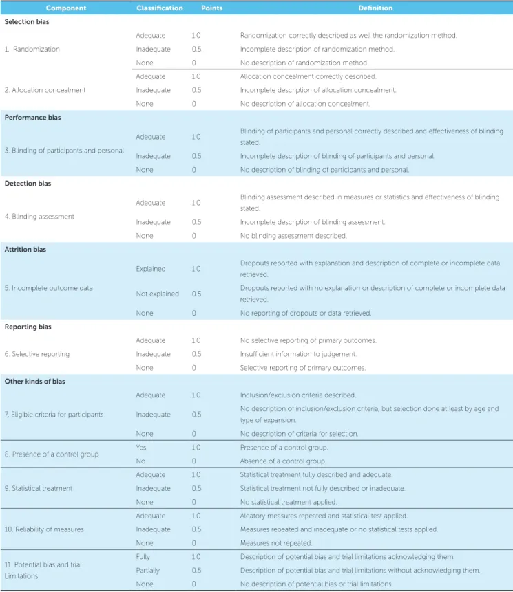
recommen-dations17 and a modiication of the methodological
qual-ity scoring scale developed by Vilani et al18 (Table 2).
Table 1 -Definition of outcome measurements.
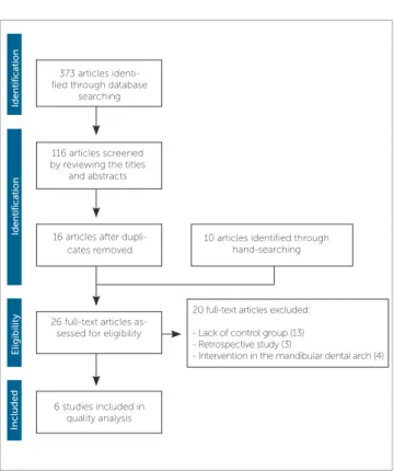
Figure 1 - Flow diagram adapted from the PRISMA statement.16
Outcome measurement Definition
Intercanine distance Linear distance between the crown tips or the midpoints of the lingual gingival margins of both mandibular canines. Inter-deciduous molar or
interpremolar distances
Linear distance between the buccal cusp tips or the midpoints of the lingual gingival margins of the left and right mandibular deciduous molars or premolars.
Inter-irst permanent molar distance Linear distance between the mesiobuccal cusp tips, the center of the fossa or the midpoints of the lingual gingival margins of both mandibular permanent irst molars.
Arch length A line measured perpendicularly in the horizontal plane connecting the mesial aspects of the mandibular permanent irst molars to the point between the mandibular central incisors.
Arch perimeter The length of a curve from the mesial surface of the mandibular permanent irst molars, bisecting the contact points of the deciduous molars or premolars and canines, and smoothly itting on the incisal edges of the anterior teeth. Tooth inclination Angle between the clinical crown axis and the occlusal plane.
RESULTS
The electronic search retrieved 373 articles. Ater examination of the titles and abstracts of these studies, 56 articles were selected, however this number was re-duced to 16 when duplicates were removed. Ten addi-tional articles were found ater hand-search on the ref-erences of the previous 16 studies found. The full-text copies of all of these articles were analyzed according to the eligibility and exclusion criteria, resulting in 6 studies qualiied for the inal analysis. The low diagram shows the process of article selection (Fig 1). Kappa sta-tistic was performed ater article selection and showed
excellent interexaminer agreement (K = 0.94).19
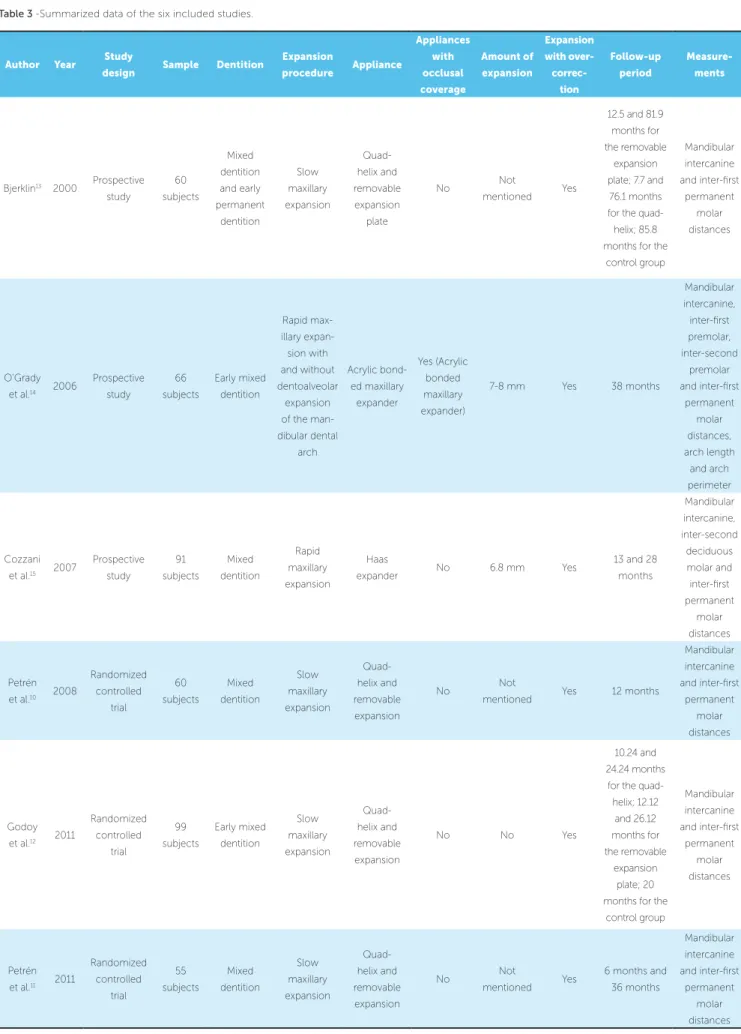
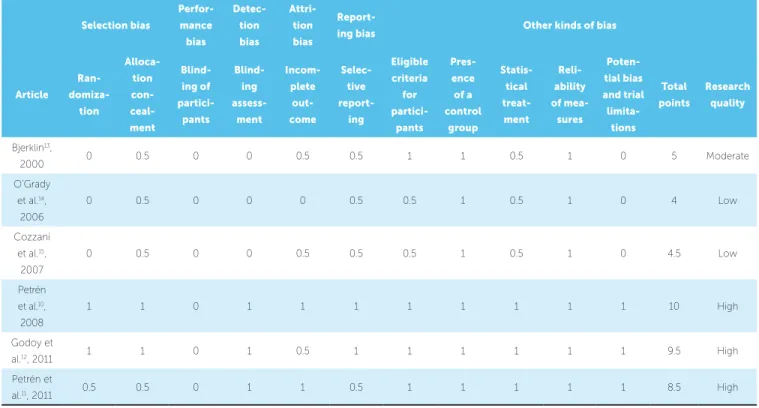
A summary of the methodological characteristics of the inal studies — such as authors, year of publication, study design, sample size, dentition stage, type of expan-sion procedure, type of appliance used, amount of maxil-lary expansion, follow-up period and the measurements performed in the mandibular dental arch — is given in Table 3. Application of the methodological quality check-list is shown in Table 4. Kappa statistics was performed ater quality assessment of the studies and showed good
interexaminer agreement (0.85).19
All the selected studies assessed the spontaneous dentoalveolar changes of the mandibular dental arch ater slow or rapid maxillary expansion, performing measurements with digital sliding caliper in conven-tional dental models. The main variables assessed in
these studies were: mandibular intercanine distance10-15
(between the crown tip or the lingual gingival margin),
inter-deciduous molar or interpremolar distance14,15
(between the center of the fossa or the lingual gingival
margin), inter-irst permanent molar distance10-15
(be-tween the mesiobuccal cusp tips, the center of the fossa
or the lingual gingival margin), arch length,14 arch
pe-rimeter14 and inclination of the irst permanent molars.14
373 articles identi-fied through database
searching
116 articles screened by reviewing the titles
and abstracts
10 articles identified through hand-searching
20 full-text articles excluded:
- Lack of control group (13) - Retrospective study (3)
- Intervention in the mandibular dental arch (4) 26 full-text articles
as-sessed for eligibility
6 studies included in quality analysis
Included
Eligibility
Identifica
tion
Identifica
tion
Component Classification Points Definition
Selection bias
1. Randomization
Adequate 1.0 Randomization correctly described as well the randomization method. Inadequate 0.5 Incomplete description of randomization method.
None 0 No description of randomization method.
2. Allocation concealment
Adequate 1.0 Allocation concealment correctly described. Inadequate 0.5 Incomplete description of allocation concealment. None 0 No description of allocation concealment.
Performance bias
3. Blinding of participants and personal
Adequate 1.0 Blinding of participants and personal correctly described and efectiveness of blinding stated.
Inadequate 0.5 Incomplete description of blinding of participants and personal. None 0 No description of blinding of participants and personal.
Detection bias
4. Blinding assessment
Adequate 1.0 Blinding assessment described in measures or statistics and efectiveness of blinding stated.
Inadequate 0.5 Incomplete description of blinding assessment. None 0 No blinding assessment described.
Attrition bias
5. Incomplete outcome data
Explained 1.0 Dropouts reported with explanation and description of complete or incomplete data retrieved.
Not explained 0.5 Dropouts reported with no explanation or description of complete or incomplete data retrieved.
None 0 No reporting of dropouts or data retrieved.
Reporting bias
6. Selective reporting
Adequate 1.0 No selective reporting of primary outcomes. Inadequate 0.5 Insuicient information to judgement. None 0 Selective reporting of primary outcomes.
Other kinds of bias
7. Eligible criteria for participants
Adequate 1.0 Inclusion/exclusion criteria described.
Inadequate 0.5 No description of inclusion/exclusion criteria, but selection done at least by age and type of expansion.
None 0 No description of criteria for selection.
8. Presence of a control group Yes 1.0 Presence of a control group. No 0 Absence of a control group.
9. Statistical treatment
Adequate 1.0 Statistical treatment fully described and adequate. Inadequate 0.5 Statistical treatment not fully described or inadequate. None 0 No statistical treatment applied.
10. Reliability of measures
Adequate 1.0 Aleatory measures repeated and statistical test applied. Inadequate 0.5 Measures repeated and inadequate or no statistical tests applied. None 0 Measures not repeated.
11. Potential bias and trial Limitations
Fully 1.0 Description of potential bias and trial limitations acknowledging them. Partially 0.5 Description of potential bias and trial limitations without acknowledging them. None 0 No description of potential bias or trial limitations.
Table 3 -Summarized data of the six included studies.
Author Year Study
design Sample Dentition
Expansion procedure Appliance Appliances with occlusal coverage Amount of expansion Expansion with over- correc-tion Follow-up period Measure-ments
Bjerklin13 2000 Prospective study 60 subjects Mixed dentition and early permanent dentition Slow maxillary expansion Quad-helix and removable expansion plate No Not mentioned Yes
12.5 and 81.9 months for the removable
expansion plate; 7.7 and
76.1 months for the quad-helix; 85.8 months for the
control group Mandibular intercanine and inter-irst permanent molar distances O’Grady et al.14 2006
Prospective study 66 subjects Early mixed dentition Rapid max-illary expan-sion with and without dentoalveolar expansion of the man-dibular dental arch Acrylic bond-ed maxillary expander Yes (Acrylic bonded maxillary expander)
7-8 mm Yes 38 months
Mandibular intercanine, inter-irst premolar, inter-second premolar and inter-irst permanent molar distances, arch length and arch perimeter Cozzani et al.15 2007
Prospective study 91 subjects Mixed dentition Rapid maxillary expansion Haas
expander No 6.8 mm Yes
13 and 28 months Mandibular intercanine, inter-second deciduous molar and inter-irst permanent molar distances Petrén et al.10 2008
Randomized controlled trial 60 subjects Mixed dentition Slow maxillary expansion Quad-helix and removable expansion No Not
mentioned Yes 12 months
Mandibular intercanine and inter-irst permanent molar distances Godoy et al.12 2011
Randomized controlled trial 99 subjects Early mixed dentition Slow maxillary expansion Quad-helix and removable expansion
No No Yes
10.24 and 24.24 months
for the quad-helix; 12.12 and 26.12 months for the removable expansion plate; 20 months for the
control group Mandibular intercanine and inter-irst permanent molar distances Petrén et al.11 2011
Randomized controlled trial 55 subjects Mixed dentition Slow maxillary expansion Quad-helix and removable expansion No Not mentioned Yes
Selection bias
Perfor-mance
bias
Detec-tion
bias
Attri-tion
bias
Report-ing bias Other kinds of bias
Article
Ran- domiza-tion
Alloca-tion
con-
ceal-ment
Blind-ing of
partici-pants
Blind-ing
assess-ment
Incom-plete
out-come
Selec-tive
report-ing
Eligible criteria
for
partici-pants
Pres-ence
of a control
group
Statis-tical
treat-ment
Reli-ability
of mea-sures
Poten-tial bias
and trial
limita-tions
Total
points
Research
quality
Bjerklin13,
2000 0 0.5 0 0 0.5 0.5 1 1 0.5 1 0 5 Moderate
O’Grady et al.14,
2006
0 0.5 0 0 0 0.5 0.5 1 0.5 1 0 4 Low
Cozzani et al.15,
2007
0 0.5 0 0 0.5 0.5 0.5 1 0.5 1 0 4.5 Low
Petrén et al.10, 2008
1 1 0 1 1 1 1 1 1 1 1 10 High
Godoy et
al.12, 2011 1 1 0 1 0.5 1 1 1 1 1 1 9.5 High
Petrén et
al.11, 2011 0.5 0.5 0 1 1 0.5 1 1 1 1 1 8.5 High
Table 4 -Quality assessment of the selected studies.
Research quality or methodological soundness: high, >8 points; moderate, 5 to 8 points; low, <5 points.
In general, the follow-up period of the spontaneous dentoalveolar changes of the mandibular dental arch af-ter SME was greaaf-ter than that of the RME. The short-term spontaneous dentoalveolar changes ater slow
maxillary expansion was evaluated in four studies,10-13
while three of them assessed the long-term efects of
SME.11-13 On the other hand, only one of the selected
studies analyzed the short-term spontaneous
dentoal-veolar changes ater rapid maxillary expansion,15 while
the long-term occlusal changes ater RME was analyzed
in two studies.14,15
Short-term spontaneous dentoalveolar changes after SME
The removable expansion plate promoted greater in-creases of the mandibular intermolar distance (0.4mm, at the lingual gingival margin level, and 1.2mm, at the mesiobuccal cusp tip level), compared to the quad-helix appliance (-0.4mm, at the lingual gingival margin
lev-el, and -0.1mm, at the mesiobuccal cusp tip level), six
months ater slow maxillary expansion.11
No significant spontaneous dentoalveolar chang-es were observed in the mandibular dental arch of
the treated and untreated groups 4.24 months12 and
7.7 months13 after slow maxillary expansion
us-ing the quad-helix appliance and 6.12 months12 and
12.5 months13 after SME with the removable
expan-sion plate. On the other hand, the quad-helix appli-ance promoted a significantly greater increase of the mandibular intercanine distance (0.2mm) compared to the control group (0.0mm), 12 months after the
expansion procedure.10
Long-term spontaneous dentoalveolar changes after SME
expansion plate (-0.36mm), 16.24 to 18.12 months ater slow maxillary expansion. However, no signiicant dif-ferences were found between the experimental groups
and the control group (-0.18mm).12
Thirty-six months ater the slow maxillary expan-sion procedure, the control group showed a signiicantly greater increase of the mandibular inter-irst permanent molar (0.5mm), compared to the quad-helix (-0.6mm)
and the removable expansion plate groups (-0.6mm).11
Seventy-six months ater SME, the quad-helix group showed a signiicantly smaller decrease (-0.6mm) of the mandibular intercanine distance compared to the
control group (-1.3mm).13
Finally, a signiicantly greater increase of the man-dibular inter-irst permanent molar distance was found for the control group (0.5mm) compared to the remov-able expansion plate group (-0.1mm), approximately
eighty-two months ater slow maxillary expansion.13
Short-term spontaneous dentoalveolar changes after RME
Signiicant increases of 0.9mm in the mandibular intercanine distance and 0.7 mm on the mandibular inter-second deciduous molar distance were found for the experimental group, 1.1 years ater rapid maxillary expansion using the Haas-type expander. However, no statistically signiicant diferences were found between the experimental and control groups in the mandibular intercanine and inter-second deciduous molar distances ater the 13 months of follow-up. No intra and inter-group diferences were found for the mandibular inter
irst-permanent molar distance.15
Long-term spontaneous dentoalveolar changes after RME
Only a signiicant increase of 0.9mm was found in the mandibular intercanine distance of the experimen-tal group, 28 months ater rapid maxillary expansion
with the Haas-type expander.15 However, the increase
amount in mandibular intercanine, inter-second decid-uous molar and in inter-irst permanent molar distances were signiicantly smaller in the experimental compared
to the control group.15
Similar results were found when spontaneous den-toalveolar changes of the mandibular dental arch were analyzed 3.2 years ater rapid maxillary expansion
us-ing the acrylic splint expander.14 Signiicant increases
of 1.0mm in the mandibular intercanine, 1.8mm in the inter-irst deciduous molar, 1.6mm in the inter-second deciduous molar and 1.9mm in the inter-irst perma-nent molar distances were found in the experimental
group.14 Additionally, signiicant decreases of 0.8mm
in arch length and 1.2mm in arch perimeter and buccal inclination of the mandibular irst permanent molars of
7.7° were found in the treated group.14 However, no
sta-tistically signiicant diferences were found between the
experimental and control group changes.14
DISCUSSION
Short and long-term dentoskeletal efects of slow and rapid maxillary expansion were already analyzed
in systematic reviews and meta-analyses.20-27 However,
none of these studies evaluated the spontaneous dento-alveolar changes of the mandibular dental arch during these follow-up periods.
A previous study28 showed that signiicant increases
of the mandibular dental arch width and perimeter tend to occur 6.1 years ater RME in adolescent patients. However, in this longitudinal study, the treatment changes were measured before the expansion procedure and ater completion of edgewise appliances therapy. Methodologically, performing orthodontic interven-tion in the mandibular dental arch represents a con-founding factor in the analysis of the spontaneous den-toalveolar changes concurrent to slow or rapid maxillary expansion. Thus, a systematic review of the literature was necessary to better evaluate this issue.
In this review, the selected randomized controlled
trials10-12 obtained greater scores in the quality assessment
compared to the prospective studies.13-15 This inding is
expected because the RCTs must be written according to the methodological requirements of the CONSORT
statement.29 From the randomized controlled trials, the
study of Petrén et al11 obtained the smallest score
be-cause the authors did not describe how the additional ten patients recruited to comprise the treated group, were randomized and allocated.
The selected prospective studies showed important methodological limitations, such as the lack of detailed
description of randomization of patients,11,13-15
alloca-tion concealment,11,13-15 blinding assessment,13-15
re-porting of outcome data,11,13-15 drop-out,14,15 eligibility
and exclusion criteria,14,15 sample size calculation,13-15 as
Thus, these studies were qualiied with low to moderate risk of bias since the absence of these criteria increases the potential risk of bias.
Neither the selected RCTs10-12 nor the prospective
studies13-15 reported double-blinding of participants
and personal. This is not a methodological concern as double-blinding would not be possible in these experi-mental studies involving maxillary expansion. Inevita-bly, both orthodontists and patients were not blinded regarding the appliance type.
The follow-up assessment of treated and untreated patients is important for comparison purposes.
How-ever, two studies,11,12 did not compare the short-term
mandibular dental arch changes of the experimental and control groups, because the untreated group was followed-up only in the long-term. Thus, only the experimental group changes could be evaluated in the short-term. The lack of a comparison between the experimental and control group changes in the short-term is a limitation, because it is unknown if the changes observed in the treated group occurred consequently to the maxillary expansion procedure, due to growth, or both.
Additionally, attention is required when
interpret-ing the short and long-term results of Cozzani et al15.
In this study, the mandibular arch widths in the short and long-term posttreatment observation stages were compared with transversely selected age matching con-trol groups at those stages. Therefore, although there were some spontaneous transverse increases in the perimental group, the mandibular dental arch of the ex-perimental group showed smaller transverse dimensions than the control groups. However, this type of compari-son is not very reliable.
The selected studies showed negligible spontane-ous dentoalveolar changes of approximately 1mm in the mandibular dental arch of patients treated with slow or rapid maxillary expansion. The clinical implication of
this is that clinically signiicant spontaneous dentoalve-olar changes should not be expected in the mandibular dental arch ater SME or RME in the short and long-term. If patients show constricted mandibular dental arch with or without incisor crowding, dentoalveolar expansion of the mandibular dental arch may be
indicat-ed.14 This clinical implication is conirmed by O’Grady
et al.14 In this longitudinal study, patients treated with
the Schwarz’s appliance showed, in the long-term, sig-niicantly greater increases of the mandibular dental arch widths, arch perimeter and buccal inclination of posterior teeth compared to the control group.
A limitation of the present systematic review was the diiculty in inding studies with adequate con-trol groups. Additionally, a great number of articles analyze the long-term changes of maxillary expansion followed by ixed orthodontic appliances in the maxil-lary and mandibular dental arches. Future randomized controlled trials with appropriate control groups should analyze the short and long-term spontaneous dentoal-veolar changes of mandibular dental arch ater SME and RME to better evaluate this issue. Improvement in the methodological quality of studies and the homogeneity of RCTs might give further information for future me-ta-analyses, which can provide higher levels of scientiic
evidence to support the orthodontic clinical practice.30
CONCLUSION
» Based on the results from this systematic review, there is enough evidence to conclude that negligible short and long-term spontaneous dentoalveolar changes occur in the mandibular dental arch ater SME or RME in the mixed and early permanent dentition.
1. McNamara JA Jr. Maxillary transverse deiciency. Am J Orthod Dentofacial Orthop. 2000 May;117(5):567-70.
2. Kutin G, Hawes RR. Posterior cross-bites in the primary and mixed dentition.
Am J Orthod. 1969;56:491-504.
3. Thilander B, Wahlund S, Lennartsson B. The efect of early interceptive treatment
in children with posterior cross-bite. Eur J Orthod. 1984 Feb;6(1):25-34.
4. Sandikcoglu M, Hazar S. Skeletal and dental changes after maxillary expansion in
the mixed dentition. Am J Orthod Dentofacial Orthop. 1997 Mar;111(3):321-7.
5. Melsen B. Palatal growth studied on human autopsy material. A histologic
microradiographic study. Am J Orthod. 1975 July;68(1):42-54.
6. Gianelly AA. Rapid palatal expansion in the absence of crossbites: added value?
Am J Orthod Dentofacial Orthop. 2003 Oct;124(4):362-5.
7. Lima AC, Lima AL, Lima Filho RMA, Oyen OJ. Spontaneous mandibular
arch response after rapid palatal expansion: a long-term study on Class I malocclusion. Am J Orthod Dentofacial Orthop. 2004 Nov;126(5):576-82.
8. Halazonetis DJ, Katsavrias E, Spyropoulos MN. Changes in cheek pressure
following rapid maxillary expansion. Eur J Orthod. 1994 Aug;16(4):295-300.
9. Küçükkeleş N, Ceylanoğlu C. Changes in lip, cheek, and tongue pressures after
rapid maxillary expansion using a diaphragm pressure transducer. Angle Orthod. 2003 Dec;73(6):662-8.
10. Petrén S, Bondemark L. Correction of unilateral posterior crossbite in the mixed dentition: a randomized controlled trial. Am J Orthod Dentofacial Orthop. 2008 June;133(6):790.e7-13.
11. Petrén, Bjerklin K, Bondemark L. Stability of unilateral posterior crossbite
correction in the mixed dentition: a randomized clinical trial with a 3-year follow-up. Am J Orthod Dentofacial Orthop. 2011 Jan;139(1):e73-81. 12. Godoy F, Godoy-Bezerra J, Rosenblatt A. Treatment of posterior crossbite
comparing 2 appliances: a community-based trial. Am J Orthod Dentofacial Orthop. 2011 Jan;139(1):e45-52.
13. Bjerklin K. Follow-up control of patients with unilateral posterior cross-bite treated with expansion plates or the quad-helix appliance. J Orofac Orthop. 2000;61(2):112-24.
14. O’Grady PW, McNamara JA Jr, Baccetti T, Franchid L. A long-term evaluation of the mandibular Schwarz appliance and the acrylic splint expander in early mixed dentition patients. Am J Orthod Dentofacial Orthop. 2006 Aug;130(2):202-13. 15. Cozzani M, Guiducci A, Mirenghi S, Mutinelli S, Siciliani G. Arch width changes
with a rapid maxillary expansion appliance anchored to the primary teeth. Angle Orthod. 2007 Mar;77(2):296-302.
16. Moher D, Liberati A, Tetzlaf J, Altman DG, The PRISMA Group. Preferred reporting items for systematic reviews and metaanalyses: the PRISMA Statement. Syst Rev. 2015;4(1):1-9.
REFERENCES
17. Higgins JPT, Green S. Cochrane handbook for systematic reviews of
interventions. Version 5.1.0 [updated March 2011 Access in: 2016 27th July]. Cochrane Collaboration. 2011. Available from:
http://www.cochrane-handbook.org.
18. Vilani GN, Mattos CT, Ruellas ACO, Maia LC. Long-term dental and skeletal changes in patients submitted to surgically assisted rapid maxillary expansion: a meta-analysis. Oral Surg Oral Med Oral Pathol Oral Radiol. 2012 Dec;114(6):689-97.
19. Viera AJ, Garret JM. Understanding interobserver agreement: the kappa statistic. Fam Med. 2005;37(5):360-3.
20. Schifman PH, Tuncay OC. Maxillary expansion: a meta analysis. Clin Orthod Res. 2001 May;4(2):86-96.
21. Petrén S, Bondemark L, Söderfeldt B. A systematic review concerning early orthodontic treatment of unilateral posterior crossbite. Angle Orthod. 2003 Oct;73(5):588-96.
22. Lagravere MO, Major PW, Flores-Mir C. Long-term skeletal changes with rapid maxillary expansion: a systematic review. Angle Orthod. 2005 Nov;75(6):1046-52.
23. Lagravere MO, Major PW, Flores-Mir C. Long-term dental arch changes after rapid maxillary expansion treatment: a systematic review. Angle Orthod. 2005 Mar;75(2):155-61.
24. Lagravere MO, Major PW, Flores-Mir C. Skeletal and dental changes with ixed slow maxillary expansion treatment: a systematic review. J Am Dent Assoc. 2005 Feb;136(2):194-9.
25. Bazargani F, Feldmann I, Bondemark L. Three-dimensional analysis of efects of rapid maxillary expansion on facial sutures and bones. Angle Orthod. 2013 Nov;83(6):1074-82.
26. Agostino P, Ugolini A, Signori A, Silvestrini-Biavati A, Harrison JE, Riley P. Orthodontic treatment for posterior crossbites. Cochrane Database Syst Rev. 2014 Aug 8;(8):CD000979.
27. Zhou Y, Long H, Ye N, Xue J, Yang X, Liao L, et al. The efectiveness of non-surgical maxillary expansion: a meta-analysis. Eur J Orthod. 2014;36(2):233-42. 28. McNamara JA Jr, Baccetti T, Franchi L, Herberger TA. Rapid maxillary expansion
followed by ixed appliances: a long-term evaluation of changes in arch dimensions. Angle Orthod. 2003 Aug;73(4):344-53.
29. Moher D, Hopewell S, Schulz KF, Montore V, Gotsche PC, Devereaux PJ, et al. CONSORT 2010 explanation and elaboration: updated guidelines for reporting parallel group randomized trials. Int J Surg. 2012;10(1):28-55.