Al and PEG Efect on Structural and Physicochemical Properties of CoFe
2O
4Fatemeh Mostaghnia*, Yasaman Abedb
Received: December 04, 2016; Revised: February 07, 2017; Accepted: February 18, 2017
In this work, pure and Alumina doped cobalt ferrite nanoparticles CoFe2−xAlxO4 (for x = 0.44)
have been synthesized by the sol gel method. The inluence of alumina doping on the morphological
and mechanical properties of CoFe2O4 nano-particles were investigated by means of X-ray powder
difraction (XRD) and rietveld analysis. XRD analysis conirmed that the single phase formation of pure nano particles with the expected cubic inverse spinel structure with Fd3m space group and without any impurity phase. Alumina doping were led to a decrease in the crystallite size, lattice parameter, elastic constants and magnitude of moduli. It is explained on the basis of the replacement of Fe ions with half-illed d-shell (3d5) and larger radius by Al3+ ions with a completely illed shell
(2p6) and smaller radius.
Keywords: Spinels, Ferrite, Cobalt ferrite, Rietveld Reinement
* e-mail: [email protected]
1. Introduction
Spinels of the type AB2X4 are one of the most interesting and important families of crystalline compounds due to their broad applications in magnetic materials, ceramics, catalysis,
etc. Spinels of the type A2+B3+
2O4 belong to a large group
of composite oxides with a cubic symmetry (space group Fd3m), where A and B are cations with variable valence. In
natural spinels usually A represents divalent cation, while B represents trivalent cation (A2+B3+2O4, so-called 2-3 spinels).1-3
Among spinel composite oxides, spinel ferrite nanoparticles
have been studied for many years due to their magnetic
and electrical properties. In particular, the CoFe2O4 has
covered a broad applications including electronic devices,
ferroluids, magnetic drug delivery, microwave devices and high density information storage.4-9 Cobalt ferrite (CoFe
2O4)
is one of the well known hard magnetic materials which has strong magnetic anisotropy, high magnetostriction, moderate magnetization, high coercivity at room temperature and high
chemical stability and good electrical insulation.10-13 There
are diferent methods which have been reported for the preparation of magnetic nanoparticles of cobalt ferrite.14-17
Cobalt ferrite is a partially inverse spinel with formula (Cox Fe1-x)[Co1-x Fe1+x]O4, formed by oxygen atoms in a closed packing structure where, Co2+ and Fe3+ occupy either
tetrahedral or octahedral sites.18-22 Several researchers have
reported on Ti, Y, Gd, Pr, Zn, Ni and Dy doped cobalt ferrite.23-28
The present work is focused on the efects of Al3+ doping on
the structural, morphological and elastic properties of aluminium
ion doped cobalt ferrite obtained via sol-gel method. The X-ray powder difraction patterns, the microstructure and the elastic
properties are discussed as a function of the Al3+ doping.
2. Materials and Methods
2.1. Material
Iron (III) nitrate nonahydrate Fe(NO3)3.9H2O, cobalt(II)
nitrate hexahydrate Co(NO3)2.6H2O and aluminium nitrate
nonahydrate-Al(NO3)3∙9H2O as source of metal ions and
polyethylene glycol (average molecular weight: 4000,
Qualigen) as a surfactant were used. The pH was controlled by amonium hydroxide NH4OH. All reactants were purchased
from Sigma-Aldrich. Deionized water served as reacting medium.
2.2. Synthesis of nanoparticles
Nanocrystalline powders of CoFe2-xAlxO4 (x = 0,
0.4) were prepared by the sol-gel method. In a typical reaction, cobalt nitrate (Co(NO3)2.6H2O), iron nitrate
(Fe(NO3)3.9H2O), and aluminum nitrate (Al(NO3)3.9H2O)
were individually dissolved in 10 ml of deionized water
in their respective stoichiometry. The solutions were then mixed and stirred for 30 minutes at room temperature. In
addition, polyethylene glycol (average molecular weight:
4000, Qualigen) served as a surfactant for this reaction and poured into the above mixture. Then, the 25% ammonia
solution was added drop by drop and stirred vigorous on
a magnetic stirrer. The inal pH of the mixture was about 8. The resulting solution was kept under stirring at 60oC
for 1 hour. The obtained gels were dried at 80ºC and
calcined at 500 oC for 3 hours.
Crystal structure of nanoparticles was determined by a
Bruker make difractometer, Cu-Kα X-rays of wavelength
(λ=1.5406 Å). The XRD patterns were recorded in the 2θ
range of 10–90o with a step width of 0.02 s-1. a Chemistry Department, Payam Noor University, Iran, P.O.Box: 19395-4697
2.3. Simulation analysis
The accelrys materials studio 6.0 visualisation package was used to create the nanoparticles. The X-ray difraction (XRD) pattern was analysed with the help of relex module by employing rietveld reinement technique. The XRD pattern
of the pure and aluminia doped CoFe2O4 was reined using
the Fd3m space group. The Castep module was employed to
calculate the structural, electronic, and elastic properties of
two samples. Generalized gradient approximation (GGA) with the Perdew–Burke–Ernzerhof was used for all calculations.
3. Results and Discussion
3.1. X-Ray difraction studies
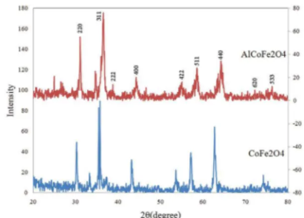
X-ray difraction pattern of synthesized aluminia doped
and pure CoFe2O4 are presented in Figure. 1.
Figure 1. XRD pattern of synthesized CoFe2O4 and Al-dopped CoFe2O4
Peak details of synthesized nanoparticles sumerised in Table 1 and 2. The sharp and broad peaks in these XRD patterns, conirming the single phase formation of pure nano particles with the expected cubic inverse spinel structure with Fd3m space group and without any impurity phase. It can
be observed that the intensities of the 220 planes increase and the intensity of 440 plane decrease with the addition of aluminium indicating the preference of the Co2+, Fe3+ and Al3+ ions by the octahedral B and tetrahedral A sites. Indeed, this may be indicated the redistribution of the cations in the
nanostructure aluminium cobalt ferrite. From this data it is
evident that the ratio of Fe3+(oct.)/Fe3+ (tet.) changes with addition of Al3+ ion. In the present case the cubic phase spinel
ferrite structure for two composition is mainly due to deicit
of Co2+ ions in the octahedral sites(<85%) that leads to the
absence of co-operative active Jahn-Teller distortion. The
crystallite size of the nano-particles were calculated from the
most intense peak (3 1 1) of XRD data using Debye-Scherer equation. The crystallite size of Al3+ doped and pure CoFe
2O4
nano-particles were 18.9 and 24.3 nm respectively. The lattice
constants of Al3+ doped and pure CoFe2O4, calculated from
F(θ) were 8.22 and 8.32 repectively.
As can be seen, the crystallite size and lattice constant
were decreased with alumina doping. Similar results have been reported in the literature.29,30 This behavior of the lattice
constant with alumina doping is due to the replacement of larger Fe3+ ions (0.645 oA) by smaller Al3+ ions (0.535 oA)
in the system.
The x-ray density (ρx) of the samples was determined
using relation given by Smith and Wijn: 31,32
( )
N a
M
8
1
x A
3
t
=
where, M is the molecular weight of the composition,
NA is the Avogadro’s number and ‘a’ is the lattice constant.
As there are 8 formula unit in the unit cell so 8 is included
in the formula. The x-ray density (ρx) of alumina doped
and pure CoFe2O4, were 3.605 and 3.746 g/cm3 repectively.
It can be observed that the x-ray density (ρx) decreased
with alumina doping, because the decrease in molecular
weight overtakes the decrease in volume of the unit cell.
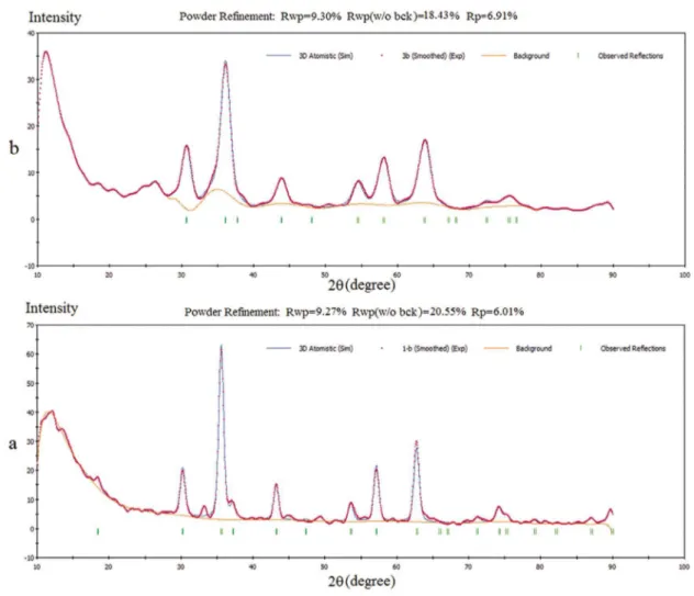
3.2. Rietveld analysis
Various structural parameters can be determined from
rietveld analysis and are found useful to explain other physical properties.33 This method is based on a least-squares
it between step-scan data of a measured difraction pattern and a simulated X-ray-difraction pattern.
In this study the crystalline structure of the pure and aluminia doped CoFe2O4 were analized in detailed by
rietveld proile reinement method in relex module. The XRD patterns for these samples have reined using the Fd3m space group (Figure 2).
The itting quality of the experimental data is assessed
by computing the parameters R-pattern factor Rp and the
weighted-proile factor Rwp.
34-36 These factors are deined as:
( )
R
I w
I
I w
2
Wp
i i
c i
i
02
0 2
2
$
=
#
Q
-
V
&
/
/
( )
R
y
y
y
3
p
io i
io ic
i
=
-/
/
Where Io and Ic are respectively observed and calculated
integrated Bragg intensities (without background). Rp and
Rwp values for pure and aluminia doped CoFe2O4 were (6.01,
9.27) and (6.91, 9.30) respectively. The calculated R factors indicated that the models were correct.
Oxygen positional parameter (u) for each composite was calculated using the formulae available in the literature.
Table 1. Peak analysis of XRD pattern of synthesized CoFe2O4
2θ [deg.] h k l FwHM [2θ] d-spacing a [A] F(θ) Cry.size [nm]
30.19 220 0.2460 2.9596 8.37 1.82961 33.4
33.22 310 0.5904 2.6964 8.52 1.62781 14.1
35.56 311 0.3444 2.5245 8.37 1.50955 24.3
43.13 400 0.2952 2.0972 8.36 1.03974 29.0
53.47 422 0.7872 1.7135 8.37 0.90145 11.3
57.08 511 0.3444 1.6133 8.31 0.82074 26.3
62.71 440 0.6000 1.4803 8.37 0.12157 15.5
Table 2. Peak analysis of XRD pattern of synthesized Al-dopped CoFe2O4
2θ [deg.] h k l FwHM [2θ] d-spacing a [A] F(θ) Cry.size [nm]
30.75 220 0.1968 2.9068 8.22178 1.77766 41.8
34.36 310 0.2952 2.6099 8.25323 1.56398 28.2
36.20 311 0.4428 2.4811 8.22898 1.47009 18.9
44.04 400 0.7872 2.0559 8.22384 1.16509 10.9
54.47 422 0.7872 1.68453 8.25248 0.87748 11.4
58.23 511 0.3936 1.58429 8.23221 0.79441 23.1
64.11 440 0.7200 1.45126 8.20957 0.68597 13.0
1/4) for which uidea = 0.250 (origin at B-site), while u
43 give
‘u’ assuming centre of symmetry at (3/8, 3/8, 3/8) for which
uidea = 0.375 (origin at A-site). These two factors are related by following formula:
M-O M-M
( )
u
43m=
u
3m+
1
8
4
( )
R
A=
a
3
S
u
-
4
1
X
-
R
05
.
( )
R
Ba
3
u
2 75
u
64
43
R
6
2
1 2
0
=
S
-
+
X
-The radius of the tetrahedral and octahedral sites in a spinel
ferrite can be calculated using the following formulae: 37,38
Where RO is the radius of the oxygen ion (1.32 oA)
and u represents the oxygen positional parameter (table 3). Using the lattice constant as well as oxygen parameter (u) of each composite, interatomic distances have been calculated from following equations.39,40 The calculated
values are presented in Table 3.
( )
Shared octahedral edge
d
a
2
2
u
2
1
7
AE
m
1 2 43
=
Q
V
S
-
X
( )
Shared tetrahedral edge
d
a
2
1
2
u
8
BE
m
1
2 43
=
Q
V
Q
-
V
( )
Octahedral bond lenght
d
a
3
u
11
4
u
64
43
9
BL
m m
43 2 43
1 2
=
#
Q
V
-
+
&
( )
Tetrahedral bond lenght
d
a
3
u
4
1
10
AL
m
43
=
S
-
X
( )
Unshared octahedral edge
d
a
4
u
3
u
16
11
11
BEu
m m
43 2 43
1 2
=
#
Q
V
-
+
&
It is observed from Table 3 that the doping with alumina
brought about a decrease in the values of RA, RB, edge lengths
and bond lengths. This could be related to the smaller radius
of Al3+ ion as compared to Fe3+ ion. In fact, the ionic radius of the Al3+ ion is 0.050 nm, while the ionic radius of the Fe3+ ions is 0.064 nm.
The interionic distances between the cations (b, c, d, e, and f) and between the cation and anion (p, q, r and s) were also calculated using the experimental values of lattice constant (a) and oxygen positional parameters (u3m) (Tables
4 and 5) by the following relations:41,42
p
a
5u
b
a
4 2
8
1 2
=
Q
-
V
=
S
X
q
=
a u
S
-
1
4 3
X
12c
=
S
a
8 11
X
12r
=
a u
S
-
1
8 11
X
12d
=
S
a
4 3
X
12s
=
a
S
1
3
u
+
1
8 3
X
12e
=
S
3
a
8 3
X
12f
=
S X
a
4 6
12Table 4 indicates that, both interatomic distances between the cation-anion (p, q, r and s) and between the cations (b, c, d, e and f) decrease with alumina doping. This result is accordance with decrease in unit cell volume.
The bond angles (θ1, θ2, θ3, θ4 and θ5) were calculated by
simple trigonometric principle using the interionic distances with the help of following formula: 41,42
(12)
(13)
(14)
(15)
(16)
( )
cos
pq
p
q
c
2
17
1 1
2 2 2
i
=
-#
+
-
&
( )
cos
pr
p
r
e
2
18
2 1
2 2 2
i
=
-#
+
-
&
( )
cos
p
p
b
2
2
19
3 1 2
2 2
i
=
-#
-
&
( )
cos
ps
p
s
f
2
20
4 1
2 2 2
i
=
-#
+
-
&
( )
cos
rq
r
q
d
2
21
5 1
2 2 2
i
=
-#
+
-
&
These values are tabulated in Table 5.
It is observed from Table 5 that angles θ3 and θ4 decrease
while θ1, θ2 and θ5 increase with alumina doping. The
observed decrease in θ3 and θ4 indicative of weakening of
the B-B interaction while increase in θ1, θ2 and θ5 suggest
strengthening of the A-B and A-A interactions on Al3+
-substitution in the system.
3.3. Elastic properties
The elastic constants of solids provide a link between the mechanical and dynamical behaviors of crystals. Cubic
crystals have three independent elastic constants: C11, C12, and
,
,
( )
B
B
B
G
G
G
B
B
C
C
2
2
3
2
22
v R v R
v R
11 12
=
+
=
+
=
=
+
,
( )
G
C
C
C
C
C
C
G
C
C
C
4
3
5
5
3
23
R v
44 11 12
44 11 12
11 12 44
=
+
-=
-
+
Q
Q
V
V
,
( )
Y
B
BG
G
v
B
G
B
G
3
9
2 3
3
2
24
=
+
=
Q
-
+
V
Young’s moduli Y and the Poisson ratio υ. Values of these
factors are computed using the following relationships:43-45
Table 3. Edge lengths [Ao], bond lengths[Ao], radius of the tetrahedral and octahedral [Ao], and anion parametes [Ao] of Al-Co-Fe-O system
compound dAe dBE dBEu dAl dBl RA RB U43 U3
CoFe2O4 2.8162 3.0352 2.9140 1.7436 2.0948 0.4236 0.7748 0.371 0.246
CoAl0.4Fe1.6O4 2.7918 3.0196 2.9071 1.7098 2.0924 0.3172 0.7724 0.370 0.245
Table 4. Inter-ionic distances [Ao], of Al-Co-Fe-O system
compound p q r s b c d e f
CoFe2O4 2.1132 1.7436 3.3322 3.5833 2.9411 3.4492 3.6025 5.403 5.094
CoAl0.4Fe1.6O4 2.0961 1.7064 3.271 3.5356 2.905 3.407 3.5592 5.338 5.033
Table 5. Bond angles (degree) of Al-Co-Fe-O system
compound θ1 θ2 θ3 θ4 θ5
CoFe2O4 125.54 165.4 88.24 124.86 84.28
CoAl0.4Fe1.6O4 126.96 167.68 87.72 124.69 85.14
Table 6. Elastic constants for Al-Co-Fe-O system
compound C11 C12 C44 B Y ʋ G
[PA] [GPA] [GPA] [GPA] [GPA] [GPA]
CoFe2O4 186.08 28.6087 50.2593 81.10 178.4627 0.1333 60.2061
CoAl0.4Fe1.6O4 157.37 12.2987 47.8372 60.65 155.5927 0.0725 56.5492
Complate set of the calculated elastic constants for two
samples are collected in Table 6.
It can be seen that magnitude of moduli were decreased on Al3+ doping. It is explained on the basis of the replacement of Fe3+ ions with half-illed d-shell (3d5) by Al3+ ions with a
noble gas outer electron shell (2p6) structure which do not
contribute to the bond formation.
However it is well known that the cations with a completely illed outer electron shell structure are more
stable and less compressible than the cations with a
half-illed or a incomplete outer shell. In other word the material is compressed easier when the radius of the ions is longer. Therefore, strength of bonding and magnitude of moduli are expected to decrease.46, 47
4. Conclusions
Pure and Al3+ doped cobalt ferrite nanoparticles were
successfully synthesized by sol-gel method and thermally
treated at 500ºC for 3 hours. XRD and the rietveld analysis
showed that two composition were formed into single
phase cubic spinel structure. Various structural parameters can be determined from x-ray powder difraction pattern analysis and are found useful to explain other physical properties. The lattice parameters and the crystallite size
were found decreasing with Al3+ doping. The strength of the A-B interaction increase while B-B interaction decreases with Al3+ substitution for Fe3+ in the system. Also magnitude of moduli were decreased on Al3+ doping.
This is due essentially to the ion size diference among
Al3+ and Fe3+ ions.
5. Acknowledgement
6. References
1. O’Handley RC. Modern Magnetic Materials – Principles and
Applications. Hoboken: John Wiley & Sons; 2000.
2. Buschow KHJ, ed. Handbook of Magnetic Materials, Vol. 19.
Amsterdam: Elsevier, North Holland; 2011.
3. Sun S, Zeng H, Robinson DB, Raoux S, Rice PM, Wang SX,
et al. Monodisperse MFe2O4 (M= Fe, Co, Mn) Nanoparticles.
Journal of American Chemical Society. 2004;126(1):273-279. 4. Amstad E, Textor M, Reimhult E. Stabilization and functionalization
of iron oxide nanoparticles for biomedical applications.
Nanoscale. 2011;3(7):2819-2843.
5. Desantis C, Siegel R, Bandi P, Jemal A. Breast cancer statistics,
2011. CA: A Cancer Journal for Clinicians. 2011;61(6):409-418.
6. Chen J. High Temperature Polyol Synthesis of Super paramagnetic
CoFe2O4 Nanoparticles for Magnetic Resonance imaging Contrast
agents. Journal of Inorganic Materials. 2009;24(5):967-972.
7. Bixner O, Lassenberger A, Baurech D, Reimhult E. Complete Exchange of the Hydrophobic Dispersant Shell on Monodisperse
Superparamagnetic Iron Oxide Nanoparticles. Langmuir.
2015;31(33):9198-9204.
8. Grunewald TA, Lassenberger A, van Ostrum PDJ, Rennhofer H, Zirbs R, Capone B, et al. Core-Shell Structure of Monodisperse Poly(ethylene glycol)-Grafted Iron Oxide Nanoparticles Studied
by Small-Angle X-ray Scattering. Chemistryof Materials.
2015;27(13):4763-4771.
9. Kurzhals S, Zirbs R, Reimhult E. Synthesis and Magneto-Thermal Actuation of Iron Oxide Core-PNIPAM Shell Nanoparticles.
ACSApplied Materials & Interfaces. 2015;7(34):19342-19352.
10. El-Shobaky GA, Turky AM, Mostafa NY, Mohamed SK. Efect
of preparation conditions on physicochemical, surface and
catalytic properties of cobalt ferrite prepared by coprecipitation.
Journal of Alloys and Compounds. 2010;493(1-2):415422. 11. Schefe JR, Allendorf MD, Coker EN, Jacobs BW, McDaniel
AH, Weimer AW. Hydrogen Production via Chemical Looping Redox Cycles Using Atomic Layer Deposition-Synthesized
Iron Oxide and Cobalt Ferrites. Chemistryof Materials.
2011;23(8):2030-2038.
12. Pervaiz E, Gul IH, Anwar H. Hydrothermal Synthesis and
Characterization of CoFe2O4 Nanoparticles and Nanorods. Journal
of Superconductivity and Novel Magnetism. 2013;26(2):415-424. 13. Kurtan U, Topkaya R, Baykal A, Toprak MS. Temperature
dependent magnetic properties of CoFe2O4/CTAB nanocomposite
synthesized by sol–gel auto-combustion technique. Ceramics
International. 2013;39(6):6551-6558.
14. Sinkó K, Manek E, Meiszterics A, Havancsák K, Vainio U, Peterlik H. Liquid-phase syntheses of cobalt ferrite nanoparticles.
Journal of Nanoparticle Research. 2012;14:894-908. 15. Jiang G, Chang Q, Yang F, Hu X, Tang H. Sono-assisted
preparation of magnetic ferroferric oxide/graphene oxide
nanoparticles and application on dye removal. Chinese Journal
of Chemical Engineering. 2015;23(3):510-515.
16. Liu Q, Sun J, Long H, Sun X, Zhong X, Xu Z. Hydrothermal
synthesis of CoFe2O4 nanoplates and nanoparticles. Materials Chemistry and Physics. 2008;108(2-3):269-273.
17. Freitas JC, Branco RM, Lisboa IGO, Costa TP, Campos MGN, Jafelicci Júnior M, et al. Magnetic Nanoparticles Obtained by Homogeneous Coprecipitation Sonochemically Assisted.
Materials Research. 2015;18(Suppl. 2):220-224.
18. Ahmed MA, El-Khawlani AA. Enhancement of the crystal size
and magnetic properties of Mg-substituted Co ferrite. Journal of
Magnetism and Magnetic Materials. 2009;321(13):1959-1963. 19. Rana S, Philip J, Raj B. Micelle based synthesis of cobalt ferrite
nanoparticles and its characterization using Fourier Transform İnfrared Transmission Spectrometry and Thermogravimetry.
Materials Chemistry and Physics. 2010;124(1):264-269. 20. Ayyappan S, Mahadevan SP, Chandramohan P, Srinivasan
MP, Philip J, Raj B. Inluence of Co2+ Ion Concentration on
the Size, Magnetic Properties, and Purity of CoFe2O4 Spinel
Ferrite Nanoparticles. The Journal of Physical Chemistry C.
2010;114(14):6334-6341.
21. Meng YY, Liu ZW, Dai HC, Yu HY, Zeng DC, Shukla S, et al.
Structure and magnetic properties of Mn(Zn)Fe2−xRExO4 ferrite
nano-powders synthesized by co-precipitation and reluxing
method. Journal of Powder Technology. 2012;229:270-275.
22. Deraz NM. Glycine-assisted fabrication of nanocrystalline cobalt
ferrite system. Journal of Analytical and Applied Pyrolysis.
2010;88(2):103-109.
23. Shobana MK, Nam W, Choe H. Yttrium-doped cobalt
nanoferrites prepared by sol–gel combustion method and its
characterization. Journal of Nanoscience and Nanotechnology.
2013;13(5):3535-3538.
24. Chae KP, Lee JG, Kweon HS, Lee YB. The crystallographic,
magnetic properties of Al, Ti doped CoFe2O4 powders grown by
sol–gel method. Journal of Magnetism and Magnetic Materials.
2004;283(1):103-108.
25. Oliveira VD, Rubinger RM, Silva MR, Oliveira AF, Rodrigues
G, Ribeiro VAS. Magnetic and Electrical Properties of MnxCu
1-xFe2O4 Ferrite. Materials Research. 2016;19(4):786-790.
26. Panda RN, Shih JC, Chin TS. Magnetic properties of
nano-crystalline Gd- or Pr-substituted CoFe2O4 synthesized by the
citrate precursor technique. Journal of Magnetism and Magnetic
Materials. 2003;257(1):79-86.
27. Kambale RC, Song KM, Koo VS, Hur N. Low temperature
synthesis of nanocrystalline Dy3+ doped cobalt ferrite:
Structural and magnetic properties. Journal of Applied Physics.
2011;110(5):053910.
28. Wang L, Li FS. Mössbauer study of nanocrystalline Ni–Zn
ferrite. Journal of Magnetism and Magnetic Materials.
2001;223(3):233-237.
29. Xu S, Shangguan W, Yuan J, Chen M, Shi J. Preparation and
Photocatalytic Properties of Magnetically Separable TiO2
Supported on Nickel Ferrite. Chinese Journal of Chemical
Engineering. 2007;15(2):190-195.
30. Yousei MH, Manouchehri S, Arab A, Mozafari M, Amiri GR,
Amighian J. Preparation of cobalt-zinc ferrite (Co0.8Zn0.2Fe2O4)
nanopowder via combustion method and investigation
of its magnetic properties. Materials Research Bulletin.
31. Raghavender AT, Pajic D, Zadro K, Milekovic T, Venkateshwar Rao P, Jadhav KM, et al. Synthesis and magnetic properties of
NiFe2-xAlxO4 nanoparticles. Journal of Magnetism and Magnetic
Materials.2007;316(1):1-7.
32. Smith J, Wijn HPJ. Ferrites. Eindhoven: Philips Technical
Library; 1959.
33. Louh R, Reynolds TG, Buchanan RC. Ferrite Ceramics. In:
Buchanan RC, ed. Ceramic Materials for Electronics. 3rd Ed.
Boca Raton: CRC Press; 2004.
34. Young RA. Introduction to the Rietveld Method. In: Young RA, ed. The Rietveld Method. Oxford: Oxford University Press; 1993.
35. Polvakov NE, Leshina TV, Meteleva ES, Dushkin AV, Konovalova TA, Kispert LD. Enhancement of the Photocatalytic Activity of
TiO2 Nanoparticles by Water-Soluble Complexes of Carotenoids.
The Journal of Physical Chemistry B.2010;114(45):14200-14204.
36. Young RA, ed. The Rietveld Method. Oxford: Oxford University
Press; 1993.
37. Toby BH. R factors in Rietveld analysis: How good is good
enough? Powder Difraction. 2006;21(1):67-70.
38. Sharma RK, Sebastian V, Lakshmi N, Venugopalan K, Reddy VR, Gupta A. Variation of structural and hyperine parameters
in nanoparticles of Cr-substituted Co-Zn ferrites. Physical
Review B. 2007;75(14):144419.
39. Goldman A. Modern Ferrite Technology. 2nd Ed. New York:
Springer; 2006.
40. Amer MA, El Hill M. Mössbauer and X-ray studies for Ni0.2ZnxMg0.8−xFe2O4 ferrites. Journal of Magnetism and Magnetic
Materials.2001;234(1):118-125.
41. Birgani AN, Niyaifar M, Hasanpour A. Study of cation distribution
of spinel zinc nano-ferrite by X-ray. Journal of Magnetism and
Magnetic Materials. 2015;374:179-181.
42. Bhatu SS, Lakhani VK, Tanna AR, Vasoya NH, Buch JU, Sharma PU, et al. Efect of nickel substitution on structural, infrared
and elastic properties of lithium ferrite. Indian Journal of Pure
and Applied Physics. 2007;45(7):596-608.
43. Sharii I, Shokrollahi H, Doroodmand MM, Sai R. Magnetic
and structural studies on CoFe2O4 nanoparticles synthesized by co-precipitation, normal micelles and reverse micelles
methods. Journal of Magnetism and Magnetic Materials.
2012;324(10):1854-1861.
44. Beer FP, Johnston ER Jr., DeWolf JT. Mechanics of Materials.
New York: McGraw-Hill Higher Education; 2006.
45. Grimvall G. Thermophysical Properties of Materials. 1st Ed.
Amsterdam: Elsevier, North Holland; 1999.
46. Bouhemadou A. Theoretical study of the structural, elastic and
electronic properties of the GeX2O4 (X = Mg, Zn, Cd) compounds
under pressure. Modelling and Simulation in Materials Science
and Engineering.2008;16(5):055007.
47. Haines J, Léger JM, Bocquillon G. Synthesis and Design of
Superhard Materials. Annual Review of Materials Research.
![Table 3. Edge lengths [A o ], bond lengths[A o ], radius of the tetrahedral and octahedral [A o ], and anion parametes [A o ] of Al-Co-Fe-O system](https://thumb-eu.123doks.com/thumbv2/123dok_br/18883679.423303/5.765.89.349.155.387/table-edge-lengths-lengths-radius-tetrahedral-octahedral-parametes.webp)
