he great saphenous vein
in situ
for the arterialization of the
venous arch of the foot
Utilização da safena magna
in situ
para arterialização do arco venoso do pé
Cesar Roberto Busato¹, Carlos Alberto Lima Utrabo², Ricardo Zanetti Gomes³, Eliziane Hoeldtke², Joel Kengi Housome²,
Dieyson Martins de Melo Costa², Cintia Doná Busato
4Abstract
Background: Critical lower limb ischemia in the absence of a distal arterial bed can be treated by arterialization of the venous arch of the foot.
Objetive: he objective of this paper was to present the technique and the results of the arterialization of the venous arch of the foot with the in situ
great saphenous vein.
Methods: Eighteen patients, 11 with atherosclerosis (AO), 6 with thromboangiitis obliterans (TO) and 1 with popliteal artery aneurysm thrombosis (TA) were submitted to venous arch arterialization. he in situ great saphenous vein was anastomosed to the best donor artery. Arterial low derived from the venous system progresses through the vein whose valves were destroyed. he collateral vessels of the great saphenous vein are linked from the anastomosis to the medial malleolus and preserved from this point onward.
Results: Limb salvage was achieved in 10 (55.6%) patients, 5 with AO and 5 with TO. Seven (38.9%) patients were amputated, 5 with AO, 1 with TO and 1 with TA. One (5.5%) patient died.
Conclusion: Arterialization of the venous system of the foot should be considered for the salvage of limbs with critical ischemia in the absence of a distal arterial bed.
Keywords: hromboangiitis obliterans; limb salvation; temporal arterialization; limb amputation.
Resumo
Contexto: O tratamento da isquemia crítica de membros inferiores sem leito arterial distal pode ser realizado por meio da inversão do luxo no arco venoso do pé.
Objetivo: O objetivo deste trabalho foi apresentar a técnica e os resultados obtidos com a arterialização do arco venoso do pé, mantendo a safena magna in situ.
Métodos: Dezoito pacientes, dos quais 11 com aterosclerose (AO), 6 com tromboangeíte obliterante (TO) e 1 com trombose de aneurisma de artéria poplítea (TA) foram submetidos ao método. A safena magna in situ foi anastomosada à melhor artéria doadora. O luxo arterial derivado para o sistema venoso progride por meio da veia cujas válvulas são destruídas. As colaterais da veia safena magna são ligadas desde a anastomose até o maléolo medial, a partir do qual são preservadas.
Resultados: Dos pacientes, 10 (55,6%) mantiveram suas extremidades, 5 com AO e 5 com TO; 7 (38,9%) foram amputados, 5 com AO, 1 com TO e 1 com TA; houve 1 óbito (5,5%).
Conclusão: A inversão do luxo arterial no sistema venoso do pé deve ser considerada para salvamento de extremidade com isquemia crítica sem leito arterial distal.
Palavras-chave: Tromboangeíte obliterante; salvamento de membro; arterialização temporal; amputação de membro.
Study carried out at the Service of Vascular Surgery of Santa Casa de Misericórdia de Ponta Grossa, Ponta Grossa (PR), Brazil.
1 Associate Professor at Universidade Estadual de Ponta Grossa (UEPG); Head of the Service of Vascular Surgery of Santa Casa de Misericórdia de Ponta Grossa, Ponta Grossa (PR), Brazil. 2 Vascular Surgeon at Santa Casa de Misericórdia de Ponta Grossa, Ponta Grossa (PR), Brazil.
3 Adjunct Professor at UEPG; Vascular Surgeon at Santa Casa de Misericórdia de Ponta Grossa, Ponta Grossa (PR), Brazil. 4 Medical Student at Faculdade Evangélica do Paraná (Fepar), Curitiba (PR), Brazil.
No conlict of interest was declared concerning the publication of this article
Presented at the VII Meeting of Vascular and Endovascular Surgery, São Paulo – São Paulo; Presented at the XXI Scientiic Conclave of Medical Students, Curitiba – Paraná Received on: Feb 24, 2010. Accepted on: May 31, 2010
Introduction
In critical ischemia without arterial run-of, one of the
ways to irrigate the ischemic limb is to turn the course of
the low reversely through the venous system to treat rest
pain or to promote healing of ulcers and amputations.
Atherosclerosis obliterans
(AO), especially associated
with diabetes mellitus,
thromboangiitis obliterans
(TO) in
most cases, and popliteal artery aneurysms with distal bed
thrombosis are conditions that justify the indication of this
procedure.
he irst experiments of therapeutic arteriovenous
is-tulas were made on the proximal portion of the lower limbs,
in the beginning of the past century, but no favorable
re-sults were obtained. Since 1970, with the pioneer work by
Lengua
1, the istulas have been extended to the foot, and
good results have been reported in many publications with
the use of reversed
2-16and
in situ
11,17-20great saphenous vein.
Objective
To describe the technique and to present the results
ob-tained ater arterializations of the venous arch of the foot
with great saphenous vein maintained
in situ
.
Methods
Angiography and arterial duplex scan were performed
as routine examinations in search of the vascular run-of for
treatment with conventional grat and of a satisfactory
do-nor artery. he venous duplex mapping studied and marked
preferentially the great saphenous vein and its extension
into the foot venous arch, as well as determining patency of
the remaining veins of the deep venous system of the foot
to assure the return of the blood hyperlow generated by
performing an arterio-venous istula.
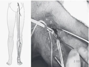
he great saphenous vein was then anastomosed
end-to-side to the best donor vein (Figure 1).
The arterial flow into the venous system progressed
through the vein whose valves had been destroyed by
the Mills valvulotome (Otemac®), which was introduced
through collateral veins until the medial malleolus
(Figure 2).
At this point, the anterior perforating vein of the
mal-leolus was invariably found, and all the other foot
collat-eral veins were then preserved. By means of phlebotomy in
the dorsal venous arch, at the level of the irst interdigital
space, the destruction of the valves was completed, thereby
allowing arterial blood low to the dorsal portion of the foot
(Figure 3). All side branches of the great saphenous vein
Figure 1– Great saphenous vein in anastomosis end-to-side to the best donor artery.
Figure 2– Arterial low into the venous system progresses through the vein, whose valves are destroyed by the valvulotome introduced through collateral veins untill the medial malleolus.
Figure 4– Ligation of collateral of the great saphenous vein, from the arterial anastomosis untill the malleolus anterior perforating vein, also shown by angiography.
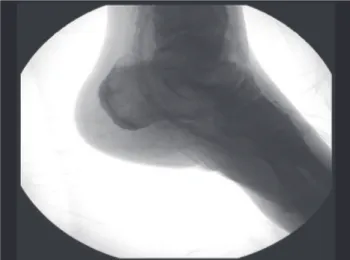
Figure 5– Angiography showing difusion of contrast into the deep system and duplex with systolic-diastolic low at the level of the dorsal venous arch of the foot.
Figure 6 – Postoperative angiography showing fulillment of the plantar
arch of the foot and of the small saphenous vein.
were ligated from the arterial anastomosis untill the
ante-rior perforating vein of the malleolus (Figure 4).
Eighteen patients with critical ischemia without
arte-rial run-of, out of whom 11 had AO, 6 had TO and 1 had
late presentation of popliteal artery aneurysm with distal
thrombosis, were submitted to the method. Among the 11
patients with AO, six had
diabetes mellitus
and, out of these,
two had renal failure and depended on hemodialysis.
Results
Among the 18 arterialized patients, 10 had foot salvage
(55.6%). Six patients achieved healing of minor
amputa-tions: two transmetatarsial, two inger and two phalanx
am-putations. Seven of them went through major amputations
(38.9%): three above the knee and four below the knee. One
patient with
diabetes mellitus
and chronic renal failure died
(5.5%) ater developing septicemia by ascending infection.
Out of the 11 patients with AO, 5 had limb salvage, 5
sufered major amputations and 1 died. Out of the six
pa-tients with TO, ive had their lower extremities maintained
and one went through a major amputation. he patient
who presented critical ischemia due to thrombosis of
pop-liteal artery aneurysm and distal arterial obstruction had
above-knee amputation.
he average follow-up of patients whose limbs were
salvaged was 695.6 days (213 to 1,006). Two patients with
AO died due to comorbidities related to patent grat. Two
patients, despite having their istulas closed, had their
ex-tremities salvaged, and a third one still presented patent
is-tula. Among patients with TO, four had patent istulas and
one presented closed istula.
Discussion
Good surgical outcomes are related to precise
indi-cation, arterial and venous preoperative investigation of
limb at risk, and details of the surgical technique. he
presence of pulse and thrill in the dorsal venous arch is
mandatory, as well as the maintenance of the foot veins
from the malleolar anterior perforating vein, and the
in-tegrity of the deep venous system, which functions as an
“escape route” for the blood hyperlow generated by the
AV istula (Figure 5).
Root and Cruz
21and Matolo
22demonstrated
experi-mentally that end-to-side istulas enabled a good reverse
blood low and better results in comparison to the terminal
ones, which lead to edema, ecchymosis, and necrosis due to
venous overload.
he malleolar anterior perforating vein drains part of
the low into the anterior tibial veins and part into the foot
proximal dorsal veins (Figure 5).
Lofgren et al.
23demonstrated that injection of blue latex
in the dorsal venous arch, between the irst and the second
metatarsal bones, drained into the proximal deep and
su-pericial veins. hey also noticed that over half of the
per-forating veins (between 6 and 12), which enable
commu-nication between the deep and supericial venous systems,
lacks valves, thus allowing blood low in both directions.
he most important perforating vein is that of the irst
in-terdigital space, measuring approximately 3 mm
24. In
post-operative angiographies, added to what was reported,
ill-ing of the plantar arch and of the small saphenous vein was
observed (Figure 6).
Although TO afects both veins and arteries, the great
and small saphenous veins are rarely afected by the
inlam-matory process
25.
Maintaining the great saphenous vein
in situ
allows the
“arterialization” of the foot venous arch with one
anasto-mosis without removing the vein of its original bed, thus
avoiding a tunnel for the venous grat. However, results
de-pend more on the characteristics of the patient than on the
technique itself.
In 2006, similar results were found in a survey of 56
publications in which the procedures were performed to
treat critical ischemia without distal run-of by diferent
techniques. A meta-analysis comprising seven papers
gath-ered a total of 228 patients with 231 treated extremities and
a 71% success rate with healing of lesions, minor
amputa-tions and improvement of pain at rest: 140 cases of OA and
91 cases of TO
26.
We conclude that distal revascularization of the limb
with critical ischemia by foot reverse low with
in situ
sa-phenous vein arterialization must be considered as an
at-tempt to salvage the afected lower extremity presenting
critical ischemia without distal arterial run-of.
References
1. Lengua F, Herrera EZ, Kunlin J. Nuevos documentos experimental-es de inversion circulatoria em miembro isquemico y de inyeccion retrograda em piezas anatomicas. Diagnostico. 1984;13:77-86.
2. Lengua F, Helfner L. Técnica de arterialization de l ares venosa del pie. Rev Sand Polic. 1984;35:203-10.
3. Lengua F, Nuss JM, Lechner R, Kunlin J. Arterialization of the ve-nous network of the foot through a bypass in severe arteriopathic ischemia. J Cardiovasc Surg. 1984; 25:357-60.
4. Lengua F, Nuss JM, Bufet JM, Lechner R. Etude comparative de deux modalités d’arterialisation des veines du pied en ischémie cri-tique. J Chir. 1993;130:12-9.
5. Lengua F. Le pontage d’artérialisation veineuse distale peut-il être bénéique au pied diabétique avec nécrose? Chirurgie. 1994-1995;120:143-52.
6. Lengua F, Cohen R, Huillier BL, Buffet JM. Arteriovenous re-vascularization for lower limb salvage in unreconstructible arterial occlusive disease (long term outcome). Vasa. 1995;24: 261-9.
7. Lengua F, Madrid A La, Acosta C, et al. L’arterialisation des veines du pied pour sauvetage de membre chez l’artéritique. Technique et resultats. Ann Chir. 2001;126:629-38.
8. Pokrovski AV, Dan VN, Khorovets AG, Chupin AV. Arterialization of venous blood low in the foot in the treatment of severe isch-aemia in patients with crural arterial occlusions and non-function-ing plantar arch. Khirurgiia. 1990;5:35-42.
9. Pokrovski AV, Dan VN, Khorovets AG, Chupin AV. Arterialization of the foot venous system in the treatment of the critical lower limb ischaemia and distal arterial bed occlusion. An Vasc Surg. 1996;4:73-93.
10. Chen XS, Lin T, Chen DL, Guan YB. Venous arterialization in the treatment of extensive arterial occlusion of lower extremities. J Surg Concepts Pract. 1998;3:219-21.
11. Taylor RS, Belli AM, Jacob S. Salvage of critically ischaemic limbs. Lancet. 1999;354:1962-5.
12. Engelke C, Morgan RA, Quarmby JW, Taylor RS, Belli AM. Distal venous arterialization for lower limb salvage: angiographic ap-pearances and interventional procedures. Radiographics. 2001;21:1239-50.
13. Rowe VL, Hood DB, Liphan J, et al. Initial experience with dorsal venous arch arterialization for limb salvage. Ann of Vasc Surg. 2002;16:187-92.
14. Ozbeck C, Kestelli M, Emrecan B, et al. A novel approach: ascend-ing venous arterialization for atherosclerosis obliterans. Eur J Vasc Endovas Surg. 2005;29:47-51.
15. Gavrilenko AV, Skrylev SI. Long-term results of venous blood flow arterialization of the leg and foot in patients with critical lower limb ischemia. Angiol Sosud Khir. 2007;13: 95-103.
16. Keshelawa G, Gigilashvili K, Chkholaria A, Pagava G, Janashia G, Beselia K. Foot venous system arterialization for salvage of non-reconstructable acute ischemic limb: a case report. J Vasc Nurs. 2009;27:13-6.
17. Busato CR, Utrabo CAL, Housome JK, Gomes RZ. Arterialização do arco venoso do pé para tratamento da isquemia crítica sem leito distal. Cir Vasc & Angiol. 1999;15:117-21.
18. Busato CR, Utrabo CAL, Gomes RZ, et al. Arterialização do arco ve-noso do pé para tratamento da tromboangeíte obliterante. J Vasc Bras. 2008;7:267-71.
19. Gasparis AP, Noor S, Da Silva MS, Tassiopoulos AK, Semel L. Distal venous arterialization for limb salvage: a case report. Vasc Endovasc Surg. 2002;36:469-72.
20. Lozano A, Melon J, Ruiz-Grande F, et al. Arterialización venosa dis-tal en cirugía de salvación de extremidad. Resultados preliminaries. Angiologia. 2002;54:204-26.
22. Matolo NM, Cohen SE, Wolfmann EF Jr. Use of an arteriovenous istula for treatment of the severely ischemic extremity: experi-mental evaluation. Ann Surg. 1976;5:622-5.
23. Lofgren EP, Myers TT, Lofgren KA, Kuster G. he venous valves of the foot and ankle. Surg Gynecol Obstet. 1968;8:289-90.
24. Garrido MBM. Anatomia médico-cirúrgica do sistema venoso dos membros inferiores. In: Mafei FHA. Doenças vasculares periféricas. 3. ed. Rio de Janeiro: Medsi; 2002. v. 1. p. 133-67.
25. Kaufman P. Tromboangeíte obliterante. In: Mafei FHA. Doenças vasculares periféricas. 3. ed. Rio de Janeiro: Medsi; 2002. v. 2. p. 1271-9.
26. Lu XW, Idu MM, Ubbink DT, Legemate DA. Meta-analysis of the clinical efectiveness of venous arterialization for salvage of criti-cally ischaemic limbs. Eur J Vasc Endovasc Surg. 2006;31:493-9.
Correspondence:
César Roberto Busato Rua Saldanha da Gama, 425 CEP 84015-130 – Ponta Grossa (PR), Brazil E-mail: crbusato@brturbo.com.br
Authors’ contributions

