Endovascular treatment of an abdominal aortic
pseudoaneurysm: a case report
Tratamento endovascular de pseudoaneurisma da aorta abdominal: relato de caso
Emerson Henrique do Nascimento1, Adaylton Aragão Correia2, Vânia Lúcia Cabral Rebouças3, Stefan de Oliveira Simões4,
Carmelo Silveira Carneiro Leão Filho5, Gustavo Tavares Collares da Penha6
Introduction
Traumatic abdominal aortic injury is considered to be one of the most fatal among all vascular injuries, accounting for 50 to 78% of the mortality rate. About 30% of the victims are already dead on arrival at the hospital1. The technical advances, the development of
new devices and the training of professionals turned the endovascular method into an excellent option for treating vascular injuries when the conventional surgi-cal access is complex or too morbid for a polytraumatic patient2. In this paper, we report a rare case of
trau-matic abdominal aortic pseudoaneurysm treated by en-dovascular approach.
Abstract
A 17-years-old male student has received several gunshots and was submitted to exploratory laparotomy. After surgery, he complained of pain in the lower limbs and a pulsatile abdominal mass. An abdominal computerized tomography (CT) scan was carried out and revealed an abdominal aorta pseudoaneurysm of about 8 cm in the larger diameter between the celiac trunk and the superior mesenteric artery. An arteriography conirmed the diagnosis and he was submitted to the lesion embolization with cotton suture wires attached to metallic guide wire fragments. After six months, an abdominal aorta Doppler ultrasonography and a new abdominal CT scan were ordered and depicted low inside the pseudoaneurysm. he patient was then submitted to a new endovascular embolization, and an 18 x 58 mm uncovered stent was placed. After six months from the last procedure, a new abdominal CT scan showed exclusion of the lesion.
Keywords: Aneurysm, false; embolization, therapeutic; aorta, abdominal.
Resumo
Um estudante de 17 anos, masculino, sofreu ferimentos por arma de fogo e foi submetido a uma laparotomia exploradora. No pós-operatório, queixava-se de dores em membros inferiores e de massa abdominal pulsátil. Realizou tomograia computadorizada (TC) de abdome, que evidenciou pseudoaneurisma de aorta abdominal de cerca de 8 cm no maior diâmetro, localizado entre o tronco celíaco e a artéria mesentérica superior. Uma arteriograia conirmou o diagnóstico e procedeu-se, então, a embolização da lesão com fragmentos de io-guia montados com ios de algodão. Após seis meses, realizou ecoDoppler de aorta abdominal e nova TC de abdome, que evidenciaram luxo no interior do saco do pseudoaneurisma. Foi, então, submetido a nova embolização endovascular e implante de stent não–revestido de 18 x 58 mm. Após seis meses do último procedimento, realizou-se nova TC de abdome que demonstrou exclusão da lesão.
Palavras-chave: Falso aneurisma; embolização terapêutica; aorta abdominal.
Study carried out at the Service of Vascular Surgery of Hospital Universitário Walter Cantídeo, Universidade Federal do Ceará (UFC), Fortaleza, CE, Brazil.
1 Vascular and Endovascular surgeon; Member of the Brazilian Society of Angiology and Vascular Surgery (SBACV); Coordinator of Endovascular Surgery at Hospital Universitário Walter
Cantídeo of UFC, Fortaleza, CE.
2 Resident physician of Vascular Surgery at Hospital Universitário Walter Cantídeo da UFC, Fortaleza, CE.
3 Vascular surgeon; Member of the SBACV; Head of the Service of Vascular Surgery at Hospital Universitário Walter Cantídeo da UFC, Fortaleza, CE.
4 Vascular surgeon; Vascular ultrasonographist; Member of SBACV; Head of the Department of Vascular Echography of the Service of Vascular Surgery; Supervisor of the Program of Medical
Residence in Vascular Surgery at Hospital Universitário Walter Cantídeo da UFC, Fortaleza, CE.
5 Vascular and Endovascular surgeon; Member of SBACV; Post-graduate student (Master degree) at the Department of Surgery of UFC; Member of the Service of Vascular Surgery of Hospital
Universitário Walter Cantídeo da UFC, Fortaleza, CE.
6 Vascular surgeon; Member of the Service of Vascular Surgery of Hospital Universitário Walter Cantídeo da UFC, Fortaleza, CE.
No conlict of interest was declared concerning the publication of this article. Received on Feb 6, 2010. Accepted on Apr 12, 2010.
Case description
On April 23, 2008, a 17-year-old male student was ad-mitted at a local Hospital with ive gunshot wounds (one on the right hand, one on the right arm, two on the occipi-tal region and one on the back). He underwent immediate exploratory laparotomy. At operation, a bilateral retro-peritoneal hematoma on zone II was found. Because there was no expansion of the hematoma and also because the patient was hemodynamically stable, the surgeon chose expectant management. In the postoperative period, the patient developed temporary paraplegia, recovering the movements of the lower limbs in a few days. However, he continued to have intense burning pain and sensation of weight on both legs. He was discharged from the hospital and referred to a specialized service for motor rehabilita-tion, where a Doppler ultrasonography of the aorta and contrast-enhanced computed tomography scan of the ab-domen showed an abdominal aortic pseudoaneurysm be-tween the celiac trunk and the superior mesenteric artery, measuring about 8 cm in diameter (Figure 1). He was then referred to our Service on June 5, 2008.
Physical examination at admission revealed a pale young man in good general condition, oriented and co-operative. Cardiac and pulmonary auscultations were normal. he abdomen was lat and symmetric, and had a previous xipho-pubic laparatomy scar, normal bowel sounds and the presence of a pulsatile mass and of epigas-tric 2+ in 6+ bruit without thrill at palpation. he upper and lower extremities presented normal pulses, without edema. Sensitivity, tonus and muscle strength were slight-ly reduced in the lower limbs.
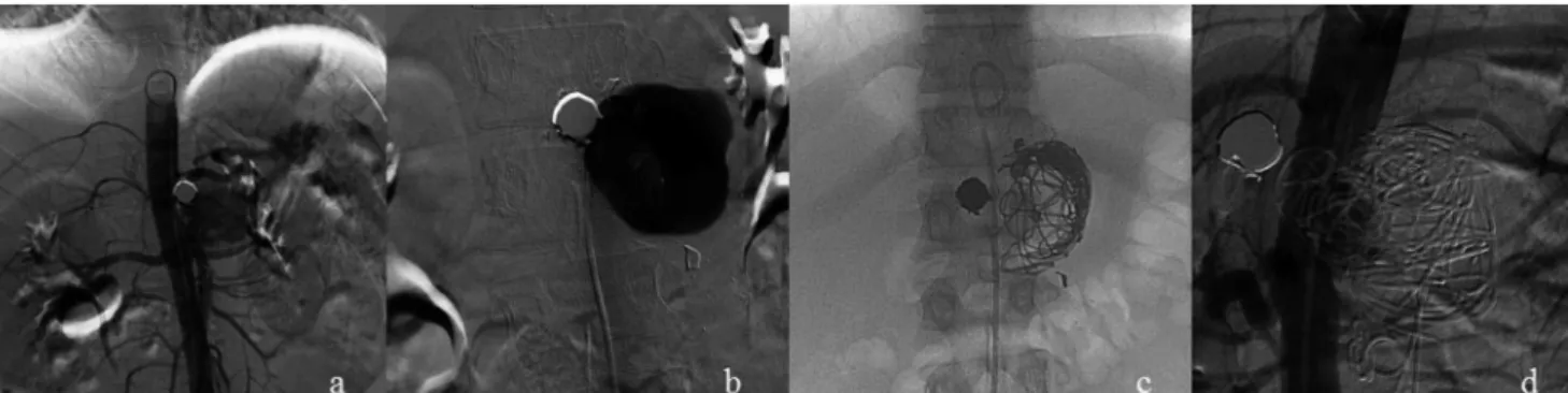
On June 6, 2008, the patient was submitted to aor-tography, which showed an abdominal aortic pseudoa-neurysm with its ostium between the origins of the ce-liac trunk and the superior mesenteric artery, as well as a metallic artifact on the L1-L2 projection (Figures 2A and 2B). On June 9, Seldinger retrograde puncture of the right femoral artery with placement of a 40-cm 6Fr sheath in the aorta was performed, besides selective catheterization of the pseudoaneurysm ostium and embolization with fragments of 0.035 guide wire, tied together with 3.0 cot-ton thread segments (Figure 3). Control aortography re-vealed signiicant reduction of the contrasted area and a
Figure 1 – Doppler ultrasound of the abdominal aorta showing interior low of the hematoma (above); abdominal CT scan showing the
Figure 2 – Aortography showing the contrast low to the interior of the pseudoaneurysm (A); selective catheterization of the pseudoaneurysm (B); embolization result (C); embolization result (oblique) (D).
Figure 3 – Aspect of the embolization material
Figure 4 – Second procedure. Arteriography pre-embolization (A);
control arteriography after embolization and stent position (B)
small region next to the ostium which maintained discrete contrast low (Figures 2C and 2D).
On the irst post-embolization day, he developed a tem-perature of 38.4°C. On the second day, laboratorial exams showed anemia (hemoglobin = 9.56 mg). As the patient was hemodynamically stable and without abdominal pain, the expectant management was the treatment of choice. On the fourth day ater embolization, he went through an evalu-ation by the Neurology Service, which disclosed pain and signs of neurological deicit, probably as a result of the gun-shot injury on the cauda equina. he patient was discharged from the hospital and referred to physical therapy and am-bulatory follow-up.
On June 30, 2008, he was submitted to a new CT scan; on October 23, to Doppler ultrasound of the abdominal aorta; on October 31, he underwent catheter aortography. All three examinations showed persistent low into the an-eurysmal sac. On October 29, he was submitted to a new embolization by means of puncture of the right and let femoral arteries and introduction of 10Fr and 6Fr sheaths, respectively. Embolization was performed with 10 ibrous coils, and guide wire segments were tied together with 3.0 cotton thread ater the insertion of an uncovered self-ex-pansible nitinol stent (18 x 58 mm) on the aorta, aiming at remodeling the ostium of the pseudoaneurysm and re-taining the embolization material within the sac. he post-procedure arteriography did not show contrast in the aneu-rysm sac (Figures 4A and 4B). On the second postoperative day, he was discharged without complaints, without pulsa-tile abdominal mass and murmur. he patient is currently on follow-up with abdominal CT scans every six months, which have shown absence of low in the pseudoaneurysm.
Discussion
Penetrating trauma is the most common cause of ab-dominal aortic injury. horacoabab-dominal vessel injuries
are associated with high pre-hospital and hospital mortal-ity rates3. In about 18% of the cases, bleeding is contained
on the retroperitoneum and the patients are normotensive at admission4. Abdominal trauma may manifest as free
in-traperitoneal hemorrhage or as a contained retroperitoneal hematoma (pseudoaneurysm)5.
aneurysm”, it arises when three layers of the artery are lac-erated or ruptured, determining blood leakage through the laceration, without low interruption inside the blood vessel. he surrounding tissues restrain the hematoma and a pseudocapsule is formed around the arterial wall. Its continuous expansion may cause complications due to the compression and inlammatory process on neighboring structures (veins, hollow organs and nerves) and, inally, rupture of the pseudoaneurysm, with severe hemorrhage, hypovolemic shock and death6.
Our patient, who sufered gunshot wounds, presented a pseudoaneurysm in the region described as zone II (ret-roperitoneal region, involving kidneys and renal vessels)4.
Hematomas in this region increase the suspicion of renal vessel lesion, which was not conirmed in this case. As a general rule, retroperitoneal hematomas resulting from penetrating traumas require exploration of the adjacent kidney (Huey Long rule)7, except when it is a stable
peri-renal hematoma in a patient whose preoperative CT scan does not show urine extravasation or in case of stable retro-hepatic hematoma5. he prognosis of penetrating
abdomi-nal aortic injuries is signiicantly better than that of thoracic aortic injuries, probably because the retroperitoneal tissues contain the hemorrhage4.
he intervention technique may be electively selected for patients who are hemodynamically stable and pres-ent with traumatic pseudoaneurysm8. In the last decades,
endovascular techniques have been more oten used for the management of traumatic lesions9. Since 1972, when
Margolies et al. performed the embolization of a traumatic hemorrhage10, the techniques and materials used in
endo-vascular embolization have shown excellent results with minimal invasion.
Catheter embolization is indicated for injuries of inac-cessible arteries11. In this case, the pseudoaneurysm was
lo-cated between the celiac trunk and the superior mesenteric artery, an area of diicult access by conventional surgical techniques, especially concerning proximal and distal ves-sel control.
he factors that inluenced the choice of endovascular technique in the reported case were:
1. young patient (18 years old); 2. hemodynamic stability;
3. topography of the injury (diicult surgical access); 4. peritoneal cavity with previous approach (with possible adhesions between organs);
5. forty-ive days with hematoma on the retroperito-neum (likely necessity of ibrous areas’ dissection12).
The embolization materials may be fluid or solid. As examples, one may mention: clots with or without additives, absorbable gelatin sponge, liofilized dura ma-ter, particles of polyvinyl alcohol(PVA), microspheres (acrylic copolymer with porcine gelatin), polymerized glue, ejectable or detachable coils covered or not with thrombogenic synthetic fibers, detachable or non-de-tachable balloons and tampon-like occlusion devices8.
In this patient, we used embolization devices (frag-ments of 0.035 guide wire + segment of 3.0 cotton wire) in order to occlude the pseudoaneurysm, which was partially achieved on the first embolization and com-pleted through embolization with fibrous coils and ap-position of uncovered stent.
Ulacker reports that the irst model of coils for em-bolization was made of guide wire segments without the inner wire, mechanically twisted, with a wadding of wool on its extremity. Ater the development of the Gianturco coils, the necessity for materials for occlusion of large-caliber vascular systems, arteriovenous istulas and an-eurysm has raised. he same author described 100% of success in the occlusion of large-caliber vessels using artisanal large springs for embolization that were 20 cm long, and Dacron® thread wadding attached to it, on 3-cm
intervals. hese modiied guide wire segments were used to treat high-debt and large-caliber arteriovenous istu-lae. his material was described as 10 to 15-cm guide wire with the lexible inner wire removed, but with its shape irregularly modiied, angled or twisted to wad in the inte-rior of the vessel13.
he stent was used to obtain a better remodeling of the pseudoaneurysm neck, which became larger due to the amount of embolization material injected in its interior. Besides that, it also had the function of avoiding the migra-tion of this material to the aortic lumen. In this patient, a stent a little larger than necessary was used (18 x 58 mm), once it was the only stent available in our hospital that was compatible with the procedure.
Hospital discharge two days ater the procedure and without complaints demonstrates the eicacy of the meth-od in managing the injury and reducing the postoperative morbidity.
References
1. Razuk-Filho A, Coimbra R. Trauma dos vasos abdominais. In: Brito CJ. Cirurgia vascular: cirurgia endovascular, angiologia. 2ª ed. Rio de Janeiro: Revinter; 2008. p. 1159-66.
2. Aun R, Puech-Leão P, Netto BM. Lesões vasculares traumáticas e iatrogênicas: tratamernto endovascular. In: Brito CJ. Cirurgia vascular: cirurgia endovascular, angiologia. 2ªd ed. Rio de Janeiro:
Revinter; 2008. p. 1461-67.
3. Costa-Val R, Campos-Christo SF, Abrantes WL, Campos-Christo MB, Marques MC, Miguel EV. Relexões sobre o trauma cardio-vascular civil a partir de um estudo prospectivo de 1000 casos atendidos em um centro de trauma de nível I.Rev Col Bras Cir. 2008;35(3):162-7.
4. Demetriades D. Abdominal vascular injuries. In: Rutherford RB. Rutheford’s vascular surgery. 6th ed. Philadelphia: WB Saunders;
2005. p. 1028-44.
5. Hirshberg A, Mattox KL. Vascular trauma. In: Haimovici H. Haimovici’s vascular surgery. 5th ed. Hoboken, NJ, USA:
Wiley-Blackwell; 2003. p. 421-36.
6. Rich NM. Revisão histórica das fístulas arteriovenosas e dos pseudoa-neurismas traumáticos. In: Rich NM, Mattox KL, Hishberg A, editores. Trauma vascular, 2ª ed., Rio de Janeiro: Dilivros; 2006. p. 483-551.
7. Feliciano DV. Lesões da aorta abdominal e das artérias viscerais. In: Rich NM, Mattox KL, Hishberg A, editores. Trauma vascular, 2ª ed., Rio de Janeiro: Dilivros; 2006. p. 315-29.
8. Araujo AP, Gomes CFA, C FM. Embolização terapêutica. In: Brito CJ. Cirurgia Vascular: cirurgia endovascular, angiologia, 2ª ed. Rio de Janeiro: Revinter; 2008. p. 1469-93.
9. Tucker S Jr, Rowe VL, Rao R, Hood DB, Harrell D, Weaver FA. Treatment options for traumatic pseudoaneurysms of the paravis-ceral abdominal aorta. Ann Vasc Surg. 2005;19(5):613-8.
10. Starnes BW, Arthurs ZM. Endovascular management of vascular trauma. Perspect Vasc Surg Endovasc her. 2006;18(2):114-29.
11. Sclafani SJA. Radiologia diagnóstica e intervencionista no trauma vascular. In: Rich NM, Mattox KL, Hishberg A, editores. Trauma vascular. 2ª ed. Rio de Janeiro: Dilivros; 2006. p. 191-218.
12. Aun R, Saliture-Neto FT, Lederman A, Waksman H. Tratamento endoluminal de aneurismas anastomóticos na aorta abdominal: relato de dois casos. J Vasc Bras. 2006;5(1):58-62.
13. Ulacker R. Técnicas e materiais de embolização – complica-ções. In: Ulacker R, editor. Radiologia intervencionista. São Paulo: Sarvier; 1987. p. 29-63.
Correspondence:
Adaylton Aragão Correia Rua Vilebaldo Aguiar, 1.580, apto. 402 – Papicu CEP 60190-780 – Fortaleza, CE E-mail: [email protected]
Authors’ contributions