Intrauterine growth restriction in monochorionic-diamniotic twins
RITA DE CÁSSIA ALAM MACHADO1, MARIA DE LOURDES BRIZOT1*, SEIZO MIYADAHIRA1, ROSSANA PULCINELI VIEIRA FRANCISCO1,
VERA LÚCIA JORNADA KREBS2, MARCELO ZUGAIB1
1Department of Obstetrics, Hospital das Clínicas, University of São Paulo’s Medical School, São Paulo, SP, Brazil 2Department of Pediatrics, Hospital das Clínicas, University of São Paulo’s Medical School, São Paulo, SP, Brazil
S
UMMARYStudy conducted at Hospital das Clínicas, University of São Paulo, São Paulo, SP
Article received: 7/19/2013
Accepted for publication: 2/3/2014
*Correspondence:
Departamento de Obstetrícia e Ginecologia, Instituto Central, Hospital das Clínicas Address: Av. Dr. Enéas de Carvalho Aguiar,
255, 10o andar, cj. 10.037 Postal Code: 05403-000 São Paulo -- SP Phone: (11) 2661-6209 Fax: (11) 2661-6445 [email protected]
http://dx.doi.org/10.1590/1806-9282.60.06.019
Conflict of interest: none
Objective: to evaluate neonatal morbidity and mortality in
monochorionic--diamniotic (MCDA) twin pregnancies complicated by selective intrauterine growth restriction (sIUGR) and non-selective intrauterine growth resctriction (nsIUGR).
Methods: neonatal morbidity parameters and mortality were analyzed in 34
twins with IUGR (< 10th percentile on twins’ growth charts): 18 with sIUGR and
16 with nsIUGR. The sIUGR group was made up of 18 pregnancies in which growth was restricted in only one fetus (n = 18). The nsIUGR group was compo-sed of 8 pregnancies in which both fetuses presented restricted growth (n = 16). Cases of twin-to-twin transfusion syndrome and fetal malformation were not included in the study.
Results: the MCDA twin pregnancies with sIUGR had a higher rate of
orotra-cheal intubation (p = 0.001) and mechanical ventilation (p = 0.0006), as well as longer than average fasting time (p = 0.014) compared to those in which the fe-tuses had nsIUGR. A higher incidence was also observed of types II and III um-bilical artery Doppler velocimetry patterns in the sIUGR cases (p = 0.002). The-re was no signiicant diffeThe-rence between the two groups as to mortality during pregnancy and the neonatal period (p = 0.09).
Conclusion: in MCDA twin pregnancies, sIUGR presents more severe umbilical
artery Doppler velocimetry abnormalities and worse morbidity than nsIUGR.
Keywords: monozygotic twins, disease in twins, fetal growth retardation, laser
Doppler lowmetry, morbidity, newborn infant.
I
NTRODUCTIONIntrauterine growth restriction (IUGR) in monochorio-nic pregnancies may affect one (selective IUGR, sIUGR) or both fetuses (non-selective IUGR, nsIUGR). However, when affecting both fetuses, IUGR morbidity, as compa-red with sIUGR morbidity in MCDA pregnancies, remains unknown.6
Selective intrauterine growth-restriction (sIUGR) af-fects 12-15% of monochorionic pregnancies.1 The
mono-chorionic placenta structure accounts for most of the morbidity associated with these pregnancies. A greater incidence of marginal or velamentous umbilical cord in-sertion, unequal sharing of the placental bed, and unba-lanced arterio-venous anastomoses, with consequent com-pensatory mechanisms, make each monochorionic placenta an individual and atypical arrangement.2,3
Gratacos et al.,4 in 2004, initiated a series of studies
demonstrating increased neonatal morbidity in mono-chorionic and diamniotic (MCDA) pregnancies affected by sIUGR with abnormalities in Doppler velocimetry. In 2007, Gratacos et al.,5 comparing groups of
sIUGR-affec-ted MCDA pregnancies with different umbilical artery Doppler velocimetry patterns (normal Doppler, persis-tent absent or reversed end diastolic velocity low, inter-mittent absent or reversed end diastolic velocity low), re-ported, in the group with the latter pattern, a higher rate of intrauterine death (IUD) in the smaller fetus and of brain lesions in the larger fetus.
Gao et al.,7 evaluated neonatal morbidity in 7
IUGR. In their study, conducted with a mixed sample of mono and dichorionic twin pregnancies, monochorioni-city turned out to be a higher risk factor for IUGR, espe-cially sIUGR (p<0.001). The groups associated with IUGR had a higher incidence of intrauterine death (IUD) and neonatal death (ND) (p<0.001).
However, there are no studies in the literature, speci-ically complicated by sIUGR compared to nsIUGR in MCDA pregnancies.
The aim of the current study was to evaluate prena-tal evolution as well as perinaprena-tal morbidity and morta-lity in MCDA twin pregnancies complicated by sIUGR in contrast with nsIUGR.
M
ETHODSThis was a retrospective cohort study involving 34 mo-nochorionic-diamniotic fetuses with IUGR (< 10th
per-centile, according to the Alexander et al.8 growth chart).
We included cases that were followed up in our twin cli-nic and delivered in our hospital between 2004 and 2010. The study was approved by the Hospital’s Ethics Com-mittee (0092/10).
The sIUGR group comprised 18 pregnancies in which only one fetus was restricted in growth (n = 18). The nsIU-GR group was composed of 8 pregnancies in which both fetuses presented restricted growth (n = 16).
Cases with twin-to-twin transfusion syndrome (TTTS) (n = 43), fetal malformation (n = 36), as well as three ca-ses of sIUGR and IUD prior to 26 weeks that did not de-liver in our institution and chorionicity not determined were not included in the study.
In our twin clinic, management of monochorionic twin pregnancies includes serial evaluations of fetal biometry, Doppler velocimetry, and biophysical proile, as well as mo-nitoring of TTTS signs and amniotic luid volume.
Ultrasound examination was performed transabdo-minally using a 3.5-5.0 MHz curvilinear-array transducer (Envisor – Phillips; Voluson Expert- GE). Fetal weight was estimated using the four-parameter formula (head and abdominal circumferences, biparietal diameter, and fe-mur length) described by Hadlock et al..9 Gestational age
was estimated based on the date of the last menstrual pe-riod and on irst-trimester or two consecutive second-tri-mester ultrasound scans. Selective IUGR was deined as estimated fetal weight below the 10th percentile in one
twin according to a nomogram for twins.8
Oligohydramnios was deined as the deepest vertical pocket (DVP) of 2 cm or less and polyhydramnios as the DVP of 8 cm or greater prior to 20 weeks and 10 cm or greater after 20 weeks.10 Diagnosis criteria for TTTS was
considered as previously described.11 Chorionicity was
determined by irst-trimester ultrasound examination and/or pathoanatomical examination in all cases.
Doppler examination of the umbilical artery (UA) was performed in a free loop of the umbilical cord near the respective fetus. Whenever the UA was altered, eva-luation was performed in three other areas of the umbi-lical cord. Abnormal UA Doppler was classiied according to the characteristics of diastolic low:5 type I (positive
with increased pulsatility index, PI); type II (persistent ab-sent or reversed end diastolic velocity low, AREDF), and type III (intermittent absent or reversed end diastolic ve-locity low, iAREDF).
Cases with IUGR but normal UA Doppler were fol-lowed every two weeks. Cases with abnormal Doppler and gestational age below 26 weeks were examined weekly. In our nursery, viability is considered after 26 weeks of ges-tation; therefore, cases with type I Doppler were monito-red weekly and cases with type II or III were admitted to the hospital and monitored daily with Doppler veloci-metry of UA, middle cerebral artery, and ductus venosus in
combination with biophysical proile and fetal heart rate measurements. All cases were managed expectantly as they were not subjected to laser therapy or selective feti-cide.
Delivery indication due to fetal deterioration was con-sidered before 28 weeks when there was AREDF in the
ductus venosus; after this period, if any of the following
pa-rameters were present: ductus venosus PI persistently
abo-ve 1.0,12 persistently abnormal fetal heart rate traces, and
maintenance of abnormal biophysical proile (< 6) in eva-luations 6 hours apart. Cases with AREDF or iAREDF not presenting fetal deterioration were delivered at 34 weeks of gestation. For type I, in accordance with our pro-tocol for monochorionic twin pregnancy, delivery was scheduled at 36 weeks or earlier if fetal deterioration was observed.
Indicated preterm delivery was carried out based on either maternal or fetal conditions. Prophylactic antena-tal corticosteroids (2 daily IM 12mg betamethasone) were administered only if preterm delivery (< 34 weeks) was anticipated. All cases were delivered in our institution and the neonates were kept in the nursery adjacent to the maternity ward. The placentas underwent pathoanato-mical examination; however, perfusion studies of the vas-cular shunts were not performed.
score < 7, length of hospital stay, length of stay in neo-natal intensive care unit (ICU), fasting time, orotracheal intubation, mechanical ventilation, and the presence of respiratory diseases (hyaline membrane, iatrogenic pneu-mothorax, emphysema, pulmonary hypertension, wet lung syndrome, pneumonia and/or bronchopulmonary dysplasia), infectious diseases (pneumonia, sepsis), neu-rological disorders (intraventricular hemorrhage, IVH; periventricular leukomalacia, PVL; multicystic leukoen-cephalomalacia), and blood disorders (anemia, neutro-penia, polycythemia). Variables of perinatal outcome were also recorded, such as hospital discharge, intra-uterine death, and death during nursery admission.
The chi-square test or Fisher exact test were used to test the homogeneity of the groups in terms of propor-tions. Mean differences between the two groups were com-pared using Student t test or Mann-Whitney test. The
le-vel of signiicance was set at 5% for all tests.
R
ESULTSEighteen twins with sIUGR and 16 with nsIUGR were analy-zed with respect to neonatal morbidity and mortality.
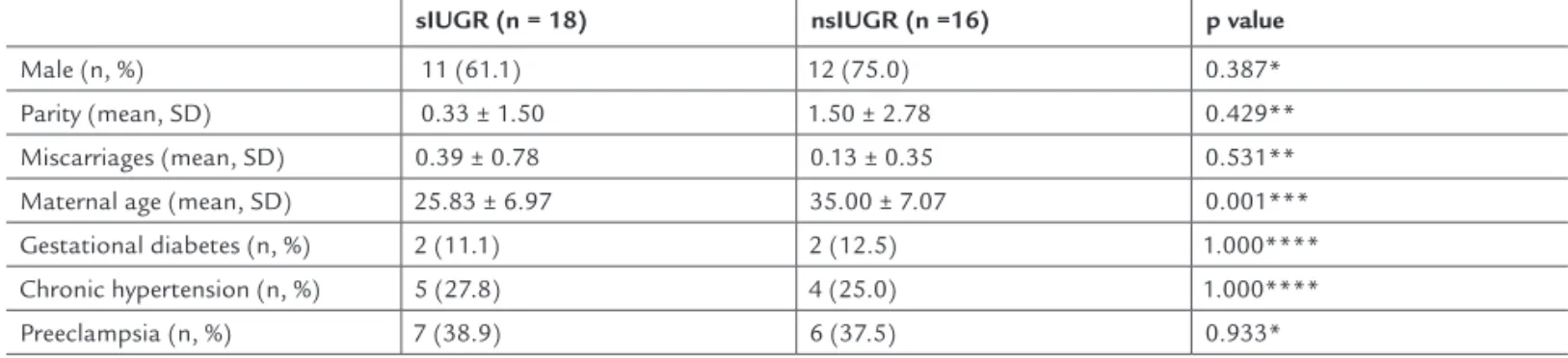
Mean maternal age was lower in the sIUGR group (p<0.001). Otherwise, groups did not differ from each other: parity, previous miscarriages and rates of gestatio-nal diabetes mellitus, chronic hypertension, and
preeclamp-sia were similar (Table 1).
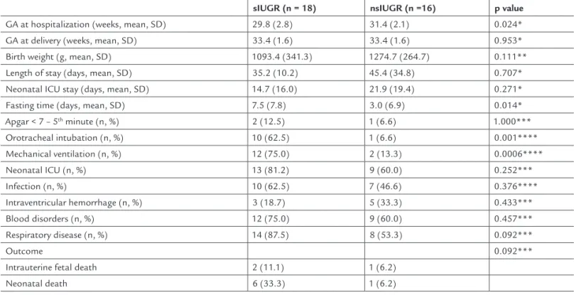
Table 2 displays the neonatal morbidity and morta-lity data. It can be seen that mean gestational age at hos-pital admittance for control of fetal surveillance was lo-wer in the sIUGR group (p = 0.024) and that there was longer time of fasting in the nursery (p = 0.014) as well as a higher need for orotracheal intubation (p = 0.001)
and use of mechanical ventilation (p = 0.0006) among the newborns with sIUGR. Mean gestational age at deli-very, mean birth weight and total length of nursery and neonatal ICU stay did not differ signiicantly between the two groups.
Three fetuses with restricted growth (2 with sIUGR and 1 with nsIUGR) progressed to IUDs; all had REDF in the umbilical artery (type II). One fetal death occurred at 26 weeks of gestation with ductus venosus PI equaling 1.44
at the last evaluation. The fetus was receiving corticothe-rapy for lung maturation; the non-restricted twin was also dead. Another fetus died at 28 weeks of gestation; the duc-tus venosus PI (0.82) and biophysical proile parameters were
normal 24 hours prior to death. The co-twin was delivered at 32 weeks of gestation (nsIUGR). In the third case, fetal death was diagnosed at 32 weeks of gestation, and, as in the previous case, the ductus venosus PI (0.85) and
biophy-sical proile parameters were normal 24 hours prior to death; delivery of the co-twin was performed soon after de-tection of the fetal death of the growth-restricted one. The-re was no diffeThe-rence in the incidence of IUD and ND between the two groups (p = 0.092; Table 2).
Table 3 shows the distribution of Doppler velocimetry patterns in the sIUGR and nsIUGR groups. Type I UA Doppler velocimetry was more frequent in the nsIUGR group, whereas type II and type III UA Doppler veloci-metry occurred more often in the sIUGR group.
D
ISCUSSIONAlthough the two study groups were no different regar-ding gestational age at delivery and birth weight, the MCDA pregnancies complicated by sIUGR had a higher incidence of neonatal morbidity than the nsIUGR group.
TABLE 1 Characteristics of the study population. sIUGR = selective intrauterine growth restriction; nsIUGR = non-selective intrauterine growth restriction
sIUGR (n = 18) nsIUGR (n =16) p value
Male (n, %) 11 (61.1) 12 (75.0) 0.387*
Parity (mean, SD) 0.33 ± 1.50 1.50 ± 2.78 0.429**
Miscarriages (mean, SD) 0.39 ± 0.78 0.13 ± 0.35 0.531**
Maternal age (mean, SD) 25.83 ± 6.97 35.00 ± 7.07 0.001***
Gestational diabetes (n, %) 2 (11.1) 2 (12.5) 1.000****
Chronic hypertension (n, %) 5 (27.8) 4 (25.0) 1.000****
Preeclampsia (n, %) 7 (38.9) 6 (37.5) 0.933*
* Chi-square test.
** Parametric Mann-Whitney test. *** Student t test.
often seen in nsIUGR twins than in sIUGR twins. Our indings led us to question whether sIUGR and nsIUGR arise from different mechanisms regarding placental an-gio-structure, the former being related to vascular com-munication, as described in previous studies,3,5,14,15 and
the latter to placental insuficiency. Also, we wondered if the older maternal age in the nsIUGR-related cases is a predictor of placental insuficiency. The non-assessment of placental vascularization in our study, however, pre-vents the conirmation of these hypotheses.
All three IUD cases (2 cases of sIUGR and 1 case of nsIUGR) reported herein were associated with UA Doppler velocimetry type II (REDF). Our data agree with those by Ishi et al.,16 who reported a higher risk for
IUD and ND in IUGR cases with persistent AREDF. No difference was found between the study groups in rela-tion to IUD or ND. However, our indings suggest a tendency towards higher mortality in the sIUGR group: a total of 8 cases of IUD and ND (44.4%) as against the 2 cases in the nsIUGR group (12.5%). A larger number of cases may be necessary to evaluate with greater cer-tainty the seemingly discrepant mortality rates of the two groups.
Selective IUGR was associated with postnatal need for in-tubation and mechanical ventilation and longer fasting time during nursery stay than that of the nsIUGR group. The reports by Gratacos et al.4,5,13 showed that sIUGR is
associated with intermittent UA low pattern (type III) and that the type of Doppler alteration is related to the number and diameter of artery-artery anastomoses. That prompted us to investigate if there were any differences between the sIUGR and nsIUGR groups regarding Doppler low pattern. In our study, type II and type III UA Doppler low pat-terns were more frequently in sIUGR twins than in nsIU-GR twins. On the other hand, type I UA pattern was more
TABLE 2 Neonatal morbidity and mortality parameters in selective and non-selective intrauterine growth restriction (sIUGR; nsIUGR). GA: gestational age; ICU: intensive care unit
sIUGR (n = 18) nsIUGR (n =16) p value
GA at hospitalization (weeks, mean, SD) 29.8 (2.8) 31.4 (2.1) 0.024*
GA at delivery (weeks, mean, SD) 33.4 (1.6) 33.4 (1.6) 0.953*
Birth weight (g, mean, SD) 1093.4 (341.3) 1274.7 (264.7) 0.111**
Length of stay (days, mean, SD) 35.2 (10.2) 45.4 (34.8) 0.707*
Neonatal ICU stay (days, mean, SD) 14.7 (16.0) 21.9 (19.4) 0.271*
Fasting time (days, mean, SD) 7.5 (7.8) 3.0 (6.9) 0.014*
Apgar < 7 – 5th minute (n, %) 2 (12.5) 1 (6.6) 1.000***
Orotracheal intubation (n, %) 10 (62.5) 1 (6.6) 0.001****
Mechanical ventilation (n, %) 12 (75.0) 2 (13.3) 0.0006****
Neonatal ICU (n, %) 13 (81.2) 9 (60.0) 0.252***
Infection (n, %) 10 (62.5) 7 (46.6) 0.376****
Intraventricular hemorrhage (n, %) 3 (18.7) 5 (33.3) 0.433***
Blood disorders (n, %) 12 (75.0) 9 (60.0) 0.457***
Respiratory disease (n, %) 14 (87.5) 8 (53.3) 0.092***
Outcome 0.092***
Intrauterine fetal death 2 (11.1) 1 (6.2)
Neonatal death 6 (33.3) 1 (6.2)
* Parametric Mann-Whitney test. ** Student t test.
*** Fisher exact test.
**** Chi-square test (orotracheal intubation, power = 0.94 and mechanical ventilation, power = 0.97).
TABLE 3 Distribution of umbilical artery Doppler patterns5 in selective and non-selective intrauterine growth
restriction (sIUGR; nsIUGR)
sIUGR nsIUGR p value
n % n %
Type I 2 11.1 11 68.8 0.002
Type II 11 61.1 4 25
Type III 5 27.8 1 6.2
Total 18 100 16 100
Our results were similar to those by Ishi et al.16 who
observed a higher risk of death in cases below the 3rd
cen-tile of the normal curve for singleton pregnancies, but instead, we chose to use a speciic twin curve in order to identify the most severe cases.8
It was thus possible to observe, in the sIUGR group, a higher rate of neonatal morbidity and of severe UA Doppler velocimetry patterns, perhaps due to the greater seriousness of placental dysfunction as characterized by UA Doppler ve-locimetry (AREDF and iAREDF pattern – types II and III ac-cording to categorization by Gratacos et al.).5 Therefore,
sIU-GR points toward the need for closer fetal surveillance given the risk of neonatal complications and IUD in such cases.
The gestational age at delivery in sIUGR reported by previous studies16 was lower (32 weeks) compared to the
present study (33.36 weeks), showing that less severe ca-ses were included in our study owing to late referral of such cases to a tertiary center. Probably, several of the se-vere cases had died in utero before reaching our center.
The present study relects data from a single tertiary Center without discrepancies in the management of the ca-ses. Therefore, we present a small number of cases as com-pared to multicenter studies.5,13,16 However, we found
signi-icant association of sIUGR with mechanical ventilation and orotracheal intubation (0.97 and 0.94 , respectively).
The present series was not submitted to laser therapy or selective feticide, showing the natural evolution of the cases. There are no studies comparing neonatal outcome in cases of sIUGR and nsIUGR in MCDA pregnancies. Gao et al.,7 when evaluating IUGR in mono and dichorionic
pregnancies, observed that monochorionicity was a risk factor for IUGR. In the present study, MCDA twin preg-nancies with sIUGR presented higher rate of severe UA Doppler abnormalities leading to a worse neonatal prog-nosis compared to MCDA twin pregnancies with nsIUGR.
R
ESUMORestrição de crescimento intrauterino em gêmeos mono-coriônicos diamnióticos.
Objetivo: avaliar a morbidade e mortalidade neonatal
em gestações monocoriônicas e diamnióticas (MCDA) acometidas pela restrição de crescimento fetal seletiva (RCFS) e não seletiva (RCFNS).
Métodos: os parâmetros de morbidade e mortalidade
neonatais foram avaliados em 34 gêmeos com RCF (abai-xo do percentil 10 de uma curva de crescimento para
gê-meos): 18 com RCFS e 16 com RCFNS. O grupo com RCFS teve origem em 18 gestações, em que somente um feto apresentava RCF. O grupo com RCFNS teve origem em 8 gestações em que ambos os fetos apresentavam RCF. Foram excluídos deste estudo casos da síndrome da trans-fusão feto-fetal e malformações fetais.
Resultados: os gêmeos de gestações MCDA com RCFS
apresentaram maior frequência de entubação orotraqueal (p=0,001), ventilação mecânica (p=0,0006) e maior tem-po em jejum durante internação (p=0,014), quando com-parados aos gêmeos de gestações MCDA com RCFNS. No grupo com RCFS, também foram observados maior fre-quência de tipos II e III de dopplervelocimetria de artéria umbilical (p=0,002). Não houve diferença signiicativa entre os grupos quanto à mortalidade neonatal (p=0,09).
Conclusão: em gestações gemelares MCDA, a RCFS
re-presenta maior frequência de alterações severas na velo-cimetria Doppler da artéria umbilical e piores resultados na morbidade neonatal.
Palavras-chave: gêmeos monozigóticos, doenças em
gê-meos, retardo do crescimento fetal, luxometria por laser
Doppler, morbidade, recém-nascido.
R
EFERENCES1. Lewi L, Van Schoubroeck D, Gratacós E, Witters I, Timmerman D, Deprest J. Monochorionic diamniotic twins: complications and management options. Curr Opin Obstet Gynecol. 2003;15:177-94.
2. Machin GA: Vascular anatomy of monochorionic twin placentas; In: Blickstein I, Keith LG, editors. Multiple pregnancy: epidemiology, gestation & perinatal outcome. 2nd ed. Andover: Thomson Publishing Services; 2005. p. 193-200.
3. Lopriore E, Pasman SA, Klumper FJ, Middeldorp JM, Walther FJ, Oepkes D. Placental characteristics in growth-discordant monochorionic twins: a matched case-control study. Placenta. 2012;33:171-4.
4. Gratacos E, Lewi L, Carreras E, Becker J, Higueras T, Deprest J, Cabero L. Incidence and characteristics of umbilical artery intermittent absent and/or reversed end-iastolic low in complicated and uncomplicated monochorionic twin pregnancies. Ultrasound Obstet Gynecol. 2004;23:456-60.
5. Gratacos E, Lewi L, Munoz B, Acosta-Rojas R, Hernandez-Andrade E, Martinez JM, et al. A classiication system for selective intrauterine growth restriction in monochorionic pregnancies according to umbilical artery Doppler low in the smaller twin. Ultrasound Obstet Gynecol. 2007;30:28-34.
6. Valsky DV, Eixarch E, Martinez JM, Crispi F, Gratacos E. Selective intrauterine growth restriction in monochorionic twins: pathophysiology, diagnostic approach and management dilemmas. Semin Fetal Neonatal Méd 2010;15:342-8. 7. Gao Y, Zhiming He, Yanmin L, Sun H, Huang L, Li M, et al. Selective and
non-selective intrauterine growth restriction in twin pregnancies: high risk factors and perinatal outcome. Arch Gynecol Obstet. 2012;285:973-8. 8. Alexander GR, Michael K, Martin J, Papiernick E. What are the fetal growth
patterns of singletons, twins and triplets in the United States? Clin Obstet Gynecol. 1998;41:115-25.
9. Hadlock FP, Harrist RB, Sharman RS, Deter RL, Park SK. Estimation of fetal weight with the use of head, body and femur measurements – A prospective study. Am J Obstet Gynecol. 1985;151:333-7.
11. Quintero RA, Morales WJ, Allen MH, Bornick PW, Johnson PK, Kruger M. Staging of twin-twin transfusion syndrome. J Perinatol 1999;19:550-55. 12. Francisco RPV, Miyadahira S, Zugaib M: Predicting pH at birth in absent
or reversed end-diastolic velocity in the umbilical arteries. Ultrasound Obstet Gynecol. 2006;107:1042-7.
13. Gratacós E, Diemert A, Hecher K, Lewi P, Depres J. Clinical outcome and placental characteristics of monochorionic diamniotic twin pairs with early and late-onset discordant twin. Am J Obstet Gynecol. 2008;199:511e1-e7. 14. Lewi L, Cannie M, Blickstein I, Jani J, Huber A, Hecker K, et al. Placental
sharing, birth weight discordance, and vascular anastomoses in
monochorionic diamniotic twin placentas. Am J Obstet Gynecol. 2007;197:587e1-e8.
15. Lewi L, Gucciardo L, Huber A, Jani J, Mieghem TV, Doné E, et al. Clinical outcome and placental characteristics of monochorionic diamniotic twin pairs with early and late-onset discordant twin. Am J Obstet Gynecol. 2008;199:511e1-e7.