PB 233
Assessment of the nasolabial angle in young Brazilian black
subjects with normal occlusion
ABSTRACT: Black individuals present craniofacial characteristics which differ from those of other races, especially the white race, whose cephalometric analyses are usually considered as the standard in routine orthodontic diag-nosis and treatment planning. Further studies are therefore needed to enable more accurate and specific diagnoses for this ethnic group. The present study was conducted in order to assess average values for the nasolabial angle in young Brazilian black individuals with normal occlusion, and to assess the occurence of sexual dimorphism. Thirty-six lateral skull, extraoral radiographs from Brazilian black individuals were selected from the archives of the Scientific Recordings Department, Orthodontics Graduate Program, School of Dentistry of Piracicaba, State University of Campinas (UNICAMP). The patients’ ages varied from 10 to 14 years, they presented normal occlu-sion upon clinical examination, and had not been submitted to orthodontic treatment. The cephalometric land-marks from which the nasolabial angle was obtained and measured were traced by a single researcher. Statistical analysis and evaluation of the results led to the conclusion that the nasolabial angle of young Brazilian black individuals is sharper, i.e., the soft tissue profile is more protruded. The average value for the whole sample was 88.14o± 12.52o. The nasolabial angle was statistically smaller among females (p < 0.05), demonstrating the
occur-rence of sexual dimorphism.
DESCRIPTORS: Cephalometry; Dental occlusion; Orthodontics; African continental ancestry group; Nasolabial angle.
RESUMO: Os indivíduos melanodermas possuem características craniofaciais diferentes das apresentadas pelas demais raças, principalmente por leucodermas que, normalmente, são considerados como padrão nas análises cefalométricas utilizadas rotineiramente no diagnóstico e planejamento dos tratamentos ortodônticos. São, portan-to, necessárias novas pesquisas que permitam um diagnóstico mais acurado e específico para esse grupo étnico. Os pesquisadores desenvolveram esta pesquisa com o objetivo de verificar valores médios do ângulo nasolabial em jovens melanodermas brasileiros com oclusão clinicamente normal e de verificar a ocorrência de dimorfismo sexual. Foram selecionadas 36 telerradiografias de cabeça, tomadas em norma lateral, de indivíduos brasileiros melanodermas, na faixa etária de 10 a 14 anos, de ambos os sexos, com oclusão clinicamente normal e que nun-ca se submeteram a tratamento ortodôntico, provenientes dos arquivos do Setor de Documentação Científinun-ca do Curso de Pós-Graduação em Ortodontia da Faculdade de Odontologia de Piracicaba da Universidade Estadual de Campinas. Sobre essas radiografias foram delimitados os pontos e as linhas que dão origem ao ângulo nasolabial, o qual foi traçado e medido por um único pesquisador.Após análise estatística e avaliação dos resultados, con-cluiu-se que o ângulo nasolabial em indivíduos jovens brasileiros melanodermas apresenta-se mais agudo, ou seja, o perfil tegumentar apresenta-se mais protruso. Os valor médio obtido para a amostra toda foi 88,14o± 12,52o. O
ângulo nasolabial foi estatisticamente menor no sexo feminino (p < 0,05), demonstrando a existência de dimorfis-mo sexual.
DESCRITORES: Cefalometria; Oclusão dentária; Ortodontia; Grupo ancestral do continente africano; Ângulo na-solabial.
* Assistant Professors, PhDs; **Chairman, Professor of Orthodontics; ****Doctorate Students – Department of Pediatric Den-tistry, Orthodontics Area, School of Dentistry of Piracicaba, State University of Campinas.
*** PhD, Coordinating Professor of the Master’s Course in Orthodontics, São Leopoldo Mandic Dentistry Research Center.
Verificação do ângulo nasolabial em jovens brasileiros
melanodermas com oclusão normal
Maria Beatriz Borges de Araújo Magnani* Darcy Flávio Nouer**
234 235
234 235
INTRODUCTION
The related literature has demonstrated that skeletal, dental and facial profile differences ex-ist when subjects from dex-istinct ethnic groups are compared. Thus, each group should be evaluated differently, considering their racial characteristics, in order to produce better diagnoses and treatment planning.
Investigations comparing ethnic groups have shown that the cranial and soft tissue dimensions of African descendents are different from those of whites. Cotton et al.6 (1951) observed that black
subjects had a facial convexity angle larger than that of whites. As for the soft tissue profile, it was demonstrated that black subjects’ faces are wid-er, their mouths are larger and widwid-er, and their nos es are flatter as compared to whites23.
Alte-mus2,3 (1968, 1963) compared the soft tissue
pro-file of black and white children of both genders. He observed that the facial profile of black children was more protruded. The anterior position of the maxilla and upper lip in African-American subjects were discoveries of Drummond7 (1968); Fonseca,
Klein10 (1978), and Alexander, Hitchcock1 (1978).
Jacobson, Oosthuizen13 (1970) also observed the
same anterior relationship of the maxilla in South African black subjects. In Brazil, Magnani, Sa-kima14 (1981) detected a higher facial prognathism
in a group of black subjects with excellent occlu-sion.
The improvement of facial aesthetics has been one of the main objectives of orthodontic treat-ment. However, criteria for evaluation seem to be influenced only by the individual aesthetic concept of the professional.
Studies on facial profile have demonstrated that facial balance should have been obtained if a proportional relationship between nose, upper lip and chin exists4,11,12. Thus, the nasolabial angle
became a clinical and cephalometric parameter for the determination of soft tissue profile alterations, and it is related to the anteroposterior position of the maxilla21. Capelozza5 (1989) showed that the
nasolabial angle indicates the actual sagittal posi-tion of the maxilla, thus being considered of great clinical importance.
Owen18 (1984) reported that an arbitrary value
for the nasolabial angle adopted by plastic sur-geons and orthodontists ranged from 90° to 110°. However, McNamara Jr.15 (1984) and McNamara,
Brudon16 (1993) used the nasolabial angle in their
cephalometric evaluations and observed a value
of 102°± 8°. The nasolabial angle, given by the relationship between the upper lip and columella nasi, has great importance even when isolated of other soft tissue points. Fitzgerald et al.9 (1992)
found a mean value of 114°± 10° in young white adults, and Scheideman et al.20 (1980) found a
mean value of 111.04°.
In order to evaluate the clinical application of the nasolabial angle in orthodontic diagnosis and treatment planning, Silva Filho et al.21 (1990) found
an angle of 104°± 11,50° in white individuals with normal occlusion, without sexual dimorphism. In addition, it was observed that craniofacial growth did not alter the nasolabial angle significantly. Prahl-Andersen et al.19 (1995) observed a small
decrease in the value of the nasolabial angle with age, but with subsequent stabilization of this value in adolescence.
Siqueira et al.22 (2003) studied the changes
of the nasolabial angle as a result of deviations in the upper incisors position induced by craniofacial growth. It was concluded that facial and dental alterations that occurred during the craniofacial growth in Brazilian white youths did not influence the nasolabial angle significantly.
The nasolabial angle is representative of the soft tissue profile and remains an excellent clinical and cephalometric parameter to reveal the antero-posterior position of the maxilla and consequently to establish the treatment planning of dental and skeletal malocclusions8, thus the concern among
authors in assessing the contour of the soft tissue profile as well as in understanding the modifica-tions that happen in this facial profile as a conse-quence of growth.
Taking into account the different ethnic groups, the objective of the present study was to determine the mean value of the nasolabial angle in a group of Brazilian black subjects of both gen-ders.
MATERIAL AND METHOD
The following protocol (number 042/2003) was approved by the Ethical Committee in Re-search, School of Dentistry of Piracicaba, State University of Campinas, and is in agreement with the Resolution 196/96 of the National Committee of Health, Ministry of Health, Brazil.
Mas-234 235
234 235
ter’s Degree Course, School of Dentistry of Piraci-caba, State University of Campinas. The selected radiographs were obtained from Brazilian black subjects residing in Piracicaba city, with ages of 10 to 14 years, of both genders, with normal oc-clusion and that had never undergone orthodontic treatment.
The lateral radiographs were divided in two groups according to gender (male or female). In a dark room, a single operator drew the cephalo-grams using the following materials: a transpar-ency viewer; “ultraphan” paper with the standard size of 17.5 x 17.5 cm and 0.07 mm of thickness; a 0.3 mm pencil case; a transparent ruler with 0.5 mm subdivisions; a protractor with an ap-proach of 0.1 degree; a template; adhesive tape. The intra-examinator error did not exceed 3%; according to Midtgard et al.17 (1974), this means
that the applied method of measuring was appro-priate.
The anatomical structures of the skull and face were delimited, and lines and planes were drawn to obtain the cephalometric landmarks used in the study.
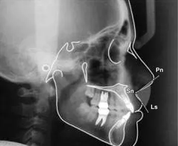
The following cephalometric landmarks were used: pronasal point (Prn), on the anterior nasal apex; subnasal point (Sn), in the middle of the in-ferior border on the anterior nasal aperture; and upper lip (Ls), the most anterior point on the ver-milion of the upper lip (Figure 1).
A cephalogram with the variables used in the present study can be seen in Figure 1.
Student’s t test analysis and a significance level of 5% were applied to determine averages, standard deviation, and minimum and maximum values of the nasolabial angle, as well as to support an evaluation of sexual dimorphism.
RESULTS
As we can observe in Table 1 and Graph 1, the mean value of the nasolabial angle for the to-tal sample of Brazilian black subjects, was 88.14°
with a standard deviation of 12.52°. For females the mean value was 85.05° with a standard devia-tion of 11.93°; for males the average was 92° with a standard deviation of 12.52°. The nasolabial angle was significantly smaller (p < 0.05) in females.
DISCUSSION
Orthodontic diagnosis should not be accom-plished based solely on hard tissue information since changes in dental positions cause alterations in the facial profile. Modern orthodontics adds ele-ments of facial analysis to the diagnosis effort.
Holdaway11,12 (1983, 1984) stated that the
orthodontic treatment plan should include the perspectives of orthodontic alterations based on soft tissue analysis, taking into account the soft tissue profile.
The nasolabial angle is an important auxiliary parameter in the diagnosis of anteroposterior max-illary discrepancies, and is a strategic area of the composition of facial profiles. As a consequence, the clinical application of this angle contributes to the differential diagnosis of skeletal malocclusions, particularly of Class II malocclusions. Burstone4
(1958) defined the nasolabial angle as representa-tive of maxillary inclination, suggesting that when this variable is increased it reflects a maxillary
TABLE 1 - Means, standard deviations (SD), minimum (Min.) and maximum (Max.) values of the nasolabial angle.
Gender
Total sample
Females Males
Mean 85.05° B 92.00° A 88.14°
SD 11.93° 12.52° 12.52°
Min. 64.00° 70.00° 64.00°
Max. 113.00° 113.00° 113.00°
Means with different letters are significantly different at p < 0.05.
FIGURE 1 - Landmarks used for measurements of the nasolabial angle. Prn: pronasal point; Sn: subnasal point; Ls: upper lip.
Sn
Pn
236 237
236 237
retrusion, and when decreased, a maxillary pro-trusion.
The related literature indicates that the value of the nasolabial angle related to harmonious faces varies considerably. Among the cited values are 114.08°± 9.58°9; 102°15,16; 105°± 8°18; 111.04°20;
104°± 11.5°21 and from 108.76° to 114.40°22.
The mean value observed in our study was 88.14°, which is smaller than that reported by authors referred in studies accomplished with white individuals. However, as our sample was composed of young black subjects, the observed nasolabial angle was smaller, suggesting that this ethnic group presents a more biprotruded pro-file and a reduced interincisive angle, which is in agreement with others studies1,2,3,6,7,10,13,14, whose
authors compared white and black subjects.
As can be observed in Table 1, the nasolabial angle presented variation according to gender; in females it was significantly smaller, confirming the existence of sexual dimorphism. This result differs from those of Silva Filho et al.21 (1990) and
Fitzger-ald et al.9 (1992), who found that black individuals
showed similar angular and linear measurements in both groups, male and female.
According to Prahl-Andersen et al.19 (1995) the
tendency of the nasolabial angle is to decrease with age, specially until adolescence, when the growth of nose, chin and lips is expressed more intensely. Some studies have demonstrated that soft tissues that vary in thickness are the main factor in the determination of full facial profile4,12,15,21.
CONCLUSION
Based on the obtained results, it was con-cluded that the mean value of the nasolabial angle found in our sample of Brazilian black youths was 88.14°± 12.52°, which is in agreement with oth-ers studies. The smaller value of the nasolabial angle suggests that black individuals present a more protruded profile and a reduced interincisive angle.
The value of the nasolabial angle was signifi-cantly smaller (p < 0.05) in females, characterizing sexual dimorphism.
Since one of the objectives of orthodontic treatment is to achieve facial aesthetics, specific soft tissue measures which respect the facial pat-tern of each race should be considered during the diagnosis phase.
Females Males
0 20 40 60 80 100 120
N
aso
la
bi
al
a
ng
le
(d
eg
re
es)
85.05 92
Mean
GRAPH 1 - Mean value of the nasolabial angle in males and females.
REFERENCES
1. Alexander TL, Hitchcock HP. Cephalometric standards for American negro children. Am J Orthod 1978;74:298-304.
2. Altemus LA. Cephalofacial relationships. Angle Orthod 1968;38:175-84.
3. Altemus LA. Comparative integumental relationships. Angle Orthod 1963;33:217-21.
4. Burstone CJ. The integumental profile. Am J Orthod 1958;44:1-25.
5. Capelozza L, de Araujo Almeida G, Mazzottini R, Cardoso Neto J. Maxillomandibular relationships in patients with dentofacial deformities: diagnostic criteria utilizing three cephalometric analyses. Int J Adult Orthodon Orthognath Surg 1989;4:13-26.
6. Cotton WN, Takano WS, Wong WMW. The Downs anal-ysis applied to three other ethnic groups. Angle Orthod 1951;21:213-20.
7. Drummond RA. A determination of cephalometric norms for the negro race. Am J Orthod 1968;54:670-82.
8. Elias AC. The importance of the nasolabial angle in the diagnosis and treatment of malocclusion. Int J Orthod 1980;18:7-12.
9. Fitzgerald JP, Nanda RS, Currier GF. An evaluation of the nasolabial angle and the relative inclinations of the nose and upper lip. Am J Orthod Dentofacial Orthop 1992;102:328-34.
10. Fonseca RJ, Klein WD. A cephalometric evaluation of American negro women. Am J Orthod 1978;73:152-60. 11. Holdaway R. A soft tissue cephalometric analysis and
its use in orthodontic treatment planning. Part I. Am J Orthod 1983;84:1-28.
236 237
236 237
13. Jacobson A, Oosthuizen L. The craniofacial skeletal pattern of the South African Bantu. J Dent Assoc S Afr 1970;25:361-5.
14. Magnani MBBA, Sakima T. Comparação cefalométrica entre negróides e caucasóides. RGO 1981;37:225-8. 15. McNamara Jr JA. A method of cephalometric
evalu-ation. Am J Orthod 1984;86(6):449-69.
16. McNamara Jr JA, Brudon WL. Orthodontic and ortho-pedic treatment in mixed dentition. Ann Arbor: Needham Press; 1993. 365 p.
17. Midtgard J, Bjork G, Linder Aronson S. Reproduc-ibility of cephalometric landmarks and errors of measure-ments of cephalometric cranial distances. Angle Orthod 1974;44(1):56-67.
18. Owen AH 3rd. Diagnostic block cephalometrics. Part
I. J Clin Orthod 1984;18:400-22.
19. Prahl-Andersen B, Ligthelm-Bakker ASWMR, Wattel E, Nanda R. Adolescent growth changes in soft tissue pro-file. Am J Orthod Dentofacial Orthop 1995;107:476-83.
20. Scheideman GB, Bell WH, Legan HL, Finn RA, Reisch JS. Cephalometric analysis of dentofacial normals. Am J Orthod 1980;78:405-20.
21. Silva Filho OG, Okada T, Tocci LFC. Avaliação ce-falométrica do ângulo nasolabial aos 7 anos, 12 anos e 19 anos, numa amostra de oclusão normal. Rev SBO 1990;1:108-13.
22. Siqueira VCV, Canuto CE, Scavone Junior H, Ne-greiros PE. O relacionamento dos ângulos nasolabial e dos incisivos superiores com o plano palatino durante a fase do “patinho feio”. Rev Dent Press Ortod Ortop Facial 2003;8(6):31-42.
23. Todd TW, Lindalla A. Dimensions of the body: whites and American negroes of both sexes. Am J Phys Anthropol 1928;7:35-101.