Andrés Felipe CARTAGENA(a) Sibelli Olivieri PARREIRAS(a) Alessandro Dourado LOGUERCIO(b) Alessandra REIS(b)
Nara Hellen CAMPANHA(c)
(a)Universidade Estadual de Ponta Grossa – UEPG, School of Dentistry, Posgraduate Program in Dentistry, Ponta Grossa, PR, Brazil.
(b)Universidade Estadual de Ponta Grossa – UEPG, School of Dentistry, Department of Operative Dentistry, Ponta Grossa, PR, Brazil.
(c)Universidade Estadual de Ponta Grossa – UEPG, School of Dentistry, Department of Prosthodontics, Ponta Grossa, PR, Brazil.
In-office bleaching effects on the pulp
flow and tooth sensitivity – case series
Abstract:Laser Doppler flowmetry (LDF) is a noninvasive method
capable of evaluating variations in pulp blood flow (PBF) and pulp vitality. This method has thus far not been used to assess changes in blood flow after in-office bleaching. The aim of this case series report was to measure changes in PBF by LDF in the upper central incisor of three patients submitted to in-office bleaching. The buccal surfaces of the upper arch were bleached with a single session of 35% hydrogen peroxide gel with three 15-min applications. The color was recorded using a value-oriented Vita shade guide before in-office bleaching and one week after the procedure. The tooth sensitivity (TS) in a verbal scale was reported, and PBF was assessed by LDF before, immediately, and one week after the bleaching session. The lower arch was submitted to dental bleaching but not used for data assessment. A whitening degree of 3 to 4 shade guide units was detected. All participants experienced moderate to considerable TS after the procedure. The PBF readings reduced 20% to 40% immediately after bleaching. One week post-bleaching, TS and PBF were shown to be equal to baseline values. A reversible decrease of PBF was detected immediately after bleaching, which recovered to the baseline values or showed a slight increase sooner than one week post-bleaching. The LDF method allows detection of pulp blood changes in teeth submitted to in-office bleaching, but further studies are still required.
Keywords:Laser-Doppler Flowmetry; Dentin Sensitivity; Tooth Bleaching.
Introduction
Dental bleaching is a widely used cosmetic protocol employed in
daily clinical practice. This technique fulills the patient’s expectations
in the search for a whiter smile. Currently, there are two main
dentist-supervised techniques: the at-home or in-ofice bleaching. The at-home bleaching is performed using a custom-itted bleaching tray illed with
a low concentration of hydrogen peroxide (HP) or carbamide peroxide. The patients are instructed to wear the bleaching tray daily, for periods ranging from 1 to 8 h from 2 to 6 weeks.1
In spite of the safety and effectiveness of this technique to whiten teeth, some patients do not want to use the trays even for shorter periods of time.
In this clinical scenario, in-ofice bleaching using higher concentrations
Declaration of Interests: The authors certify that they have no commercial or associative interest that represents a conflict of interest in connection with the manuscript.
Corresponding Author: Alessandra Reis
E-mail: reis_ale@hotmail.com
DOI: 10.1590/1807-3107BOR-2015.vol29.0026
Submitted: May 13, 2014
of HP is the alternative protocol. This protocol has been shown to produce color changes more rapidly.2
Fortunately, as long as the in-ofice bleaching is
performed more than once, bleaching of approximately 5 to 8 shade guide units is possible.2,3,4 Stable results
were also demonstrated after 9 and 24 months.4,5
Even though professional-assisted dental bleaching is considered a safe procedure, tooth sensitivity (TS) is a remarkably common side effect reported by patients.1,3,4 More than 70% of patients who undergo
in-ofice bleaching complain of TS,6 which leads some
patients to forego treatment.7
The uncomfortable and painful bleaching-induced TS is likely the result of pulp insult by the rapid diffusion of HP molecules. Pulp damage can trigger
an inlammatory reaction that leads to the release of
cell-derived factors such as adenosine triphosphate8
neuropeptides, and prostaglandins, which excite or sensitize pulpal nociceptors.9,10,11 This inlammatory
reaction also induces vasodilatation and increased
pulpal blood low (PBF).12,13,14
In a recent histological study, Costa et al.15
evaluated the effects of HP on pulp cells and showed notable damage to the pulp tissue, with mild inflammatory changes as well as sites of pulp necrosis in lower incisors. This study has raised concerns about pulp vitality of teeth soon after in-office bleaching. Although there are many clinical studies on tooth bleaching, the pulp vitality of bleached teeth has not been assessed thus far. This may be due to the fact that most of the current methods employed to assess pulp vitality may give a high percentage of false-positive results.16,17
An interesting approach is noninvasive laser Doppler, which can monitor PBF changes through measuring the velocity of red blood cells in capillaries. Additionally, it is an objective, semiquantitative, and painless method. This method was first used on human teeth by Gazelius et al.18 in 1986. The value
of this method for assessing pulp vitality has been well documented,19 but its high costs and dificulty
of use in clinical practice have delayed its wide-scale introduction.16,20 Therefore, the aim of this preliminary
case series report was to assess the viability of using the laser Doppler to monitor PBF in teeth submitted
to in-ofice bleaching.
Methodology
The Scientific Review Committee and the Committee for the Protection of Human Beings at the local university approved this clinical investigation (protocol no. 172.988).
Three patients from 18 to 24 years old sought clinical assistance at the School of Dentistry of the Universidade
Estadual de Ponta Grossa– UEPG (Paraná, Brazil). All
patients were not satisied with the yellowing color of
their teeth. The participants should have caries-free maxillary teeth without restorations. Participants who had previously undergone tooth-whitening procedures, presenting internal tooth discoloration, taking any medicine, or presenting any pulpal pathology or participants whose teeth had enamel cracks were not included in the study. After anamnesis and a clinical evaluation, the patients decided to
have in-ofice bleaching performed because they did
not complain about using the trays. All participants were informed via written consent about possible sensitivity and other conditions or likely side effects such as gingival burning.
Two weeks before the bleaching procedures, all patients received a dental screening and prophylaxis with pumice and water in a rubber cup. During this visit, possible TS due to cold air was assessed using air spray on all teeth to be bleached for 3 s. None of the patients complained about TS or reported a history of trauma or endodontic treatment on any tooth.
During the in-office bleaching session, the gingival tissue of the teeth from the upper arch was isolated with a light-curing resin dam (Top Dam, FGM, Joinville, Brazil, batch 260712). The 35% HP gel (Whiteness HP Maxx, FGM, batch 260712)
was manipulated according to the manufacturer’s
directions and applied on the buccal surfaces of all
upper anterior teeth and premolars. The in-ofice
bleaching agent was refreshed every 15 min during the 45-min application period. The lower arch was also submitted to bleaching one week after the end of the upper arch treatment but was not used for data assessment.
shade evaluation was done in a single room with artificial lighting by a single calibrated operator.
For this examination, the shade guide’s 16 tabs
were arranged from the highest (B1) to lowest (C4) value, making the color A3, number 9. This allowed for the calculation of the variation of the
shade guide units (∆SGU). The measurement area
of interest for shade matching was the middle one-third of the buccal surface of the central upper left incisor (tooth 21). The patients were instructed to record their perception of TS on a 5-point verbal rating scale during bleaching up to 1 week after the bleaching session. They kept a daily record of whether they experienced sensitivity using the following criteria: 0 = none, 1 = mild, 2 = moderate, 3 = considerable, and 4 = severe.
The PBF was recorded at baseline, immediately, and 1 week after the bleaching session in the central upper left incisor with LDF (MoorLAB/FloLAB; Moor Instruments Ltd., Axminster, England).20 The
device was calibrated against a Brownian motion
medium and zeroed against a static relector.
The laser source was 780 nm, with 0.5 mm of
iber separation in the MoorLAB probe P13, with 3.1 kHz as the primary bandwidth for a ilter set
to a 0.1-s time output constant.
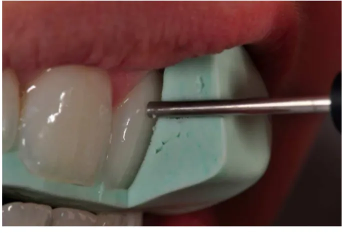
The patients rested in a supine position for 3–5 minutes before starting the LDF test. To ensure accurate and reproducible positioning of the probe at each examination, a matrix of high-viscosity silicone was prepared for each patient (Zetalabor, Zhermack, Polesine Badia, Italy). This silicone matrix was fashioned with appropriate holes with the
corresponding diameter of the lowmeter probe tip
(Figure 1). The probe was positioned so that it was perpendicular to the enamel surface, with its center 2 mm from the gingival margin and over the central long axis of the crown of the tooth.
The lux signal from the blood low monitor
was transferred to a computer, analyzed using the Moorsoft program (Moor Instruments, Axminster, England), and transformed in arbitrary perfusion units. Recordings of PBF were made for 90 s at each period. The perfusion units measured at the different periods were relative to the perfusion unit at baseline, considered to be 100%.
Results
A whitening of approximately 3 to 4 SGU was detected for the three patients after the in-office bleaching (Table 1). Some changes were found in red
blood low and TS after the in-ofice bleaching. The
incidence of TS ranged from moderate to considerable (Table 1) after bleaching. One week after the procedure, the patients reported only mild TS. A 20% to 40% reduction of the PBF was observed immediately after
in-ofice bleaching (Figure 2). One week post-bleaching,
the values of PBF either reached the baseline values or showed a slight increase.
Discussion
HP is the active molecule that causes tooth
bleaching. This study demonstrated a signiicant
improvement in shade compared with the baseline color. Previous studies have demonstrated a higher whitening degree2,3 than the current one. This lower
whitening effect is probably due to the fact that only one bleaching session was performed in this study.
Table 1. TS scores and SGU at different assessment points for each participant.
Time Assessment
Patients
TS ∆SGU
P1 P2 P3 P1 P2 P3
Baseline 0 0 0 5 6 5
After bleaching 2 3 3 - -
-1 week after bleaching 1 0 1 2 2 1
Unfortunately, HP action is not limited to dental hard tissues. HP can also reach the pulp chamber, where it was shown to reduce cell proliferation, metabolism, and viability.21 Additionally, it may also
reduce the pulp-reparative capacity22 and induce pulp
inlammation, which may be responsible for the TS experienced by the patients during in-ofice bleaching.1
All three participants from this study experienced moderate to considerable TS that decreased to a mild level one week after bleaching. The literature usually reports that TS normally persists for up to four days after bleaching,2 but durations of up to 39 days have been
reported.7 The inlammatory process produced by tooth
bleaching usually lasts for approximately 2 weeks. During
this time, proinlammatory cytokine release continues,
which, in some cases, could perpetuate substance P release for longer periods of time and therefore cause postbleaching symptoms in some cases.10,11
The optical method herein used enables the recording of the number and velocity of particles
conveyed by a luid low.16 As red blood cells represent
the vast majority of moving cells within the dental structure tooth, measurement of the Doppler-shifted, backscattered light serves as an index of PBF. This device is capable of evaluating the dynamic changes in
blood low in a small volume of tissue (about 1 mm3).23
As most studies on LDF have been undertaken on anterior teeth, including central and lateral incisors and also canines,16,19 this preliminary case report followed the
literature trend. Additionally, we opted to measure it on the central incisor as most of clinical trials on bleaching employ this tooth as a reference for assessment of the
whitening eficacy. Besides that, blood low varies in
different teeth from the same patient;19 therefore, when
the aim is to compare different procedures, a single tooth should be selected for evaluation to avoid adding
intertooth variability on blood low response.
There is a great variability in PBF among patients,19
and this is the reason that the variation in the pulp
low in the present study was reported relative to
the flowmeter at baseline for each patient in an intraindividual comparison. This reduces the variability of the measurement and is a viable method to assess the effect of some restorative and cosmetic protocols on pulp vitality, including bleaching protocols.
When pulp is directly injured, such as during pulp exposure, neuropeptide levels increase in the pulp and trigeminal ganglia,24 These neuropeptides may
be released because of the stimulation from dental procedures, causing vasodilatation and increased PBF.14,25 However, in this study, a decrease in the PBF
in the patients was detected, which is in agreement
with the indings of Chen and Abbott.17 Contrary to
other conjunctive tissues, the inlammatory vascular
reactions in the dental pulp take place in a rigid,
enclosed dentin chamber. Any increase in blood low
and vascular permeability can cause major changes in the pressure inside the pulp chamber, which may compress blood vessels and lead to the decrease in
the pulp low14 detected by the laser Doppler device.
However, one week after tooth bleaching, the values either reached baseline values or remained slightly higher. This variability may be explained based on individual variation of the same injury. Perhaps theslight increase in PBF detected in one patient explains changes in the microcirculatory and micromorphology dynamics of the pulp tissue after
the inlammatory process.26,27,28 Further studies with a
larger sample size should be conducted to investigate
whether the dynamics of pulp low are affected by
dental bleaching and other dental procedures.
Baseline 140
120
100
80
60
40
20
0
After
P3 P2 P1
1 week
The dificulties of such a device should also be
mentioned. Variables such as the calibration of the
lowmeter, the position of the probe and patient, the patient’s systemic pressure, the patient’s use of drugs, and ambient temperature can all inluence PBF, but they
can be controlled clinically. However, there are other factors that are not under clinical control. For instance, the individual variations in the PBF and neurovascular response, the differences in the optical properties of
the dental structure (luorescence, opalescence, etc.), tooth morphology, and the surrounding tissues can cause variability in the measurements.16
Another limitation of the present study is that LDF was not compared to other methods. Unfortunately, there is no noninvasive gold standard in the literature for evaluation of pulp blood changes. Pulse oximetry and electric tests are other options to assess pulp vitality; however, previous studies suggest that LDF
is more sensitive and speciic in human teeth.29
The objective assessment of pain perception or sensitivity is limited due to its nature. There are many simple and direct methods to measure this perception of pain, as the VAS scale used in this study has done.
However, these methods are considered subjective, so accurate assessment of pain remains a challenge.30 With the
results of this preliminary study, we observed a relationship between the changes in PBF and the perceived sensitivity reported by patients. Thus, despite the limitations of LDF, it could be used to increase the reliability of pain scales as instruments to assess the perception of TS.
Finally, future studies should be conducted to evaluate PBF between teeth of the same patient and between individuals as well as to correlate PBF with TS. The preliminary results of this study suggest that the use of LDF is a viable alternative to assess the changes in pulp tissue that may occur due to tooth bleaching in shorter periods; however, its current use requires further study.
Conclusion
LDF is a viable alternative to assess blood low
changes in teeth submitted to vital tooth bleaching; however, it requires further study using a larger sample size. Immediately after bleaching, reduced
blood low was observed, but this was reversed one
week after of intervention.
1. Basting R, Amaral F, Franca F, Florio F. Clinical comparative study of the effectiveness of and tooth sensitivity to 10% and 20% carbamide peroxide home-use and 35% and 38% hydrogen peroxide in-office bleaching materials containing desensitizing agents. Oper Dent. 2012 Sep-Oct;37(5):464-73. 2. Bernardon JK, Sartori N, Ballarin A, Perdigao J, Lopes GC,
Baratieri LN. Clinical performance of vital bleaching tech-niques. Oper Dent. 2010 Jan-Feb;35(1):3-10.
3. Kossatz S, Dalanhol AP, Cunha T, Loguercio A, Reis A. Effect of light activation on tooth sensitivity after in-office bleach-ing. Oper Dent. 2011 May-Jun;36(3):251-7.
4. Tay LY, Kose C, Herrera DR, Reis A, Loguercio AD. Long-term efficacy of in-office and at-home bleaching: a 2-year double-blind randomized clinical trial. Am J Dent. 2012 Aug;25(4):199-204. 5. Giachetti L, Bertini F, Bambi C, Nieri M, Scaminaci Russo D.
A randomized clinical trial comparing at-home and in-office tooth whitening techniques: a nine-month follow-up. J Am Dent Assoc. 2010 Nov;141(11):1357-64.
6. Reis A, Dalanhol AP, Cunha TS, Kossatz S, Loguercio AD. Assessment of tooth sensitivity using a desensitizer before light-activated bleaching. Oper Dent. 2011 Jan-Feb;36(1):12-7.
7. Leonard RH Jr, Haywood VB, Phillips C. Risk factors for develop-ing tooth sensitivity and gdevelop-ingival irritation associated with night-guard vital bleaching. Quintessence Int. 1997 Aug;28(8):527-34. 8. Cook SP, McCleskey EW. Cell damage excites nociceptors
through release of cytosolic ATP. Pain. 2002 Jan;95(1-2):41-7. 9. Markowitz K. Pretty painful: why does tooth bleaching hurt?.
Med Hypotheses. 2010 May;74(5):835-40.
10. Bowles WR, Withrow JC, Lepinski AM, Hargreaves KM. Tis-sue levels of immunoreactive substance P are increased in pa-tients with irreversible pulpitis. J Endod. 2003 Apr;29(4):265-7. 11. Patel T, Park SH, Lin LM, Chiappelli F. Huang GT. Substance P induces interleukin-8 secretion from human dental pulp cells. Oral Surg Oral Med Oral Pathol Oral Radiol Endod. 2003 Oct;96(4):478-85.
12. Caviedes-Bucheli J, Ariza-Garcia G, Restrepo-Mendez S, Rios-Osorio N, Lombana N, Munoz HR. The effect of tooth bleaching on substance P expression in human dental pulp. J Endod. 2008 Dec;34(12):1462-5.
14. Kim S. Neurovascular interactions in the dental pulp in health and inflammation. J Endod. 1990 Feb;16(2):48-53. 15. Costa CA, Riehl H, Kina JF, Sacono NT, Hebling J. Human
pulp responses to in-office tooth bleaching. Oral Surg Oral Med Oral Pathol Oral Radiol Endod. 2010 Apr;109(4):e59-64. 16. Jafarzadeh H. Laser Doppler flowmetry in endodontics: a
review. Int Endod J. 2009 Jun;42(6):476-90.
17. Chen E, Abbott PV. Evaluation of accuracy, reliabil-ity, and repeatability of five dental pulp tests. J Endod. 2011 Dec;37(12):1619-23.
18. Gazelius B, Olgart L, Edwall B, Edwall L. Non-invasive re-cording of blood flow in human dental pulp. Endod Dent Traumatol. 1986 Oct;2(5):219-21.
19. Norer B, Kranewitter R, Emshoff R. Pulpal blood-flow char-acteristics of maxillary tooth morphotypes as assessed with laser Doppler flowmetry. Oral Surg Oral Med Oral Pathol Oral Radiol Endod. 1999 Jan;87(1):88-92.
20. Roy E, Alliot-Licht B, Dajean-Trutaud S, Fraysse C, Jean A, Armengol V. Evaluation of the ability of laser Doppler flow-metry for the assessment of pulp vitality in general dental practice. Oral Surg Oral Med Oral Pathol Oral Radiol Endod. 2008 Oct;106(4):615-20.
21. Min KS, Lee HJ, Kim SH, Lee SK, Kim HR, Pae HO, et al. Hydrogen peroxide induces heme oxygenase-1 and dentin sialophosphoprotein mRNA in human pulp cells. J Endod. 2008 Aug;34(8):983-9.
22. Goldberg M, Smith AJ. Cells and extracellular matrices of dentin and pulp: a biological basis for repair and tissue en-gineering. Crit Rev Oral Biol Med. 2004 Jan;15(1):13-27.
23. Vongsavan N, Matthews B. Some aspects of the use of laser Doppler flow meters for recording tissue blood flow. Exp Physiol. 1993 Jan;78(1):1-14.
24. Buck S, Reese K, Hargreaves KM. Pulpal exposure al-ters neuropeptide levels in inflamed dental pulp and tri-geminal ganglia: evaluation of axonal transport. J Endod. 1999 Nov;25(11):718-21.
25. Heyeraas KJ, Kim S, Raab WH, Byers MR, Liu M. Effect of electrical tooth stimulation on blood flow, interstitial fluid pressure and substance P and CGRP-immunoreactive nerve fibers in the low compliant cat dental pulp. Microvasc Res. 1994 May;47(3):329-43.
26. Matheny JL, Abrams H, Johnson DT, Roth GI. Microcircu-latory dynamics in experimental human gingivitis. J Clin Periodontol. 1993 Sep;20(8):578-83.
27. Mesaros SV, Trope M. Revascularization of traumatized teeth assessed by laser Doppler flowmetry: a case report. Endod Dent Traumatol. 1997 Feb;13(1):24-30.
28. Núñez SC, Nogueira GE, Ribeiro MS, Garcez AS, Lage-Marques JL. He-Ne laser effects on blood microcirculation during wound healing: a method of in vivo study through laser Doppler flowmetry. Lasers Surg Med. 2004;35(5):363-8. 29. Karayilmaz H, Kirzioğlu Z. Comparison of the reliability of
laser Doppler flowmetry, pulse oximetry and electric pulp tester in assessing the pulp vitality of human teeth. J Oral Rehabil. 2011 May;38(5):340-7.