Article
ISSN 0102-695X http://dx.doi.org/10.1590/S0102-695X2011005000145
Received 30 Sep 2010 Accepted 17 Jan 2011 Available online 12 Aug 2011
induced allergic conjunctivitis
Rosemary Jorge de Mendonça Albuquerque,
1L. Kalyne A. M.
Leal,
3Mary Anne Bandeira,
3Glauce Socorro de Barros Viana,
*,2Lusmar Veras Rodrigues
11Departamento de Cirurgia, Universidade Federal do Ceará, Brazil,
2Departmento de Fisiologia e Farmacologia, Universidade Federal do Ceará, Brazil,
3Departmento de Farmácia, Universidade Federal do Ceará, Brazil.
Abstract: The objective of the study was to verify the effects of dimeric chalcones (urundeuvines A, B, and C) from Myracrodruon urundeuva Allemão, Anacardiaceae (a Brazilian anti-inflammatory species), on an allergic conjunctivitis model. Male
guinea-pigs were sensitized with two intraperitoneal injections of ovalbumin (10 μg
dissolved in 0.5 mL saline and emulsified in 0.5 mL Freund’s adjuvant), at days 0 and 7. At day 24, the animals were submitted to an ocular instillation (right eyes) with ovalbumin. At the next day, the animals were treated with chalcones (0.5 mg, three times a day for 7 days), 0.1% fluormetalone acetate (0.05 mg, as the reference drug) or saline. After anesthesia of the animals, enucleations of their corneas and conjunctivas were carried out for morphometric and histological analyses, at days 1, 3 and 7. Their radical scavenging activity and action on myeloperoxidase were also determined. We demonstrated that chalcones from M. urundeuva stem barks presented anti-inflammatory and antioxidant effects, and drastically inhibited the MPO activity, pointing them as candidates for the treatment of allergic conjunctivitis and other inflammatory conditions.
Keywords:
allergic conjunctivitis chalcones
Myracrodruon urundeuva
ovalbumin
Introduction
Several experimental models of allergic conjunctivitis are described in the literature. Among those used for studying the pathophysiological and therapeutic aspects of this pathological process, that using ovalbumin as the sensitizing agent is the most popular one (Lundberg et al., 1987; Sompolinsky et al., 1992; Nitzan et al., 1996).
Myracrodruon urundeuva Allemão,
Anacardiaceae, is a Brazilian medicinal plant largely used in popular medicine as an anti-inflammatory and wound healing agent. Earlier works carried out with crude extracts from the plant stem bark revealed its anti-inflammatory, antiulcer, wound healing, antihistaminic and antibradykinin properties (Menezes & Rao, 1988; Viana et al., 1997; Rodrigues et al., 2002). The chemical fractionation of the ethylacetate extract, followed by the pharmacological monitoring, demonstrated the presence of at least two classes of bioactive compounds: one represented by dimeric chalcones and the other by condensed tannins (Viana et al., 1995).
Chalcones, a subclass of the flavonoid family, are widely known for their anti-inflammatory
and antioxidant properties. Licochalcones from
Glycyrrhiza inflata were shown to exert a potent anti-inflammatory effect, possibly through NF-kappaB inhibition (Furusawa et al., 2009a; Furusawa et al., 2009b). It has been recently reported that a synthesized chalcone derivative suppresses NO production in LPS-stimulated RAW 264.7 macrophages, via induction of heme-oxygenase-1 (HO-1) expression and blockade of the activation of activator protein-1 (Park et al., 2009a; Park et al., 2009b). The induction of HO-1 expression, as well as suppressions of LPS-induced NO, IL-1beta and TNF-alpha production, have been also implicated in the anti-inflammatory activity of a natural chalcone, isoliquiritigenin, from Dalbergia odorifera, in RAW 264.7 macrophages (Lee et al., 2009).
Previously (Rodrigues et al., 2002) we reported that similarly to what occurred with the reference drug, 5-aminosalicylic acid (5-ASA), rats treated with the aqueous extract of M. urundeuva
the effects of the dimeric chalcones isolated from the stem bark of M. urundeuva, for the fist time on an experimental model of ovalbumin-induced allergic conjunctivitis in guinea pigs from a histological point of view. Besides, the antioxidant and radical scavenging activities of these compounds were assessed by the DPPH assay, as well as their ability to inhibit the release of myeloperoxidase (MPO), a biomarker for inflammation.
Materials and Methods
Plant material
The plant Myracrodruon urundeuva Allemão, Anacardiaceae, was collected in the city of Iguatu, state of Ceará, Brazil, and the exsiccata is deposited at the Prisco Bezerra Herbarium, of the Federal University of Ceará, under the number 14.999.
Isolation of chalcones
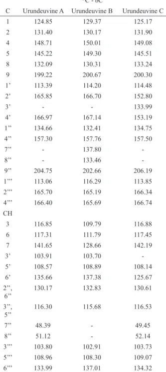
The chalcone-enriched fraction was obtained from the ethyl acetate extract prepared with 5 kg M. urundeuva ground stem bark, as described previously (Viana et al., 1995). The material was previously treated with hexane for removing lipid-type substances. The chromatographic fractionation of the ethyl acetate extract on a silica gel column resulted in seven fractions, after elution with chloroform, chloroform-acetone (9:1; 8:2; 7:3; 1:1), acetone, and methanol. Two fractions presented anti-inflammatory activity, according to the pharmacological monitoring. Preliminary chemical tests showed that one fraction presented a predominance of chalcone type compounds, and the other one mainly catechic tannins. The isolation of the fraction containing chalcone type compounds was performed using chromatographic techniques developed for that purpose (Bandeira et al., 1994) and the utilization of corn starch as the column support. The elution was carried out with a mixture of dichloromethane/methanol (95:5, v/v). Through chromatographic procedures, three dimeric chalcones, named urundeuvines A (1), B (2) and C (3) were isolated from that fraction (Viana et al., 2003). Their structural identification was carried out by 13C-
NMR spectroscopy analyses whose data are shown in Table1.
Drugs
Chloral hydrate, ovalbumin and Freund's complete adjuvant were purchased from Sigma, U.S.A. Fluormetolone acetate 0.1% was from Alcon, Brazil. All other reagents were of analytical grade.
Table 1. 13C-NMR spectroscopy data from dimeric chalcones
(urundeuvins A (1), B (2) and C (3)) of Myracrodruon urundeuva.
13C - δC
C Urundeuvine A Urundeuvine B Urundeuvine C
1 124.85 129.37 125.17
2 131.40 130.17 131.90
4 148.71 150.01 149.08
5 145.22 149.30 145.51
8 132.09 130.31 133.24
9 199.22 200.67 200.30
1’ 113.39 114.20 114.48
2’ 165.85 166.70 152.80
3’ - - 133.99
4’ 166.97 167.14 153.19
1’’ 134.66 132.41 134.75
4’’ 157.30 157.76 157.50
7’’ - 137.80
-8’’ - 133.46
-9’’ 204.75 202.66 206.19
1’’’ 113.06 116.29 113.85
2’’’ 165.70 165.19 166.34
4’’’ 166.40 165.69 166.74
CH
3 116.85 109.79 116.88
6 117.31 111.79 117.45
7 141.65 128.66 142.19
3’ 103.91 103.70
-5’ 108.57 108.89 108.14
6’ 135.66 137.38 125.67
2’’, 6’’
130.17 132.83 130.61
3’’, 5’’
116.30 115.68 116.53
7’’ 48.39 - 49.45
8’’ 51.12 - 52.14
3’’’ 103.80 102.91 103.73
5’’’ 108.96 108.30 109.07
6’’’ 133.99 137.01 134.32
Induction of allergic conjunctivitis
Fifty-four male guinea pigs, weighting 400 g on average, were used. The animals were maintained under a 12 h light/dark cycle at 25 °C, relative humidity of 55%, and free access to water and food. They were sensitized with two intraperitoneal injections of 10 µg ovalbumin each, dissolved in 0.5 mL saline. This
solution was emulsified with 0.5 mL the Freund adjuvant solution, and the injections were made at days 0 and 7. At day 24, an ocular instillation was made with 5 mg ovalbumin diluted in 10 µL saline into the right eyes of all animals. After the induction of allergic conjunctivitis, the animals were distributed into three groups: 1. treated with 0.5 mg chalcones; 2. treated with 0.05 mg of 0.1% fluormetolone acetate, a corticoid used as a standard drug) and 3. saline-treated group. The treatment started 24 h after the induction, and the drugs were administered three times a day for seven days. All animals were treated through ocular instillation, and evaluated from a histological point of view, at days 1, 3 and 7 after the induction of the conjunctivitis. At days 1, 3 and 7, the enucleation was carried out under anesthesia with chloral hydrate (400 mg/kg, i.p.), and the animals eyes were fixed into formalin and processed for histological studies (hematoxylin/eosin and toluidine blue), as previously described (Sompolinsky et al., 1992). All experiments were performed according to the Guide for the Care and Use of Laboratory Animals, from the US Health and Human Services Department.
Histological evaluation
It was performed with a 25-point integrated ocular objective. The ocular was coupled to an optical microscope, synchronized with the 40x objective (Weibel et al., 1966; Underwood, 1968) and used for neutrophil, eosinophyl, mastocyte, lymphocyte, plasmocyte and vessels counting.
Determination of chalcones DPPH radical scavenging activity in vitro
The method was that described previously (Saint-Cricq de Gaulejac et al., 1999). Briefly, an aliquot
(0.1 mL) of the chalcones solution (1-50 μg/mL) or α-tocopherol (5 and 50 μg/mL) was mixed with 3.9 mL
DPPH (0.3 mM in a 1:1 methanol:ethanol solution). The mixture was vortexed for 1 min, left standing at room temperature for 30 min, and the absorbance determined
at 517 nm. The percentage inhibition was calculated according to the following equation: % inhibition = [(Ao-Ac)/Ao] x 100, where Ao was the absorbance of the control (without chalcone) and Ac was the absorbance in the presence of the test substance.
Myeloperoxidase (MPO) release from human neutrophils
Following the method previously described (Lucisano & Mantovani, 1984), 2.5 x 10⁶ human
leukocytes (mainly neutrophils) were suspended in a buffered Hank’s solution, containing calcium and magnesium. The cells were incubated with chalcones
(0.1, 0.01 and 0.001 μg/mL) for 15 min at 37 °C, and
stimulated by the addition of phorbol myristate acetate
(PMA, 0.1 μg/mL) for 15 min at 37 °C. The suspension
was centrifuged for 10 min at 2000 x g at 4 °C, and 50
μL of supernatants were added to phosphate-buffered saline (100 μL), phosphate buffer (50 μL, pH 7.0) and
H2O2 (0.012%). After 5 min at 37 °C, TMB (1.5 mM, 20
μL) was added, the reaction stopped by 30 μL sodium
acetate (1.5 M, pH 3.0), and the absorbance determined at 620 nm.
Statistical analyses
All values are expressed as means. The data were analyzed by ANOVA, followed by the Dunnet' s test. The
results were considered statistically signiicant at p<0.05.
Results
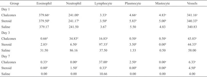
Table 2 presents the number of cells (eosinophils, neutrophils, lymphocytes, plasmocytes and mastocytes) and vessels in guinea pig corneas, after ovalbumin-induced allergic conjunctivitis. Figure 1 shows representative microphotographies (A,B,C,D) from corneas and conjunctivas of guinea pigs submitted to ovalbumin-induced allergic conjunctivitis, at day 1, in both right and left eyes. Figure 2 shows representative microphotographies from corneas (A,B,C,D) and conjunctivas (E, F, G, H), in the right eyes
HO
OH O
OH OH
O OH
OH HO
1
HO
OH O
OH OH
O OH
OH HO
2
HO
OH O
OH OH
O OH
OH HO
3
HO
H H
Table 2. Numbers of cells and vessels in guinea pig corneas, after ovalbumin-induced allergic conjunctivitis.
Group Eosinophil Neutrophil Lymphocyte Plasmocyte Mastocyte Vessels
Day 1
Chalcones 379.66a 241.00a 3.33a 4.66a 4.83a 341.16a
Steroid 379.50b 241.17b 3.50b 5.83b 5.00b 340.33b
Saline 379.17 241.50 3.67 5.50 4.83 340.83
Day 3
Chalcones 0.66d 34.83d 16.83a 0.50a 0.50a 43.83a
Steroid 2.83c 6.50c 97.33f 3.50b 0.00b 44.33b
Saline 31.50 86.16 37.50 1.33 0.50 58.00
Day 7
Chalcones 0.33a 0.00a 37.00e 2.50a 0.00a 6.33a
Steroid 0.00b 1.50f 0.33b 0.00b 0.00b 4.50b
Saline 0.00 0.00 10.66 0.00 0.00 4.00
Values are means from thirty ields studied. The data were analyzed by ANOVA (F) followed by the Dunnet 's test (D). S: Saline; A: Chalcones; C:
Steroid. aS=A; bS=C ; cS>C; dS>A; eS<A; fS<C; F(5%)= 3.68. Eosinophil: Day 1: F=0.00; Day 3: F=60.36; Day 7: F=2.50; Day 3: D(5%)=4.58; S>A (ŷ =30.84); S>C(ŷ=28.67). Neutrophil: Day 1: F= 0.00; Day 3: F= 21.30; Day 7: F= 45.00; Day 3: D(5%)=18.11; S>A (ŷ =51.33); S>C(ŷ=79.66); Day 7: D(5%)=0.26; S=A (ŷ=0.00); S<C (ŷ=1.50). Lymphocyte: Day 1: F=0.004; Day 3: F=17.08: Day 7: F=3.73; Day 3: D(5%)=29.80; S=A (ŷ=20.67); S<C(ŷ=59.83); Day 7: D(5%)=20.25; S<A (ŷ=26.34); S=C(ŷ=10.33). Plasmocyte: Day 1: F=0.16; Day 3: F=0.56: Day 7: F=0.26. Mastocyte: Day 1:
F=0.03; Day 3: F=0.50. Vessels: Day 1: F=0.00; Day 3: F=0.27: Day 7: F=1.22.
Figure 1. Photomicrographies of HE stained slices from corneas and conjunctivas of guinea pigs submitted to ovalbumin-induced allergic conjunctivitis, at day 1. A: cornea, left eye; normal epithelium and proper lamina (100x). B: conjunctiva, left eye; hydropic degeneration of some cells of the basal epithelium layer; slight edema and polimorphonuclear exudation with predominance of neutrophils over eosinophils (200x). C: cornea, right eye; hydropic degeneration in the epithelium basal layer cells; moderate edema and diffuse exudation of neutrophils and eosinophils (100x). D: conjunctiva, right eye; hydropic degeneration in some cells of the epithelium basal layer; slight intercellular edema; moderate polimorphonuclear exocytosis; intense and diffuse exudation of neutrophils and eosinophils in the proper lamina; besides, edema and prominent capillaries due to the increased vascularization and endothelial tumefaction (100x).
exocitosis. C shows occasional hydropic degenerated cells from the epithelium basal layer, a slight edema, and neutrophil, eosinophil and lymphocytic exsudations in the proper lamina, besides no vessels. G shows occasional polimorphonuclear exocytose, scanty and difuse neutrophil, eosinophil and lymphocytic exsudations in the proper lamina, and also moderate edema and congestion. D shows hydropic degeneration in some cells of the epithelium basal layer, moderate neutrophil and eosinophil exsudations, and some congested vessels in the proper lamina, besides a moderate edema. H shows hydropic degeneration in a few cells from the basal epithelium layer, a slight polimorphonuclear exocitosis, and moderate and diffuse
Figure 3. Representive HE stained photomicrographies from corneas (A, B, C, D) and conjunctivas (E, F, G, H) in the right eyes of guinea pigs submitted to ovalbumin-induced allergic conjunctivitis, at day 7. A and B (200 and 400x, respectively) show chalcone-treated corneas, with a normal epithelium, absence of exudation in the proper lamina, and a slight edema. E and F (200 and 400x, respectively) show chalcone-treated conjunctivas, with normal epithelium, occasional polimorphonuclear exudation in the proper lamina, and a slight edema. In C (200x), the treated cornea shows an epithelium with hydropic degeneration in very few cells of the basal layer. In G (100x), the corticoid-treated conjunctive shows hydropic degeneration in rare cells of the epithelium basal layer, the absence of polimorphonuclear exocitosis, slight neutrophyl and eosinophil exudations, as well as slight edema and congestion in the proper lamina. In D (200x), the saline-treated cornea shows hydropic degeneration in a very few cells of the epithelium basal layer, a slight polimorphonuclear exudation with predominance of neutrophils over eosinophils, besides a slight edema. In H (100x), the saline-treated conjunctiva shows hydropic degeneration in some cells of the epithelium basal layer, occasional polimorphonuclear exocitosis, and diffuse and scanty neutrophil and eosinophil exudations.
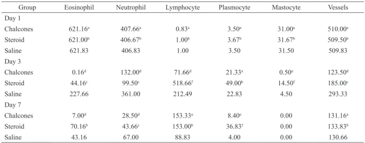
Table 3. Numbers of cells and vessels in guinea pig conjunctives, after ovalbumin-induced allergic conjunctivitis.
Group Eosinophil Neutrophil Lymphocyte Plasmocyte Mastocyte Vessels
Day 1
Chalcones 621.16a 407.66a 0.83a 3.50a 31.00a 510.00a
Steroid 621.00b 406.67b 1.00b 3.67b 31.67b 509.50b
Saline 621.83 406.83 1.00 3.50 31.50 509.83
Day 3
Chalcones 0.16d 132.00d 71.66d 21.33a 0.50a 123.50d
Steroid 44.16c 99.50c 518.66f 49.00b 14.50f 185.00c
Saline 227.66 361.00 212.49 22.83 4.50 293.33
Day 7
Chalcones 7.00d 28.50d 153.33a 8.40e 0.00 131.16a
Steroid 70.16b 43.66c 153.00b 36.83f 0.00 133.83b
Saline 43.16 67.00 88.83 4.00 0.00 130.66
Values are means in thirty ields studied. The data were analyzed by ANOVA (F) followed by the Dunnet 's test (D). S: Saline; A: Chalcones; C: Steroid.
a S=A; b S=C; c S>C; d S>A; e S<A; f S<C; F(5%)=3.68. Eosinophil: Day 1: F=0.00; Day 3: F=28.61: Day 7: F=4.21; Day 3: D (5%)=42.94; S>A
of guinea pigs submitted to ovalbumin-induced allergic conjunctivitis, at day 3, after chalcones (A, B, E and F), corticoid (C and G) and saline (D and H) treatments. Figure 3 shows representative microphotographies from corneas (A, B, C and D) and conjunctivas (E, F, G and H), in the right eyes of guinea pigs submitted to ovalbumin-induced allergic conjunctivitis, at day 7, after chalcones (A, B, E and F), corticoid (C and G) and saline (D and
H) treatments. Although no signiicant differences among
groups (chalcones, steroid and saline) were demonstrated at day 1, the numbers of eosinophils and neutrophils decreased drastically, at day 3 (mainly in the chalcones and steroid groups) and 7. The number of lymphocytes
signiicantly increased (mainly in the steroid group), at
day 3 as compared to day 1. The numbers of plasmocytes, mastocytes and vessels decreased in all groups, at day 3 as compared to day 1. Except for the high and low numbers of lymphocytes seen in the chalcone and steroid groups, respectively, as compared to the saline group, no other
signiicant alterations were observed among groups, at day
7.
Table 3 presents the number of cells and vessels in the guinea pig conjunctivas, after ovalbumin-induced allergic conjunctivitis. Significant decreases in the numbers of eosinophils (mainly with the chalcones group) and neutrophils were observed in all three groups, at day 3 as compared to day 1. Changes observed in the eosinophils number were greater in the chalcone group, as related to the steroid group. Significant increases were observed in the numbers of lymphocytes and plasmocytes in all groups, at day 3 as compared to day 1, mainly in the steroid group. On the other hand, the numbers of mastocytes and vessels decreased significantly in all groups, mainly in the chalcone group, at day 3 as related to day 1. Except for the lower number of eosinophils seen in the chalcones group and the significantly higher number of plasmocytes in the steroid group, as related to the other groups, no other changes were observed at day 7.
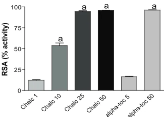
Chalcones (1, 10, 25 and 50 µg/mL) presented a significant and dose-dependent radical scavenging activity, as assessed by the DPPH assay (Figure 4). At the 25 µg/mL concentration, the effect was similar
to that observed with 50 µg/mL of α-tocopherol, used
as the reference antioxidant drug. Furthermore, a significantly decreased inhibition of MPO release from human neutrophils was demonstrated with chalcones, at concentrations as low as 0.001, 0.01, 0.1 and 1.0 µg/mL. However, no significant difference among the highest doses was observed, indicating that a maximum effect was reached (Figure 5).
Cha lc 1
Cha lc 10
Cha lc 25
Cha lc 50
alph a-toc 5 alph a-toc 50 0 25 50 75 100 a
a a a
R SA (% a c ti v ity )
Figure 4. Chalcones (Chalc, µg/mL) are potent antioxidant agents, as demonstrated by the DPPH assay in vitro. The data are means±SEM from 3 to 6 samples. The test was performed on human neutrophils, according to the method described by Saint-Cricq de Gaulejac et al. (1999). a. p<0.05 vs. controls (without chalcones). The radical scavenging activity was measured at 517 nm.
Cha lc 1
Cha lc 0
.1
Cha lc 0
.01
Cha lc 0
.001 0 10 20 30 40 50 60 70 80 90 a
a a a
MPO r e le a s e (% i n h ib iti o n ) Discussion
Myracrodruon urundeuva Allemão,
Anacardiaceae, is a medicinal plant popularly used in
Brazil, due to its anti-inlammatory and wound healing
properties (Menezes & Rao, 1988; Viana et al., 1997; Rodrigues et al., 2002). From its stem bark, several compounds were isolated, including dimeric chalcones, named urundeuvines A, B and C and matosine. As a
matter of fact, we were the irst to report on peripheral and central analgesic effects and anti-inlammatory activities
of chalcones isolated from the stem bark of M. urundeuva
(Viana et al., 2003).
Chalcones are lavonoid analogues documented
to possess a number of activities, such as antimicrobial, antiviral, antioxidant, antiprotozoal, anticancer, gastroprotective, and anti-inflammatory and analgesic properties (Devia et al., 1998; Opletalová et al., 2000; Calliste et al., 2001; Liu et al., 2001; Machala et al.,
Figure 5. Chalcones (Chalc, µg/mL) inhibit the myeloperoxidase release from human neutrophils in vitro.
2001). De León et al. (2003) demonstrated the inhibitory effects of synthetic chalcones on various mediators responsible for pain and inflammation, suggesting the effectiveness of those compounds as potent analgesic, antioxidant and anti-inflammatory agents.
A recent study evaluated the effects of the topical gel from M. urundeuva plus Lippia sidoides
(a medicinal herb used in Brazil for its antimicrobial effects), in a periodontal disease model in rats (Botelho et al., 2007). Their data showed that the alveolar bone loss was significantly inhibited by the treatment, as well as the tissue lesion. Other interesting findings were the decreased myeloperoxidase activity, as well as significant inhibitions of TNF-alpha and IL-1beta productions in the gingival tissue, indicative of anti-inflammatory activities (Lee et al., 2007). Others (Lee et al., 2006) showed that cardamomin, a chalcone derivative isolated from Alpinia conchigera, blocked
the NF-κB signaling pathway, and rescued C57BL/6
mice from LPS-induced mortality, in conjunction with decreased serum level of TNF-alpha, suggesting that compound to be a potential candidate as an anti-inflammatory agent. Recently (Furusawa et al., 2009a), chalcones derived from the species Glycyrrhiza inflata
have been shown to present a potent anti-inflammatory
activity, possibly due to their NF-κB inhibition. The
authors also reported that these chalcones effectively inhibited LPS-induced activation of PKA, which
is required for the phosphorylation of NF-κB p65,
reducing the LPS-induced production of NO, TNF-alpha and MCP-1.
In the present work, we used a model described by Nitzan et al. (1996) to evaluate from a morphometric point of view the possible effect of chalcones from M. urundeuva, on the allergic conjunctivitis induced by ovalbumin, in guinea pigs. Experimental models of allergic conjunctivitis have been already used with the objective of studying the therapeutic effect of several substances, such as lodoxamide, dissodium cromoglicate, benzodiazepine, nedocromil and cholera toxin (Goldschmidt & Luyckx, 1996; Kubota et al., 2009; Kawata et al., 1996; Calonge et al., 1996; Hoyos et al., 2000; Saito et al., 2001).
The present morphometric study of guinea pig corneas suggests that chalcones treated group showed a significantly smaller number of eosinophils, as compared to saline and even to the steroid treated group, at day 3. As far as the number of neutrophils is concerned, chalcones were less efficacious as compared to the saline group, at day 3 (Table 1). The significant increase in the number of lymphocytes, at day 3, may be explained by the later hypersensitivity reaction mediated by T lymphocytes (Metz et al., 1996). The increase in the number of lymphocytes in the steroid group, as related to the other ones, was probably due to
the induction of lymphocytes observed in the presence of the anti-inflammatory dose of steroid (Terr, 2000). Surprisingly, this was not observed in the chalcone group that showed the smallest number of lymphocytes, among all three groups studied.
As related to eosinophils and neutrophils from the conjunctive, chalcones presented a greater effect as compared to the saline group (Table 2), suggesting that these constituents may have allergic and
anti-inlammatory effects. The decrease in the number of vessels
in the chalcone group, at day 3, suggests the probable induction of an anti-angiogenic action. The histological analyses were based on blood cells counting, since they
have an important role in both allergic inlammatory and immunological responses. The inluxes of eosinophils and
mastocytes are essential to allergic processes, and produce alterations in the conjunctival mucosa. The mastocytes
are able to produce inlammatory mediators, and their
membranes have receptors for IgE and complement.
Neutrophils are iniltrating cells, important in the later
phase of the allergic process in the conjunctive, and the presence of lymphocytes and plasmocytes indicates the participation of the immune system in this allergic process (Metz et al., 1996).
It has been reported (Lee et al., 2007) that a synthesized chalcone derivative could ameliorate diseases characterized by mucosal inflammation. Thus, these authors showed that the treatment of mice with that compound significantly protected against TNBS-induced colitis. Moreover, this chalcone suppressed the expression of the intercellular adhesion molecule-1, interleukin 1 beta (IL-1beta) and TNF-alpha, in mice treated with TNBS. The pretreatment of human intestinal epithelial HT-29 cells with this chalcone also significantly inhibited the IL-8 and extracellular matrix metalloproteinase-7 levels induced by TNF-alpha.
A significant decrease in the number of vessels in the chalcone and steroid groups, as compared to the saline group, was seen in guinea pig conjunctivas, at day 3, suggesting an anti-angiogenic effect. Angiogenesis, defined as the development of new blood vessels from the existing vasculature, is essential in normal developmental processes, and uncontrolled angiogenesis is a major contributor to a number of disease states, including those were inflammation plays a significant role. Plant polyphenols are known to inhibit angiogenesis and metastasis, through regulation of multiple signaling pathways. Specifically, flavonoids and chalcones regulate the expressions of VEGF, matrix metalloproteinases, EGFR, and inhibit signaling pathways, thereby causing strong anti-angiogenic effects (Mojzis et al., 2008).
its stem bark aqueous extract is related to the anti-inflammatory as well as antihistaminic properties (Menezes & Rao, 1988; Viana et al., 1997; Rodrigues et al., 2002; Bandeira et al., 1994). In the present work, morphometric analyses showed that chalcones present potent anti-inflammatory and anti-allergic effects, in conjunctivitis induced by ovalbumin in guinea pigs. The effects of chalcones on eosinophils and mastocytes indicate that these compounds are potentially useful in cases of allergic conjunctivitis.
We also showed that chalcones significantly decrease the absorbance of DPPH, indicating a potent radical scavenging activity. The percentage inhibitions ranged from 12 to 96%, with the chalcone concentrations of 1, 10, 25 and 50 µg/mL, and the maximum effect was already reached at 25 µg/mL, as shown in Figure 1. Synthetic chalcones were recently demonstrated (Gacche et al., 2008a) to possess a radical scavenging activity, as well as the ability to inhibit polyphenol oxidase and formation of diene conjugates in vitro. Furthermore, the in vitro anti-inflammatory effects of chalcones were demonstrated by performing inhibition
assays of trypsin and β-glucuronidase activities, as well
as of diene conjugates (Gacche et al., 2008b). These results indicate that the synthetic chalcones studied are effective reducing agents, and are reactive towards stabilizing the OH and DPPH radicals. Recently (Kubota et al., 2009), resveratrol an antioxidant polyphenol as chalcones, was shown to prevent the associated cellular and molecular inflammatory responses by inhibiting oxidative damage and redox-sensitive NF-KB activation, in the model of endotoxin-induced uveitis in mice.
In the present study, chalcones produced a drastic inhibition of MPO release from human neutrophils, ranging from 68 to 80%, at the concentrations ranging from 0.001 to 1 µg/mL. Others (Maria et al., 2006) showed that synthetic chalcones present an in vitro ability to inhibit various enzymes involved in the arachidonic acid
cascade, and possess antioxidant and anti-inlammatory
activities. Thus, some of the tested compounds presented a high inhibitory activity on lipid peroxidation, and a potent inhibitory effect on superoxide anion formation.
These authors concluded that the anti-inlammatory
effects of the chalcones tested were partially mediated by their antioxidant activity, what could also be the case in the present work.
Chalcone derivatives have been recently shown
to inhibit ocular inlammation, as evaluated by models of posterior uveitis in rats and anterior ocular inlammation in rabbits (Chiou et al., 2009). Anterior ocular inlammation
may be due to the up-regulation of COX-2 (Oka et al., 2004), and certain chalcone derivatives were reported to potently inhibit iNOS-catalyzed NO production by iNOS down-regulation and/or iNOS inhibition, depending
on their chemical structure (Kim et al., 2007). Besides,
quercetin, also a lavonoid as chalcones, was shown to
relieve ocular symptoms caused by cedar pollinosis in japanese patients (Hirano et al., 2009).
All together, our results indicate, for the irst
time, that the chalcones isolated from the stem bark of M. urundeuva presented an anti-inlammatory activity, and
were eficacious in allergic conjunctivitis. Furthermore,
their potent radical scavenging and antioxidant activities, associated to the drastic inhibition of MPO release, a
marker for inlammation, point them as safe and potential candidates for the treatment of several inlammatory
processes.
Acknowledgments
The authors thank the Brazilian National
Research Council (CNPq) for the inancial support and
to Prof. M.O.L. Viana for the orthographic revision of the manuscript.
References
Bandeira MAM, Matos FJA, Braz-Filho R 1994. New chalconoid dimers from Myracrodruon urundeuva. Nat Prod Res 4: 113-120.
Botelho MA, Rao VS, Carvalho CBM, Bezerra-Filho JG, Vale ML, Montenegro D, Cunha F, Ribeiro RA, Brito GA 2007. Lippia sidoides and Myracrodruon urundeuva
gel prevents alveolar bone resorption in experimental periodontitis in rats. J Ethnopharmacol 113: 471-478.
Calliste CA, Le Bail JC, Trouillas P, Pouget C, Habrioux G, Chulia AJ, Duroux JL 2001. Chalcones: structural requirements for antioxidant, estrogenic and antiproliferative activities. Anticancer Res 21: 3949-3956.
Calonge M, Montero JA, Herreras JM, Ramón JJ, Pastor JC 1996. Efficacy of nedocromil sodium and cromolyn sodium in a experimental model of ocular allergy. Ann Allerg Asth Immunol 77: 124-130.
Chiou G, Yao QS, Varma RS 2009. Inhibition of ocular inflammation by chalcone derivatives and prednisolone. J Ocular Pharmacol Ther 8: 213-223. De León EJ, Alcaraz MJ, Dominguez JN, Charris J,
Terencio MC 2003. 1-(2,3,4-trimethoxyphenyl)-3-(3-(2-chloroquinolinyl))-2-propen-1-one, a chalcone
derivative with analgesic, anti-inlammatory and
immunomodulatory properties. Inlamm Res 52: 246-257.
Devia CM, Pappano NB, Debbatista NB 1998. Structure-biological activity relationship of synthetic trihydroxylated chalcones. Rev Microbiol 29: 307-310. Furusawa J, Funakoshi-Tago M, Mashino T, Tago K, Inoue
Furusawa J, Funakoshi-Tago M, Tago K, Mashino T, Inoue H, Sonoda Y, Kasahara T 2009b. Licochalcone A significantly suppresses LPS signaling pathway through the inhibition of NF-kappaB p65 phosphorylation at serine 276. Cell Signal 21: 778-785. Gacche R, Khsirsagar M, Kamble S, Bandgar B, Dhole N,
Shisode K, Chaudhari A 2008a. Antioxidant and anti-inflammatory related activities of selected synthetic chalcones: structure-activity relationship studies using computational tools. Chem Pharm Bull 56: 897-901.
Gacche RN, Dhole NA, Kambie SG, Bandgar BP 2008b. In vitro evaluation of selected chalcones for antioxidant activity. J Enz Inhib Med Chem 23: 28-31.
Goldschmidt P, Luyckx J 1996. Effects of lodoxamide, dissodium cromoglicate and N-acetyl-aspartyl-glutamate sodium salt on ocular active anaphylaxis.
Allergy and Immunol 28: 124-126.
Hirano T, Kawai M, Arimitsu J, Ogawa M, Kuwahara Y, Hagihara K, Shima Y, Narazaki M, Ogata A, Koyanagi M, Kai T, Shimizu R, Moriwaki M, Suzuki Y, Ogino S, Kawase I, Tanaka T 2009. Preventive effect of a flavonoid, enzymatically modified isoquercitrin on ocular symptoms of Japanese cedar pollinosis.
Allergology Internat 58: 73-382.
Hoyos L, Norris A, Vargaftig BB 2000. Effects of nedocromil sodium on antigen-induced conjunctivitis in guinea pigs. Adv Ther 17: 1-6.
Kawata E, Mori J, Ishizaki M 1996. Effect of benzodiazepine on antigen-induced eosinophl infiltration into guinea pig conjunctiva. Arerugi 45: 478-484.
Kim YH, Kim J, Park H, Kim HP 2007. Anti-inflammatory activity of the synthetic chalcone derivatives: inhibition of inducible nitric oxide synthase-catalyzed nitric oxide production from lipopolysaccharide-treated RAW 264.7 cells. Biol Pharm Bull 30: 1450-1455.
Kubota S, Kurihara T, Mochimaru H, Satofuka S, Noda K, Ozawa Y, Oike Y, Ishida S, Tsubota K 2009. Prevention of ocular inflammation in endotoxin-induced uveitis with reveratrol by inhibiting oxidative damage and nuclear factor-KB activation. Invest Ophtalmol Visual
Sci 50: 3512-3519.
Lee JH, Jung HS, Giang PM, Jin X, Lee S, Son PT, Lee D, Hong YS, Lee K, Lee JJ 2006. Blockade of nuclear factor-kappaB signaling pathway and anti-inflammatory activity of cardamomin, a chalcone analog from Alpinia conchigera. J Pharmacol Exp
Ther 316: 271-278.
Lee SH, Kim JY, Seo GS, Kim YC, Sohn DH 2009. Isoliquiritigenin, from Darbergia odorifera, up-regulates anti-inflammatopry heme oxygenase-1 expression in RAW 264.7 macrophages. Inflamm Res
58: 257-262.
Lee SH, Sohn DH, Jin XY, Kim SW, Choi SC, Seo GS 2007. 2´,4´,6´-tris (methoxymethoxy) chalcone protects against trinitrobenzene sulfonic acid-induced colitis and blocks tumor necrosis factor-alpha-induced intestinal epithelial inflammation via heme oxygenase 1-dependent and independent pathways. Biochem Pharmacol 74: 870-880.
Liu M, Wilairat P, Go ML 2001. Antimalarial alkoxylated and hydroxylated chalcones: structure-activity relationship analysis. J Med Chem 44: 4443-4452.
Lucisano YM, Mantovani B 1984. Lysosomal enzyme release from polymorphonuclear leukocytes induced by immune complexes of IgM and of IgG. J Immunol 32: 2015-2020.
Lundberg L, Bertelsen C, Zavaro A, Samra Z, Sompolinsky D 1987. Experimental allergic conjunctivitis in inbred guinea pig strains with high and low bronchial allergic reactivity. Allergy 42: 262-271.
Machala M, Kubinova R, Horavova P, Suchy V 2001. Chemoprotective potentials of homoisoflavonoids and chalcones of Dracaena cinnabari: modulations of drug-metabolizing enzymes and antioxidant activity.
Phytother Res 15: 114-118.
Maria K, Dimitra HL, Maria G 2006. Synthesis and anti-inflammatory activity of chalcones and related Mannich bases. Med Chem 4: 586-596.
Menezes AMS, Rao VS 1988. Effect of Astronium urundeuva
(aroeira) on gastrointestinal transit in mice. Braz J Med Biol Res 21: 531-533.
Metz DP, Bacon AS, Holgate S, Lighman SL 1996. Phenotypic characterisation of T-cells infiltrating the conjunctiva in chronic allergic eye disease. J Allerg Clin Immunol 88: 686-696.
Mojzis J, Varinska L, Mojzisova G, Kostova I, Mirossay L 2008. Antiangiogenic effects of flavonoids and chalcones. Pharmacol Res 57: 259-265.
Nitzan Y, Boldur I, Afgin Y, Barishak YR, Malik Z, Sompolinsky D 1996. The dynamics of inflammation of anterior eye in a novel experimental model for hypersensitivity. Cytobios 88: 105-117.
Oka T, Shearer T, Azuma M 2004. Involvement of cyclooxygenase-2 in rat models of conjunctivitis.
Curr Eye Res 29: 27-34.
Opletalová V, Oieieaacová P, Sedivy D, Meltrová D, Koiváková J 2000. Chalcones and their heterocyclic analogues as potential medicaments. Folia Pharmaceutica
Universitatis Carolinae 25: 21-33.
Park PH, Kim HS, Hur J, Jin XY, Jin YL, Sohn DH 2009a. YL-I-108, a synthetic chalcone derivative, inhibits lipopolysaccharide-stimulated nitric oxide production in RAW 264.7 murine macrophages: involvement of heme oxygenase-1 induction and blockade of activator protein-1. Arch Pharm Res 32: 79-89.
Park PH, Kim HS, Jin XY, Jin F, Hur J, Ko G, Sohn DH 2009b. KB-34, a newly synthesized chalcone derivative, inhibits lipopolysaccharide-stimulated nitric oxide production in RAW 264.7 macrophages via heme oxygenase-1 induction and blockade of activator protein-1. Eur J Pharmacol 606: 215-224.
Rodrigues LV, Ferreira FV, Regadas FSP, Matos D, Viana GSB 2002. Morphologic and morphometric analyses of acetic acid-induced colitis in rats after treatment with enemas from Myracrodruon urundeuva Fr. All. (Aroeira-do-Sertão). Phytother Res 16: 267-272. Saint-Cricq de Gaulejac NS, Provost C, Vivas N 1999.
Chem 47: 425-431.
Saito K, Shoji J, Inada N, Iwasaki Y, Sawa M 2001. Immunosuppressive effect of cholera toxina B on allergic conjunctivitis model in guinea pig. Japn J
Ophthalmol 45: 332-338.
Sompolinsky D, Lundberg L, Prause U 1992. Immunologically induced purulent anterior segment inflammation of the guinea pig eye. Allergy 47: 234-242.
Terr AI 2000. Inflamação. In Stites DP, Terr A, Parslow TG (eds.) Imunologia médica. 9 ed. Rio de Janeiro: Guanabara Koogan, p. 141.
Underwood EE 1968. Stereology, or the quantitative evaluation of microstructures. J Microsc 89: 161-190.
Viana GSB, Bandeira MAM, Matos FJA 2003. Analgesic and antiinflammatory effects of chalcones isolated from
Myracrodruon urundeuva Allemão. Phytomedicine 10: 189-195.
Viana GSB, Bandeira MAM, Moura LC, Souza-Filho MVP, Matos FJA, Ribeiro RA 1997. Analgesic and
antiinflammatory effects of the tannin fraction from
Myracrodruon urundeuva Fr. All. Phytother Res 11: 118-122.
Viana GSB, Matos FJA, Bandeira MAM, Rao VSN 1995.
Aroeira-do-sertão (Myracrodruon urundeuva Allemão): estudo botânico, farmacognóstico, químico e farmacológico. Fortaleza: EUFC.
Weibel ER, Kristler GS, Scherle WF 1966. Practical stereological methods for morphometric cytology. J Cell Biol 30: 23-38.
*Correspondence
Glauce Socorro de Barros Viana
Departmento de Fisiologia e Farmacologia, Universidade Federal do Ceará
Rua Barbosa de Freitas, 130/1100, 60170-020 Fortaleza-CE, Brazil


