Article
ISSN 0102-695X http://dx.doi.org/10.1590/S0102-695X2011005000165 Received 18 Oct 2010 Accepted 27 Dec 2010 Available online 9 Sep 2011
chemotherapeutic properties of the essential
oils from two
Origanum
species growing in
Pakistan
Abdullah I. Hussain,
1,2Farooq Anwar,
2Shazia Rasheed,
3Poonam
S. Nigam,
3Omar Janneh,
4Satyajit D. Sarker
*,51Department of Chemistry, GC University, Pakistan, 2School of Biomedical Sciences, University of Ulster, UK,
3Department of Chemistry and Biochemistry, University of Agriculture, Pakistan, 4Department of Biomolecular and Sport Sciences, Coventry University, UK,
5Department of Pharmacy, School of Applied Sciences, University of Wolverhampton,
UK.
Abstract: The GC-MS analyses of Origanum majorana L. (OME) and Origanum vulgare L. (OVE), Lamiaceae, essential oils helped identification of 39 (96.4% of the total oils) and 43 (92.9% of the total oils) components, respectively. The major constituents of OME were terpinene-4-ol (20.9%), linalool (15.7%), linalyl-acetate
(13.9%), limonene (13.4%) and α-terpineol (8.57%), whereas, thymol (21.6%),
carvacrol (18.8%), o-cymene (13.5%) and α-terpineol (8.57%) were the main
components of OVE. In the disc diffusion and the resazurin microtitre assays, OME showed better antibacterial activity than OVE with larger zones of inhibition (16.5-27.0 mm) and smaller MIC (40.9-1250.3 µg/mL) against the tested bacterial strains. Only OVE displayed anti-heme biocrystallization activity with an IC50 at 0.04 mg/mL. In the DPPH assay, OVE showed better radical-scavenging activity than OME (IC50=65.5 versus 89.2 µg/mL) and both OME and OVE inhibited lionleic
acid oxidation. However, in the bleaching β-carotene assay, OVE exhibited better
antioxidant activity than OME. In the MTT assay, OME was more cytotoxic than OVE against different cancer cell types, such as MCF-7, LNCaP and NIH-3T3, with IC50s of 70.0, 85.3 and 300.5 µg/mL, respectively. Overall, some components of OME and OVE may have antiparasitic and chemotherapeutic activity.
Keywords:
antibacterial antimalarial antioxidant cytotoxicity Lamiaceae
Origanum
Introduction
The genus Origanum L., Lamiaceae, comprises
ca. 38 species of annual, perennial and shrubby herbs, most of which are native to or restricted to the eastern part of the Mediterranean area, Europe, Asia and North Africa (GRIN Taxonomy Database, 2010). This genus includes some important culinary herbs and medicinal plants, including Origanum vulgare L. (common name ‘oregano’) and Origanum majorana L. (common name ‘sweet marjoram’). The antioxidant and other biological properties of the Origanum essential oils and extracts have recently been of great interest in both academia and food industries because of their antioxidant and antimicrobial potentials. Even though a few reports on the antimicrobial and antioxidant activities of the
Origanum essential oils are available to date (Daferera et al., 2000; Esen et al., 2007; Busatta et al., 2008), the potential antimalarial activity of O. vulgare and O.
majorana essential oils have never been investigated before. In continuation of our phytochemical and bioactivity studies on plants of Pakistani flora (Anwar et al., 2009), we now report on the potential antibacterial, antimalarial, antioxidant activities and cytotoxicities of the essential oils of O. vulgare and O. majorana using various in vitro assays. Composition of these essential oils determined by GC and GC-MS has also been reported here.
Material and Methods
Plant material
identified and authenticated by Dr Mansoor Hameed (Taxonomist), Department of Botany, University of Agriculture, Faisalabad, Pakistan, where the voucher specimens (O. majorana voucher no. 10473 and O. vulgare voucher no. 10476) were deposited. The plant materials were dried at the temperature not exceeding 35 °C for essential oil isolation.
Extraction of essential oils
The dried and finely ground (80 mesh) plant materials were subjected to hydro-distillation for 3 h using a Clevenger-type apparatus (Hussain et al., 2008). Distillates of essential oils were dried over anhydrous sodium sulfate (Merck, Darmstadt, Germany), filtered and stored at -4 °C until analyzed.
Gas chromatography (GC) analysis
The essential oils were analyzed using a Perkin-Elmer gas chromatograph model 8700, comprising flame ionization detector (FID) and HP-5MS capillary column (30 m x 0.25 mm, film thickness 0.25 µm). The injector and detector temperatures were set at 220 and 290 °C, respectively. The column oven temperature was programmed from 80 °C to 220 °C at the rate of 4 °C/min; initial and final temperatures were held for 3 and 10 min, respectively. Helium was used as carrier gas with a flow of 1.5 mL/min. A sample of 1.0 µL was injected, using the slit mode (split ratio, 1:100). All quantifications were done by a built-in data-handling program of the equipment used (Perkin-Elmer, Norwalk, CT, USA). The composition was reported as a relative percentage of the total peak area.
Gas chromatography-mass spectrometry (GC-MS) analysis
The GC-MS analysis of the essential oils was performed on an Agilent-Technologies (Little Falls, California, USA) 6890N Network gas chromatographic (GC) system, equipped with an Agilent-Technologies 5975 inert XL Mass selective detector and Agilent-Technologies 7683B series auto-injector. Compounds were separated on HP-5 MS capillary column (30 m x 0.25 mm, film thickness 0.25 µm; Little Falls, CA, USA). A sample of 1.0 µL was injected in the split mode with split ratio 1:100. For GC/MS detection, an electron ionization system, with ionization energy of 70 eV, was used. The column oven temperature program was the same as in the GC analysis. Helium was used as a carrier gas at a flow rate of 1.5 mL/min. Mass scanning range was 50-550 m/z while the injector and MS transfer line temperatures were set at 220 and 290 °C, respectively.
Identification of compounds
The components of the essential oils were identified by comparison of their retention indices relative to (C9-C24) n-alkanes either with those of published data or with authentic compounds (Massada, 1976; Adams, 2004). Compounds were further identified using their MS data compared with those from the NIST02.L and WILEY7n.L mass spectral libraries and published mass spectra and, wherever possible, by co-injection with authentic standards (Mimica-Dukic et al., 2003; Vagionas et al., 2007; Anwar et al., 2009).
Antibacterial assays
The essential oils of O. majorana and O. vulgare were individually tested against a panel of pathogenic and clinically isolated bacterial strains, including: Staphylococcus aureus NCTC 6571,
Bacillus cereus NCTC 7464, B. subtilis NCTC 10400,
Pseudomonas aeruginosa NCTC 1662, Salmonella poona NCTC 4840, Escherichia coli ATCC 8739 and ampicillin-resistant E. coli NCTC 10418. The bacterial strains were obtained from Northern Ireland Public Health Laboratory, Belfast City Hospital, Belfast, and Microbiology Laboratory, School of Biomedical Sciences, University of Ulster, Coleraine, Northern Ireland, UK.
Disc diffusion assay
The antibacterial activity of the essential oils was assessed by the disc diffusion method (Kelen & Tepe, 2008). Briefly, 100 µL of suspension containing approx 5×10⁵ colony-forming units (CFU)/mL of
bacteria cells on nutrient agar was used. The sterile filter discs (6 mm in diameter) were separately impregnated with 10 µL of essential oils and placed on the agar which had previously been inoculated with the test microorganism. Ciprofloxacin (25 µg/disc) was used as positive control, while discs without oil were used as a negative control. The plates were incubated at 37 °C for 24 h. Antibacterial activity was assessed by measuring the diameter of the zone of inhibition in millimeters (including disc diameter of 6 mm) for the test organisms, compared to the controls.
Resazurin microtitre-plate assay
For the measurement of minimum inhibitory concentration (MIC) of essential oils of O. majorana
and O. vulgare, a modified resazurin microtitre-plate assay, as reported by Sarker et al. (2007), was used.
Briefly, a volume of 100 μL essential oils solutions (2.5
mL in 10% DMSO) and standard antibiotic (1.0 mg/mL in 10% DMSO) was pipetted into the first row of the 96
well plates. To all other wells 50 μL of nutrient broth was
added. Two fold serial dilutions were performed using
a multichannel pipette such that each well had 50 μL of
the test material in serially descending concentrations.
A volume of 30 μL of 3.3x strength isosensitized broth and 10 μL of resazurin indicator solution (prepared by
dissolving 270 mg tablet in 40 mL of sterile distilled
water) were added in each well. Finally, 10 μL of
bacterial suspension was added to each well to achieve a concentration of approx 5×10⁵ CFU/mL. Each plate was wrapped loosely with cling film to ensure that bacteria did not become dehydrated. Each plate had a set of controls: a column with a ciprofloxacin as positive control, a column with all solutions with the exception of the test compound, a column with all solutions with
the exception of the bacterial solution adding 10 μL of
nutrient broth instead and a column with 10% DMSO (v/v) solution as a negative control. The plates were prepared in triplicate, and incubated at 37 °C for 24 h. The color change was then assessed visually. The growth was indicated by color changes from purple to pink or colorless. The lowest concentration at which color change occurred was taken as the MIC value.
Heme biocrystallization and inhibition assay for potential antimalarial activity
The potential antimalarial activity of plant extracts was evaluated by the method described by Fitch et al. (1999) with some modifications (Tripathi
et al., 2004). Briefly, 100 μL of essential oils at a
concentration of 0.01-10 mg/mL in 10% DMSO were
incubated with 100 μL of 3 mM hematin (freshly dissolved in 0.1 M NaOH), 10 mM oleic acid, 10 μL
of 1M HCl. After adding the test samples at varying concentrations, the reaction volume was adjusted to 1000 µL using 500 mM sodium acetate buffer of pH 5. Chloroquine diphosphate was used as a positive control with the negative control containing buffer without test compounds. The samples were incubated for 4 h with gradual shaking/inverting of each tube. After incubation, samples were centrifuged (14,000 rpm, 10 min, at 21 °C) and the hemozoin pellets were repeatedly washed with 2% (w/v) sodium dodecyl sulfate (SDS) in 0.1 M sodium bicarbonate, pH 9.0, with sonication (30 min, at 21°C; FS100 bath sonicator; Decon Ultrasonics Ltd.) until the supernatant was clear (usually 3-5 times). After the final wash, the supernatant was removed and the pellets were re-suspended in 1 mL of 0.1 M NaOH and incubated for an additional hour at r.t. Thereafter, the samples were vortexed and the hemozoin content was determined by measuring the absorbance at 400 nm (Beckmann DU640 spectrophotometer) using a 1
cm quartz cuvette. The concentration of drug required to produce 50% inhibition of polymerization (IC50) was determined graphically (Baelmans et al., 2000).
Antioxidant activity
The following assays were employed to assess the antioxidant properties of the essential oils.
The DPPH radical scavenging assay
The free radical scavenging activity of the
Origanum essential oils was assessed by measuring
their ability to scavenge 2,2׳-diphenyl-1-picrylhydrazyl
stable radicals (DPPH). The DPPH assay was performed as described by Mimica-Dukic et al. (2003). The samples (from 0.5 to 500.0 µg/mL) were mixed with 1 mL of 90 µM DPPH solution and made up with 95% methanol, to a final volume of 4 mL. Synthetic antioxidant, butylated hydroxytoluene (BHT) was used as control. After 1 h incubation period at room temperature, the absorbance was recorded at 515 nm. Percent radical scavenging concentration was calculated using the following formula:
Radical scavenging (%) = 100 x (Ablank - Asample/Ablank)
where Ablank is the absorbance of the control (containing all reagents except the test essential oils), and Asample is the absorbance of the test essential oils/compounds. IC50 values, which represented the concentration of essential oil that caused 50% scavenging, were calculated from the plot of percent scavenging against concentration.
Percent inhibition in linoleic acid system
inhibition of linoleic acid oxidation was calculated as follows:
Percent inhibition of linoleic acid oxidation = 100 – [(increase in absorbance of sample at 175 h/increase in absorbance of control at 175 h) x 100]
Bleachability of β-carotene in linoleic acid
system
The antioxidant activity of the essential oils
was further assessed by the bleaching of β-carotene/
linoleic acid emulsion system as reported by Hussain et
al. (2008). A stock solution of β-carotene-linoleic acid mixture was prepared by dissolving 0.1 mg β-carotene,
20 mg linoleic acid and 100 mg Tween 40 in 1.0 mL of chloroform (HPLC grade). The chloroform was removed under vacuum in a rotary evaporator at 50 °C. Then, 50 mL of distilled water saturated with oxygen (30 min, 100 mL min-1) were added with vigorous
shaking. An aliquot (5.0 mL) of this reaction mixture was dispensed to test tubes with 200 µL of the essential oils or main components solution, prepared at 4.0 g/L concentrations and the absorbance immediately (t = 0) measured at 490 nm against a blank, consisting
of an emulsion without β-carotene. Then emulsion
was incubated for 50 h at room temperature and the absorbance recorded at different time intervals. The same procedure was repeated with BHT and blank.
Anticancer assay: cytotoxicity
The human breast cancer cell line MCF-7 was maintained in Dulbecco’s Minimum Essential Medium (DMEM), while hormone dependent prostate carcinoma LNCaP was cultured in RPMI 1640 medium. Both media were supplemented with 10% heat-inactivated fetal calf serum, 1% L-glutamine, and 1% penicillin/ streptomycin. Cells of MCF-7 (104/well) and LNCaP
(105/well) were cultivated in 96 well plates for 24
h before the Origanum essential oils were added. Essential oils were solubilized in DMSO then diluted in culture media for use. The essential oils dilutions (0 to 0.50 mg/mL) were added to triplicate wells and cells incubated for further 24 h. DMSO was tested as solvent control while doxorubicin was used as a reference standard. Cell viability was assessed by MTT assay and the percent inhibition of cell viability was calculated using cells treated with DMSO as control (Mosmann, 1983). The IC50 values (concentrations at which 50% of cells were killed) were calculated from % inhibition of cell viability versus drug concentration graph.
Statistical analysis
All the experiments were conducted in three replicate and the data are presented as mean values±standard deviation of triplicate determinations. Statistical analysis of the data was performed by Analysis of Variance (ANOVA) using Statistica 5.5 (Stat Soft Inc, Tulsa, Ok, USA) software and a probability value of p≤0.05 was considered to represent a statistical
significance difference among mean values.
Results and Discussion
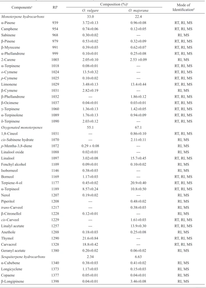
The yields of the essential oils of O. majorana
and O. vulgare were 17.2 and 22.0 g/kg, respectively (Table 1). It can be noted that Busatta et al. (2008) reported 1.20 % essential oils yield from O. majorana. However, variations in the essential oils content and profiles within a single species growing in different geographical locations are not uncommon. In the
Origanum oils, 43 compounds in all were identified (Table 1): a total of 39 individual compounds were identified from the essential oils of O. majorana
(96.4 % of the total oil), and 43 were identified from the essential oils for O. vulgare (92.9 % of the total oil). The retention indices, percentage composition and identification methods are presented in Table 1, where the compositions are listed in order of elusion from HP-5MS column. The major components in O. majorana essential oils were identified as terpiene-4-ol (20.9%), linaloterpiene-4-ol (15.7%), limonene (13.4%), linalyl
acetate (13.9%) and α-terpineol (10.8%), and the most
abundant constituents (>5%) in the essential oils of O. vulgare were found to be thymol (21.6%), carvacrol (18.8%), o-cymene (13.5%) and α-terpineol (8.57%).
Essential oils of these two Origanum species mainly consisted of oxygenated monoterpenes and monoterpene hydrocarbons. Terpiene-4-ol and thymol were the main oxygenated monoterpenes identified from the essential oils of O. majorana and O. vulgare, respectively. These findings were in good agreement with reports published previously (Vagi et al., 2005; Busatta et al., 2008), where the reported main constituent of O. majorana essential oils was terpinen-4-ol. The oils from O. vulgare, collected in Turkey, showed varied composition (Esen et al., 2007). According to Vagi et al. (2005), terpinen-4-ol was the main component of O. majorana essential oil, collected from Hungary, with contribution of 30.3%.
The antibacterial activity of the essential oils of
Componentsa RIb Composition (%) c
Mode of
Identiicationd
O. vulgare O. majorana
Monoterpene hydrocarbons 33.0 22.4
α-Pinene 939 3.72±0.13 0.96±0.08 RT, RI, MS
Camphene 954 0.74±0.06 0.12±0.05 RT, RI, MS
Sabinene 968 0.30±0.02 --- RI, MS
β-Pinene 979 0.53±0.02 0.32±0.09 RT, RI, MS
β-Myrecene 991 0.39±0.03 0.62±0.07 RT, RI, MS
α-Phellandrene 999 0.10±0.01 0.25±0.08 RT, RI, MS
2-Carene 1003 2.05±0.10 2.53 ±0.09 RI, MS
α-Terpinene 1018 0.08±0.01 --- RT, RI, MS
o-Cymene 1024 13.5±0.32 --- RT, RI, MS
p-Cymene 1025 0.10±0.02 --- RT, RI, MS
Limonene 1029 3.48±0.13 13.4±0.44 RT, RI, MS
β-Cymene 1031 2.82±0.19 --- RI, MS
β-Phellandrene 1032 --- 1.86±0.12 RT, RI, MS
β-Ocimene 1037 0.04±0.01 0.03±0.01 RT, RI, MS
γ-Terpinene 1060 1.36±0.13 1.42±0.05 RT, RI, MS
α-Terpinolene 1089 1.76±0.13 0.94±0.09 RT, RI, MS
δ-Terpinene 1090 2.03±0.12 --- RT, RI, MS
Oxygenated monoterpenes 55.1 67.1
1,8-Cineol 1031 --- 0.86±0.10 RT, RI, MS
cis-Sabinene hydrate 1070 --- 2.11±0.11 RI, MS
p-Mentha-3,8-diene 1072 0.29 ± 0.08 --- RI, MS
Linalool oxide 1088 0.02±0.01 --- RI, MS
Linalool 1097 3.02±0.08 15.7±0.45 RT, RI, MS
Fenchyl alcohol 1109 0.09±0.01 0.10±0.02 RI, MS
Isoborneol 1146 0.38±0.03 --- RI, MS
Borneol 1169 1.17±0.03 --- RT, RI, MS
Terpiene-4-ol 1177 0.45±0.02 20.9±0.40 RT, RI, MS
α-Terpineol 1189 8.57±0.24 10.8±0.50 RT, RI, MS
Nerol 1207 0.19±0.02 --- RI, MS
Piperitol 1208 --- 0.48±0.02 RI, MS
trans-Carveol 1217 --- 0.38±0.03 RI, MS
β-Citronellol 1228 0.12±0.01 --- RI, MS
cis-Carveol 1229 --- 1.61±0.03 RT, RI, MS
Linalyl acetate 1257 --- 13.9±0.30 RT, RI, MS
Anethole 1288 0.18±0.03 0.25±0.08 RI, MS
Thymol 1290 21.6±0.84 --- RT, RI, MS
Carvacrol 1328 18.8±0.42 --- RT, RI, MS
Geranyl acetate 1380 0.20±0.02 0.06±0.02 RI, MS
Sesquiterpene hydrocarbons 2.34 6.63
α-Cubebene 1340 0.38±0.03 0.41±0.02 RI, MS
Longicyclene 1373 1.17±0.03 0.15±0.03 RI, MS
Copaene 1377 0.05±0.01 0.04±0.01 RI, MS
β-Longipinene 1398 0.04±0.01 3.46±0.08 RI, MS
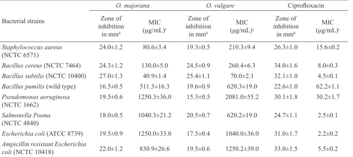
zones of inhibition (16.5-27.0 mm) and smaller MIC values (40.9-1250.3 µg/mL) against test bacterial strains. However, the antibacterial activity of the essential oils of
O. vulgare was also good (>15 mm zones of inhibition) with the zones of inhibition within the range of 15.3-25.4 mm and the MIC values in the range of 70.0-2081.1 µg/ mL. Both essential oils were active, although at relatively high concentrations, against ampicillin-resistant E. coli.
The greater resistance of Gram-negative bacteria to essential oils, as observed in this study (Table 2) might be owing to the great complexity of the double membrane-containing cell envelope of these bacterial in contrast to the single membrane structures of Gram-positive bacteria (Kalemba & Kunicka, 2003; Bagamboula et al., 2004).
Positive control, ciproloxacin, showed much better activity than essential oils as expected. The indings of
this study were in line with the antibacterial properties of several Origanum species reported previously (Bouchra et al., 2003; Bendahou et al., 2008; Ozcan & Chalchat, 2009; Al-Kalaldeh et al., 2010; Eng & Norman, 2010; Gonzalez & Marioili, 2010). Essential oils of Origanum species generally contain monoterpenes, carvacrol, thymol, terpinene-4-ol and linalool. The biological activity of these oils is often attributed to the occurrence of such bioactive compounds (Soylu et al., 2006; Esen et al., 2007).
There are many methods reported in
Longifolene 1409 0.07±0.02 --- RI, MS
β-Caryophyllene 1421 0.45±0.03 1.64±0.03 RT, RI, MS
Aromadendrene 1430 --- 0.07±0.02 RI, MS
α-Humulene 1450 0.08±0.02 0.09±0.02 RI, MS
β-Farnesene 1458 0.02±0.01 0.05±0.01 RI, MS
Alloaromadendrene 1461 --- 0.06±0.01 RI, MS
α-Selinene 1470 --- 0.06±0.02 RI, MS
ar-Curcumene 1475 0.08±0.01 0.03±0.01 RI, MS
Germacrene D 1480 --- 0.27±0.02 RI, MS
Valencene 1482 --- 0.18±0.02 RI, MS
α-Muurolene 1499 --- 0.06±0.02 RI, MS
α-Farnesene 1500 --- 0.06±0.01 RI, MS
Oxygenated sesquiterpenes 2.50 0.19
Caryophyllene oxide 1583 0.21±0.02 0.19±0.02 RT, RI, MS
Spathulenol 1586 0.14±0.02 --- RI, MS
Cadrol 1596 1.04±0.04 --- RI, MS
α-Cadinol 1654 1.11±0.02 --- RI, MS
Total 92.9 96.4
Yield (g/kg) 22.0±1.5 17.2±0.90
Table 1. (cont.)
aCompounds are listed in order of elution from a HP-5MS column; bRetention indices relative to C9-C24 n-alkanes on the HP-5MS column; cValues are mean±standard deviation of three different samples of essential oils from each Origanum species, analyzed individually in triplicate.
Mean followed by different superscript letters in the same row represent signiicant difference (p<0.05). Compounds present in trace amounts (<0.1%) were not registered; dRT, identiication based on retention time; RI, Identiication based on retention index; MS, identiication based on
comparison of mass spectra.
literature to evaluate the antimalarial activity of crude plant extracts (Kurosawa et al., 2000; Ncokazi & Egan, 2005; Huy et al., 2006; Busatta et al., 2008). Almost all of them use live malaria parasites and or parasite extracts. Although the tests give an indication of the direct effects of the agents against the parasites, specialist care is needed to grow the parasites and the evaluation process may be costly and time consuming. On the other hand, the haem biocrystallisation assay is less costly and easy to perform. It is based on observation that during the intraerythrocytic development, malaria parasites degrade large amounts of hemoglobin within a specialized organelle, the digestive vacuole (Goldberg & Slater, 1992) to supply amino acids for growth and development. A toxic by-product, ferriprotoporphyrin IX (FPIX), is produced during the
catabolism of hemoglobin. The parasite detoxiies this
FPIX through the formation of insoluble hemozoin crystals. Thus, agents that inhibit the conversion of FPIX to hemozoin may cause death from the lytic effects of haem or the haem drug complex may mediate parasite death (Egan & Marques, 1999). Moreover, in vitro, hematin at
acidic pH, leads to β-hematin, a compound presumed to
antimalarial activity of the Origanum essential oils. Of the two Origanum essential oils tested, the O. vulgare
essential oils showed anti-heme biocrystallization activity (Table 3). The O. vulgare essential oils at a concentration of 10 mg/mL exhibited 91.7% inhibition (IC50=0.04 mg/mL). However, O. majorana showed only 49.2% inhibition at the same concentration. The positive control chloroquine showed 90% inhibition at 0.005 mg/mL. The ability of any compound to inhibit the biocrystallization of ferriprotoporphyrin IX (FPIX) is believed to be directly connected to their antimalarial activity (Raynes et al., 1996).
The antioxidant activity of Origanum essential oils was assessed by different in-vitro tests. In the DPPH assay, the radical scavenging capacity of the tested essential oils increased in a concentration dependent manner. The values for 50% scavenging (IC50) are given in Table 3. The O. vulgare essential oils showed better radical scavenging activity (IC50: 65.5 µg/mL) than that of O. majorana essential oil (IC50: 89.2 µg/mL). However, the activity of both oils was less than that of the positive control, BHT (IC50: 9.9 µg/mL), a well known free radical scavenger. Table 3 also shows the level of percent inhibition of linoleic acid oxidation as exhibited by the tested essential oils. Both O. vulgare
and O. majorana essential oils depicted statistically similar inhibition, i.e., 77.3 and 72.8%, respectively. The activity was slightly less than that of the positive control BHT (90.9%). The antioxidant activity of the essential oils of O. vulgare and O. majorana was
also assessed qualitatively by bleaching β-carotene in
linoleic acid system assay. In this assay, the greater
Table 2. Antibacterial activity of O. majorana and O. vulgare essential oilsa.
aValues are mean±standard deviation of three samples of each Origanum species, analyzed individually in triplicate; bZone of inhibition in mm
including the disc diameter (6 mm); cMIC, minimal inhibitory concentration (µg mL-1).
Bacterial strains
O. majorana O. vulgare Ciproloxacin
Zone of inhibition
in mmb
MIC
(μg/mL)c
Zone of inhibition
in mmb
MIC
(μg/mL)c
Zone of inhibition
in mmb
MIC
(μg/mL)c
Staphylococcusaureus (NCTC 6571)
24.0±1.2 80.6±3.4 19.3±0.5 210.3±9.4 26.3±1.0 15.6±0.2
Bacillus cereus (NCTC 7464) 24.3±1.2 130.0±5.0 24.5±0.9 260.4±6.3 34.0±1.6 8.0±0.3
Bacillus subtilis (NCTC 10400) 27.0±1.3 40.9±1.4 25.4±1.1 70.0±2.1 32.1±1.0 4.5±0.1
Bacillus pumilis (wild type) 16.5±0.5 511.3±16.3 19.6±0.9 620.3±19.0 22.6±1.0 62.2±1.1
Pseudomonas aeruginosa
(NCTC 1662)
19.5±0.6 1250.3±36.0 15.3±0.5 2081.0±55.2 30.1±1.8 30.2±1.7
Salmonella Poona
(NCTC 4840)
18.0±0.5 1040.3±21.2 20.5±0.7 620.2±19.0 24.7±1.1 2.5±0.1
Escherichia coli (ATCC 8739) 19.5±0.9 1250.0±33.0 17.5±0.4 1040.0±36.0 31.0±1.7 2.2±0.2
Ampicillin resistant Escherichia
coli (NCTC 10418) 22.0±1.2 830.9±26.6 19.5±0.6 1250.2±39.0 33.0±1.5 5.5±0.2
the effectiveness of an antioxidant, the slower will be the color depletion, and less decrease in absorbance indicates a lower rate of oxidation of linoleic acid reflecting higher antioxidant activity. The O. vulgare
essential oil exhibited better antioxidant activity than
O. majorana oil. The control showed the highest rate of color depletion and thus least antioxidant activity.
Lipid peroxidation is an important issue in food industries and while comparing the present findings with the literature, not even a single report showing the percent inhibition of peroxidation by Origanum
essential oils could be found. However, there are some reports available in the recent literature on the radical scavenging activity of other Lamiaceae essential oils (Puertas-Mejiia et al., 2002; Ruberto et al., 2002; Hussain et al., 2008; Kelen & Tepe, 2008; Hussain et al., 2010). The antioxidant activity of essential oils may be attributed to the presence of phenolic compounds as was observed in Salvia officinalis (Lu & Foo, 2000). Singh & Marimuthu (2006) found that essential oils effectively suppress the peroxide formation in linoleic acid system during incubation.
Origanum essential oils showed prominent cytotoxicity against both the cancer cell lines. The cytotoxicity of O. majorana essential oil against MCF-7, LNCaP and NIH-3T3 cell lines in terms of IC50 values were 70.0, 85.3 and 300.5 µg/mL, respectively, and were notably stronger than those of O. vulgare essential oil (Table 3). Doxorubicin was used as the positive control in the case of cytotoxicity test against MCF-7 and LNCaP cell lines, and the IC50 values were 28.8 and 33.3 µg/mL, respectively. These essential oils were comparatively less toxic against fibroblast (NIH-3T3) cell line. The analysis of variance showed significant (p<0.05) variation in the toxicity of both essential oils tested. Generally, it is believed that the major bioactive components of essential oils determine their biological properties. According to published guidelines, the IC50<10 µg/mL represents potentially very toxic; IC50 10-100 µg/mL represents potentially toxic; IC50 100-1000 µg/mL represents potentially harmful and IC50>1000 µg/mL represents potentially non toxic (Gad-Shayne, 1999). There are only a few reports in literature on the cytotoxicity of some Lamiaceae essential oils (Sivropoulou et al., 1996; Sivropoulou et al., 1997; Legault et al., 2003; DeSousa et al., 2004).
Finally, the composition and profile of the essential oils of O. majorana and O. vulgare as observed in the present study were comparable but not exactly the same with other published data. The bioassay results demonstrated that the essential oils of both O. majorana and O. vulgare had antioxidant, antibacterial and cytotoxic potentials. However, only O. vulgare
essential oils exhibited potential antimalarial activity.
Acknowledgements
Grants supports by the Higher Education Commission, Islamabad, Pakistan under the
International Research Initiative Support Program and the Indigenous PhD Fellowship Program schemes are gratefully acknowledged.
References
Adams RP 2004. Identification of essential oil component by gas chromatography/quadrupole mass spectroscopy.
Illinois: Allured Publishing Corporation.
Al-Kalaldeh JZ, Abu-Dahab R, Afifi FU 2010. Volatile oil composition and antiproliferative activity of Laurus nobilis, Origanum syriacum, Origanum vulgare, and
Salvia triloba against human breast adenocarcinoma cells. Nutr Res 30: 271-278.
Anwar F, Ali M, Hussain AI, Shahid M 2009. Antioxidant and antimicrobial activities of essential oil and extracts of fennel (Foeniculun vulgare Mill.) seeds from Pakistan.
Flavour Frag J 24: 170-176.
Baelmans R, Deharo E, Bourdy G, Munõz V, Quenevo C, Sauvaind M, Ginsburg H 2000. A search for natural bioactive compounds in Bolivia through a multidisciplinary approach Part IV. Is a new haem polymerisation inhibition test pertinent for the detection of antimalarial natural products? J Ethnopharmacol 73: 271-275.
Bagamboula CF, Uyttendaele M, Debevere J 2004. Inhibitory effect of thyme and basil essential oils, carvacrol, thymol, estragol, linalool and p-cymene towards
Shigella sonnei and Shigella flexneri. Food Microbiol 21: 33-42.
Bendahou M, Muselli A, Grignon-Dubois M, Benyoucef M, Desjobert JM, Bernardini AF, Costa J 2008. Antimicrobial activity and chemical composition of
Origanum glandulosum Desf. essential oil and extract obtained by microwave extraction: comparison with hydrodistillation. Food Chem 106: 132-139.
Bouchra C, Achouri M, Hassani LMI, Hmamouchi M 2003. Chemical composition and antifungal activity of essential oils of seven Moroccan Labiatae against
Botrytis cinerea Pers. Fr. J Ethnopharmacol 89:
165-Table 3. Antimalarial, antioxidant and cytotoxic activities of O. majorana and O. vulgare essential oilsa
1Values are mean±standard deviation of three samples of each Origanum species, analyzed individually in triplicate. NT=not tested.
Materials
Heme biocyrstallization assay Antioxidant activity Cytotoxicity assay (IC50, μg/mL)
Inhibition (%) at
10 mg/mL (IC50, mg/mL)
DPPH, IC50, (µg mL-1)
Inhibition of linoleic acid peroxidation (%)
Breast cancer (MCF-7)
Prostate cancer (LNCaP)
Fibroblast (NIH-3T3)
Origanum
majorana 49.2±1.8 > 10.0 89.2±2.4 72.8±4.9 70.0±2.5 85.3±2.8 300.5±12.0
Origanum
vulgare 91.7±5.3 0.04±0.01 65.5±2.0 77.3±3.5 100.0±4.5 90.1±3.2 320.3±9.7
Chloroquine diphosphate
90% with
0.005 mg/mL <0.005 NT NT NT NT NT
BHT NT NT 9.9±0.2 90.9±2.7 NT NT NT
169.
Busatta C, Vidal RS, Popiolski AS, Mossi AJ, Dariva C, Rodrigues MRA, Corazza FC, Corazza MI, Oliveira JV, Cansian RI 2008. Application of Origanum majorana L. essential oil as an antimicrobial agent in sausage. Food Microbiol 25: 207-211.
Daferera DJ, Ziogas BN, Polissiou MG 2000. GC/MS analysis of essential oils from Greek aromatic plants and their fungitoxicity on Penicillum digitatum. J Agr Food Chem 48: 2576-2581.
De Sousa AC, Alviano DS, Blank AF, Alves PB, Alviano CS, Gattass CR 2004. Melissa officinalis L. essential oil: antitumoral and antioxidant activities. J Pharm Pharmacol 56: 677-681.
Egan TJ, Marques HM 1999. The role of haem in the activity of chloroquine and related antimalarial drugs. Coordin Chem Rev 192: 493-517.
Eng W, Norman R 2010. Development of an oregano-based ointment with anti-microbial activity including activity against methicillin-resistant Staphlococcus aureus. J Drugs Dermatol 9: 377-380.
Esen G, Azaz AD, Kurkcuoglu M, Baser KHC, Tinmaz A 2007. Essential oil and antimicrobial activity of wild and cultivated Origanum vulgare L. subsp. Hirtum
(Link) letswaart from the Marmara region, Turkey.
Flavour Frag J 22: 371-376.
Fitch CD, Cai GZ, Chen YF, Shoemaker JD 1999. Involvement of lipids in ferriprotoporphyrin IX polymerization in malaria. Biochim Biophys Acta 1454: 31-37.
Gad-Shayne C 1999. Alternatives to in vivo studies in toxicology. In Balantyne B, Marrs T, Syversen T (eds)
General and applied toxicology. Vol 1. USA: Grove’s Dictionari’s Inc., p. 178.
Goldberg DE, Slater A 1992. The pathway of haemoglobin degradation in malaria. Parasitol Today 8: 280-283. Gonzalez MJ, Marioli JM 2010. Antibacterial activity of
water extracts and essential oils of various aromatic plants against Paenibacillus larvae, the causative agent of American foulbrood. J Invertebr Pathol 104: 209-213.
GRIN Taxonomy Database 2010. USDA, ARS, National Genetic Resources. http://www.ars-grin.gov/cgi-bin/ npgs/html/tax_search.pl, accessed in June 2010. Huy NT, Uyen DT, Sasai M, Trang DT, Shiono T, Harada
S, Kamei K 2006. A simple and rapid colorimetric method to measure hemozoin crystal growth in vitro. Anal Biochem 354: 305-307.
Hussain AI, Anwar F, Sherazi STH, Przybylski R 2008. Chemical composition, antioxidant and antimicrobial activities of basil (Ocimum basilicum) essential oils depend on seasonal variations. Food Chem 108: 986-995.
Hussain AI, Anwar F, Shahid M, Ashraf M, Przybylski R 2010. Chemical composition, antioxidant and antimicrobial activities of essential oil of spearmint (Mentha spicata
L.) from Pakistan. J Essent Oil Res 22: 78-84. Kalemba D, Kunicka A. 2003. Antibacterial and antifungal
properties of essential oils. Curr Med Chem 10: 813-829.
Kelen M, Tepe B 2008. Chemical composition, antioxidant and antimicrobial properties of the essential oils of
three Salvia species from Turkey flora. Bioresource Technol 99:4096-4104.
Kurosawa Y, Dorn, A, Kitsuji-Shirane M, Shimada H, Satoh T, Matile H, Hofheinz W, Masciadri R, Kansy M, Ridley RG 2000. Hematin polymerization assay as a high-throughput screen for identification of new antimalarial pharmacophores. Antimicrob Agents Chem 44: 2638-2644.
Legault J, Dahl W, Debition E, Pichette A, Maldemont JC 2003. Antitumor activity of balsam fir oil: production
of reactive oxygen species induced by α-Humulene as
possible mechanism of action. Planta Med 69: 402-407.
Lu F, Foo LY 2001. Antioxidant activities of polyphenols from sage (Salvia officinalis). Food Chem 75: 197-202.
Massada Y 1976. Analysis of essential oils by gas chromatography and mass spectrometry. New York: John Wiley & Sons.
Mimica-Dukic N, Bozin B, Sokovic M, Mihajlovic B, Matavulj M 2003. Antimicrobial and antioxidant activities of three Mentha species essential oils.
Planta Med 69: 413-419.
Mosmann T 1983. Rapid colorimetric assay for cellular growth and survival - application to proliferation and cytotoxicity assays. J Immunol Methods 65: 55-63. Ncokazi KK, Egan TJ 2005. A colorimetric high-throughput
betahematin inhibition screening assay for use in the search for antimalarial compounds. Anal Biochem 338: 306-319.
Ozcan MM, Chalchat JC 2009. Chemical composition and antimicrobial properties of the essential oil of
Orgiganum saccatum L. J Food Safety 29: 617-628. Puertas-Mejia M, Hillebrand S, Stashenko E, Winterhalter P
2002. In vitro radical scavenging activity of essential oils of Columbian plants and fractions from oregano (Origanum vulgare L) essential oil. Flavour Frag J 17: 380-384.
Raynes K, Foley M, Tilley L, Deady LW 1996. Novel bisquinoline antimalarials - synthesis, antimalarial activity, and inhibition of haem polymerization.
Biochem Pharmacol 52: 551-559.
Ruberto G, Barrata MT, Sari M, Kaabexhe M 2002. Chemical composition and antioxidant activity of essential oils from Algerian Origanum glandulosum Desf. Flavour Frag J 17: 251-254.
Sarker SD, Nahar L, Kumarasamy Y 2007. Microtitre plate-based antibacterial assay incorporating resazurin as an indicator of cell growth, and its application in the
in vitro antibacterial screening of phytochemicals.
Methods 42: 321-324.
Singh G, Marimuthu P 2006. Antioxidant and biocidal activities of Carum nigrum (seed) essential oil, oleoresin, and their selected components. J Agr Food Chem 54: 174-181.
Sivropoulou A, Nikolaou C, Papanikolaou E, Kokkini S, Lanaras T, Arsenakis M 1997. Antimicrobial, cytotoxic, and antiviral activities of Salvia fruticosa
essential oil. J Agr Food Chem 45: 3197-3201. Sivropoulou A, Papanikolaou E, Nikolaou C, Kokkini S,
cytotoxic activities of Origanum essential oils. J Agr Food Chem 44: 1202-1205.
Soylu EM, Soylu S, Kurt S 2006. Antimicrobial activity of the essential oils of various plants against tomato late blight disease agent Phytophthora infestans. Mycopathologia 161: 119-128.
Tripathi AK, Khan SI, Walker LA, Tekwani BL 2004. Spectrophotometric determination of the novo
hemozoin/β-hematin formation in an in vitro assay.
Anal Biochem 325: 85-91.
Vagi E, Simandi B, Suhajda A, Hethelyi E 2005. Essential oil composition and antimicrobial activity of Origanum majorana L. extracts obtained with ethyl alcohol and supercritical carbon dioxide. Food Res Int 38: 51-57.
Vagionas K, Graikou K, Ngassapa O, Runyoro D, Chinou I 2007. Composition and antimicrobial activity of the essential oils of three Satureja species growing in Tanzania. Food Chem 103: 319-324.
*Correspondence
Satyajit D. Sarker
Department of Pharmacy, School of Applied Sciences, University of Wolverhampton
MM Building, Molineux Street, Wolverhampton WV1 1SB, West Midland, UK