VII
Radiol Bras. 2008 Set/Out;41(5):VII–VIII
Renato Tavares Daher1
, Sérgio Daher2
, Murilo Tavares Daher3
, Ricardo Tavares Daher4
, Marcelo Eus-táquio Montandon Júnior5
, Cristiano Montandon5
Study developed at the Spine Unit of Hospital de Acidentados de Goiânia and at Clínica Multimagem, Goiânia, GO, Brazil. 1. MD, Resident in Radiology and Imaging Diagnosis at Hospital das Clínicas, Universidade Federal de Goiás (HC-UFG), Goiânia, GO, Brazil. 2. MD, Orthopedist, Head for the Unit of Vertebral Column Surgery, Hospital das Clínicas of Universidade Federal de Goiás (HC-UFG) and Hospital de Acidentados de Goiânia, Goiânia, GO, Brazil. 3. MD, Orthopedist, Trainee at the Spine Unit – Universidade Estadual de Campinas (Unicamp), Campinas, SP, and Associação de Assistência à Criança Deficiente (AACD), São Paulo, SP, Brazil. 4. Graduate Student of Medicine, Escola Superior de Ciências da Saúde do Distrito Federal (ESCS-DF), Brasília, DF, Brazil. 5. Titular Members of Colégio Brasileiro de Radiologia e Diagnóstico por Imagem (CBR) (Brazilian College of Radiology and Imaging Diagnosis), MDs, Radiologists at Clínicas da Imagem e Multimagem de Goiânia, Goiânia, GO, Brazil. E-mail: [email protected]
0100-3984 © Colégio Brasileiro de Radiologia e Diagnóstico por Imagem
Which is your diagnosis?
•
Qual o seu diagnóstico?
Daher RT, Daher S, Daher MT, Daher RT, Montandon Jr ME, Montandon C. Which is your diagnosis? Radiol Bras. 2008;41(5):VII–VIII.
A female, Caucasian, 76-year-old patient reported the onset, approximately three weeks ago, of an intense, diffuse lumbar pain, irradiating over the lower limbs up to the knees. The pain did not decrease with rest and presented only a mild improvement with analgesic and non-hormonal anti-inflammatory drugs. The patient reported a previous history of rectal neoplasm treated with surgery and radiotherapy six months ago. At clinical examination, the patient presented pain at palpation and movement of the lumbar region, negative Lasègue’s sign, with no motor or sensitivity deficit. Plain radiography (unshown), computed tomography (Figure 1) magnetic resonance imaging (Figures 2, 3 and 4) and, subsequently, open biopsy and histology of the sacrum were performed.
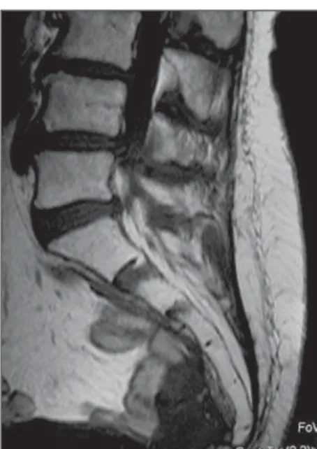
Figure 1. Computed tomography – axial view. Figure 2. Magnetic resonance imaging – sagit-tal, T1-weighted image.
VIII Radiol Bras. 2008 Set/Out;41(5):VII–VIII Images description
Figure 1. Computed tomography – axial view demonstrating anterior cortical fracture of the left sacral wing associated with osteocondensation with adjacent cor-tical thickening.
Figure 2. Magnetic resonance imaging – sagittal, T1-weighted image demonstrat-ing small fracture traces characterized by hypointense signal subjacent to disk spaces S1-S2 and S2-S3.
Figure 3. Magnetic resonance imaging – axial T2-weighted image demonstrating fracture traces on the spongeous bone of the sacral wings, parallel to the sacroiliacal joints associated with adjacent medullary bone edema.
Figure 4. Magnetic resonance imaging – coronal T1-weighted fat-saturated image acquired after intravenous contrast injec-tion. Note the intense, asymmetrical con-trast uptake in the sacral bone marrow be-side the fracture traces, most noticeable at left, additionally to another horizontal com-ponent subjacent to the disk S1-S2, char-acterizing the typical H-shaped fracture.
Diagnosis: Sacral insufficiency frac-tures.
COMMENTS
Stress fractures affect patients with no history of trauma, and are classified into subgroups as follows: fatigue fractures – in a normal bone submitted to a repetitive ef-fort; or insufficiency fracture – occurring during normal stress on a generally osteo-penic bone, likewise in the present case(1).
Firstly described in 1982 by Lourie, sacral insufficiency fracture is a frequent complication of osteoporosis, affecting 2% to 4% of caucasian women with > 60 years, constituting a relevant cause for pelvic and/ or lumbar pain(2). Postmenopausal
os-teoporotic women submitted to pelvic ra-diotherapy are even more prone to this complication, like in the present case, de-spite the absence of actinic alterations as demonstrated by biopsy(3). The load exerted
by the body over the sacrum through the spine, associated the mechanical strength transferred by the deambulation to the sacrum through the sacroiliacal joints, cause bilateral fractures on the sacral wings, parallel to these joints, and a third fracture on the same bone interconnecting them and forming the typical H-shaped fracture(4). Hip fractures, particularly in the
pubic branches, are frequently associated, which has not been observed in the present case.
Main clinical characteristics are pelvic and lumbar pain, impaired deambulation and, rarely, neurological and medullary compression symptoms(2,3), likewise in the
case presently described.
The diagnostic suspicion is based on an association of clinical data, physical exami-nation and imaging findings. Plain radiog-raphy of the pelvis can hardly demonstrate the fracture trace. The most frequent radio-graphic finding is osteopenia(3). The most
easily noticeable tomographic findings are radiolucent lines and sclerotic bands on the sacral wings, besides anterior cortical frac-tures of the sacrum, typically found in these cases(1,2,5). Bone scintigraphy demonstrates
the H-shaped radiopharmaceutical uptake. Magnetic resonance imaging presents 100% sensitivity and 83% specificity(3),
with T1-weighted sequences demonstrat-ing hypointense signal on bilateral fracture traces parallel to the sacroiliacal joints, and a third perpendicular line interconnecting them, forming a H-shaped fracture(5,6).
T2-weighted sequences demonstrate medul-lary edema, which may hide the fracture trace early in the first months(2,3,5).
Para-magnetic contrast-enhanced T1-weighted fat-suppressed sequences have shown to be more sensitive for identifying fracture traces(1,3,5).The evaluation of coronal,
ob-lique images of the sacrum provide useful information for the diagnosis.
In patients with a known neoplasm, sec-ondary implant is the primary differential diagnosis to be initially considered, since disabling pain is also a typical symptom of this entity. Typically in these cases, multi-focal, infiltrative solid lesions can be ob-served affecting adjacent soft tissues, dif-ferently from sacral insufficiency frac-tures(5).
FINAL CONSIDERATIONS
With the increased longevity and the consequent prevalence of osteoporosis, particularly amongst women, sacral insuf-ficiency fractures tend to become an in-creasingly frequent diagnosis. In the present report, the authors demonstrate the relevance of recognition of the typical im-aging findings by radiologists to avoid unnecessary invasive procedures.
REFERENCES
1. Keogh C, Torreggiani WC, Al-Ismail K. Muscu-loskeletal case 21. Insufficiency fracture of the sacrum. Can J Surg. 2002;45:92, 153.
2. Grangier C, Garcia J, Howarth NR, et al. Role of MRI in diagnosis of insufficiency fractures of the sacrum and acetabular roof. Skeletal Radiol. 1997;26:517–24.
3. Peh WC, Khong PL, Yin Y, et al. Imaging of pel-vic insufficiency fractures. Radiographics. 1996; 16:335–48.
4. Mammone JF, Schweitzer ME. MRI of occult sac-ral insufficiency fractures following radiotherapy. Skeletal Radiol. 1995;24:101–4.
5. Blake SP, Connors AM. Sacral insufficiency frac-ture. Br J Radiol. 2004;77:891–6.