Arq Neuropsiquiatr 2009;67(4):1023-1028
CliniCal behavior of Streptococcus
pneumoniae meningoenCephalitis
Raisa Bu-Coiiu Fanego
1, Alberto J. Dorta-Contreras
1, Bárbara Padilla-Docal
1,
Martha O’ Farril-Sanchez
2, Isabel Lopez-Hernandez
2abstract – Objective: There was an increased number of cases of meningoencephalitis caused by Streptococcus pneumoniae, after the successful vaccination campaigns against Neisseria meningitidis and Haemophilus influenzae. This paper aims at describing the clinical characteristics, the laboratory findings, the complications, and the therapeutic management of these patients, who have been suffering from this disease since 1993 to 2006. Method: Twelve children with Streptococcus pneumoniae meningoencephalitis admitted to the pediatric hospital of San Miguel del Padron, City of Havana in this period were assessed. Results: Children under one year are the most frequently affected. Septic shock and brain edema were the most severe complications. Three patients died, implying that this disease has a serious course. Early treatment of brain edema is very important to reduce mortality. The elective drugs for treatment of these cases of Streptococcus pneumoniae meningoencephalitis were vancomycin combined with cephalosporin, cefotaxime or ceftriaxone type. Conclusion: Patients with Streptococcus pneumoniae meningoencephalitis show clinical characteristics, complications, and sequels that are different to other bacterial meningoencephalitis, meaning that they could be helpful for physicians considering the differential diagnosis of meningoencephalitis.
KEY WORDS: streptococcus pneumoniae, meningoencephalitis, treatment.
Comportamiento clinico y terapéutico de la meningoencefalitis por streptococcus pneumoniae
resumo – Objetivo: Existe un incremento de la meningoencefalitis producida por Streptococcus pneumoniae, después de las campañas exitosas de vacunación contra Neisseria meningitidis y Haemophilus influenzae. El objetivo de este trabajo es describir las caracteristicas clinicas, los hallazgos de laboratorio, las complicaciones y el manejo terapéutico de los pacientes que sufrieron esta enfermedad desde 1993 a 2006. Método: Se estudiaron doce niños con meningoencefalitis por Streptococcus pneumoniae ingresados en el Hospital Pediátrico de San Miguel del Padrón, Ciudad de La Habana en este periodo. Resultados: Los niños menores de un año son los más frecuentemente afectados. El shock séptico y el edema cerebral las mayores complicaciones. Tres pacientes fallecieron. Esta enfermedad ha tenido un curso serio. El tratamiento temprano del edema cerebral es muy importante para reducir la mortalidad. Los medicamentos de elección para tratar la meningoencefalitis por
Strepcococcus pneumoniae en los casos estudiados fueron la vancomicina combinada con cefalosporina del tipo de la cefatoxima o la ceftriaxona. Conclusion: Los pacientes con meningoencefalitis por Streptoccocus pneumoniae exhibieron características clínica, complicaciones y secuelas las cuales se diferencian de otras meningoencefalitis bacterianas. Por eso estos elementos pueden ayudar a los médicos en el diagnóstico diferencial PALAVRAS-CLAVE: streptococcus pneumoniae, meningoencefalitis, tratamiento.
1Central Laboratory for Cerebrospinal Fluid (LABCEL). Medical School “Dr. Miguel Enríquez” Ramón Pintó 202, Luyanó. Apartado 10049 CP 11000
Ciu-dad Habana, Cuba; 2Pediatric Hospital San Miguel del Padrón. Calzada de San Miguel del Padrón 1615 entre Balear y Santa Francisca. CP 11000 Ciudad
Habana, Cuba.
Received 22 July 2009, received in inal form 23 July 2009. Accepted 4 August 2009.
Dr. Alberto Juan Dorta-Contreras – Laboratorio Central de Líquido Cefalorraquídeo (LABCEL) / Facultad de Ciencias Médicas “Dr. Miguel Enríquez” - Ramón Pintó 202, Luyanó / Apartado 10049 - CP 11000 Ciudad Habana - Cuba. E-mail: [email protected]
Pneumococcus causes the death of 18 000 children × 105 inhabitants annually, meaning two children per hour, and it is responsible for four important disease, otitis,
cases of acute otitis that may lead to deafness, 330,000 pneumonia cases, 1,200 cases of pneumococcal sepsis and 3,900 pneumonia cases. Streptococcus pneumoniae causes more than 1 million of deaths in children worldwide annu-ally, 90% of them occurring in developing countries2.
Lately, Neisseria meningitidis and Haemophilus inlu-enzae meningoencephalitis cases have dropped drastical-ly due to the successful vaccination campaign carried out in Cuba by the national health system. As a result, Strep-tococcus pneumoniae has taken the position of the pre-viously mentioned germs3.
Streptococcus pneumoniae affects mainly children un-der one year of age, specially new-born infants. However both the average age and the age range for this infection are increasing4. Thus, it is necessary to produce a vaccine with the most frequent serotypes of this bacteria in Cuba. We describe clinical characteristics, laboratory ind-ings, complications, therapeutic management and sequels found in a group of patients affected by the disease from 1993 to 2006.
method
A descriptive, transversal and prospective study was per-formed through direct observation of patients since the time of their irst lumbar puncture.
Sick children form peripheral municipalities, comprising both urban and suburban areas, were consulted in this hospital. Patients enrolled in this study were diagnosed with Streptococ-cus pneumoniae meningoencephalitis, its biological agent de-tected at the microbiology laboratory of the pediatric hospital of San Miguel del Padrón from 1993 to 2006. Twelve children ad-mitted with the presumptive diagnosis of bacterial meningoen-cephalitis were studied.
Patients were admitted in the Intensive Care Unit of the hos-pital. The sera and CSF samples were simultaneously collected at the moment of admission when the symptoms started. The anti-biotic and symptomatic treatment was initiated immediately af-ter, according to the clinical characteristics of each patient. Sam-ples were divided in aliquots; subsequently they were properly labeled and kept at –80oC for further use. The laboratory
vari-ables were processed, according to international standards. Universally accepted routine methods were used for the chemical study. Cellular and differential counting of CSF was expressed according to the number of cells × 10–6/L, and in
per-centage (respectively). Normal cellular counting for children was considered to be up to 20 cells × 10–6/L. The predominance of
cells does not deine etiology in bacterial meningoencephalitis.
Latex agglutination test
A modiied latex agglutination test was employed for the rapid detection of Streptococcus pneumoniae made by Bio Merieux (Slidex Meningite Kit). It contains sensitizing latex par-ticles with capsular antigens of S. pneumoniae (83 serotypes), A
and C serogroup of N. meningitidis and type b H. inluenzae. The latex agglutination test is rapid and simple to perform, yielding
S. pneumoniae data directly by testing of CSF. The sensibility for
S. pneumoniae is 0.1–0.5 {Jg/mL).
Microbiological cultures and bacterial identiication Isolation of pneumococci by inoculation of CSF and blood into solid culture media is very eficient if the patients had not been previously treated before with antibiotics. It allows for de-termination of the presence of S. pneumoniae in the initial ma-terial. The appearance and the size of pneumococcal colonies depend upon the composition of the culture medium, the bio-logical properties and the amount of S. pneumoniae and other microorganisms in the material undergoing the assessment. The identiication of pneumococci is based on their cultural and mor-phological properties, and it is carried out by a variety of tests.
S. pneumoniae is a Gram positive capsulated cocci. Its shape is of the lanceolate type, and it measures 0.5 to 1.2 µm, appear-ing in pairs or diplococci. They are facultative anaerobics, requir-ing protein and hematologic supplements.
CSF and blood samples were cultured in Oxoid chocolate agar in an environment containing 8–10% CO2. In this type of culture, the bacteria grows as non pigmented round colonies of 1–3 mm diameter. After 48 hours in the cultures, it takes an um-bilical like shape due to a progressive cellular autolysis, and the colonies usually have a greenish ring. To characterize the strain of the colony, different biochemical tests were used. Bacteriol-ysis with bile salts was used for the phenotypic specie identii-cation. The optoquine susceptibility also was employed by cul-ture of blood agar, by putting an Oxoid disk with optoquine on the surface of culture in order to measure the inhibition ring. If the ring became soluble in presence of bile salts, it is a determi-nant that the strain is indeed S. pneumoniae.
results
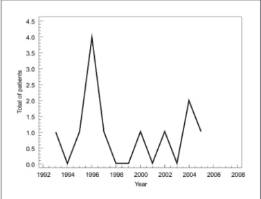
The incidence of meningoencephalitis by Streptococ-cus pneumoniae is shown in Fig 1.
The microorganism grew in all studied cases and Gram staining showed Gram-positive lanceolate diplococci.
Bacterial meningitis was suspected by cytological ex-amination of CSF. Latex agglutination test was done di-rectly on untreated CSF samples, showing positive reac-tions for S. pneumoniae
The etiology was deinitively established by culture and by smear examination.
Gram-positive coccal bacteria were demonstrated in chocolate agar culture.
Morphological appearance of bacteria using optical microscopy and positive detection for biochemical tests suggested the infection with Streptococcus pneumoniae.
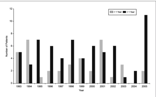
The high incidence of bacterial meningoencephalitis cases of non-determined cause is shown in Fig 2.
More frequent signs and symptoms are shown in Table 1. Fig 2. Incidence of unknown etiology meningoencephalitis.
Table. Streptococcus pneumoniae meningoencephalitis. Clinical and laboratory characteristics.
Characteristics
Average time of incubation Range
4.25 days 2–6 days Symptoms and signs
ARI Vomiting Headache Fever
Enlarged fontanel Irritability Somnolence Food rejection
No. patients 12
5 1 12
5 5 5 6
% 100
41 8 100
41 41 41 50 Laboratory
Average differential cell count CSF (%) diagnostic lumbar puncture
Polymorphonuclear cells=84.7 Lymphocytes=36.7
Average total cells in CSF 653 cells × 10–6L
CSF protein Increased in 100% of patients
CSF/serum glucose Index 50% decreased of all patients
Average globular sedimentation rate 92.7
Average differential cell count in blood Polymorphonuclear cells=65
All patients showed brain edema and 40% had a con-vulsive status. Septic shock and multi organ failure affect-ed fourth of the patients.
Computed tomography was performed in two pa-tients, showing diffused cortical atrophy and ventriculo-megaly. Cerebral edema was observed in both cases.
There were three cases of death (25%), two of them un-der one year of age, the other child in the group 1–4 years. Vancomycin was the drug of choice, in combination with cephalosporin, cefotaxime or ceftriaxone to treat Strep-tococcus pneumoniae meningoencephalitis in these cases. Short-term sequels were brain paralysis in two of the 12 patients (16%).
disCussion
Low incidence of Streptococcus pneumoniae may lead to the mistaken belief that this microorganism is not a pub-lic health problem in our area. However, sometimes there is no bacterial growth due to the early treatment with an-tibiotics these patients, who receive these drugs when at-tending the primary care units. Therefore, the real number of affected patients due this bacterium may be higher5,6.
In fact, the etiological diagnosis of 106 patients was not possible, meaning that there might be a hidden in-cidence of Streptococcus pneumoniae that could not be veriied.
Fast diagnostic methods are an alternative to improve the diagnosis since they do not need the germ to be in-tact, since they can recognize rests of the microorganism. For example, latex agglutination tests or polymerase chain reaction (PCR), can be used for faster diagnosis7.
All of the patients in this study had respiratory infec-tions prior to admission. Streptococcus pneumoniae is an airborne bacterium, carrying a high spreading risk due to the little drops of < 10 µm that remain suspended for more than 30 min. These minute drops may easily reach the alve-olus, carrying the risk of infection in the lung parenchyma. Its polysaccharide capsule allows it to avoid phago-cytosis, creating an invasive risk that may occur frequent-ly in children, especialfrequent-ly in synergy with other viral respi-ratory diseases. Respirespi-ratory viruses have a cytopathic ef-fect on the cilium of the respiratory mucosa either by destroying it or altering its genetic code. The alteration of the barrier mechanisms in this situation favors the in-crease of Streptococcus pneumoniae previously inoculat-ed in colonizinoculat-ed children. Children with tubaric dysfunc-tion or respiratory allergy are at a higher risk of suffer-ing acute otitis media or sinusitis. Infants from develop-ing countries are colonized earlier with higher rates of na-sopharyngeal carriers. Streptococcus pneumoniae carries an extreme risk of invasive infections in immunocompro-mised hosts, including children with functional or surgi-cal asplenia, AIDS or any other severe
immunocompro-mised disease: diabetes, nephritis, cardiopathy or chron-ic respiratory syndrome8.
The main sign was fever in all cases, with weakness, food rejection or irritability. Enlarged fontanel was pres-ent in children under one year of age. Studies conduct-ed in Spain found that 93.4% of children had fever equal to or higher than 37.5oC in the moment o admission, the mean duration was 3.25±3.71 days (mean 2 days) and the most frequent presentation forms were hidden bacter-emia (45.6%), bacteremic pneumonia (27.5%), meningitis (14.6%) and bacteremic otitis (9.4%). Bacteremic cellulitis, arthritis and mastoiditis were also present (5.9%)9. Som-nolence and vomits were other reported signs. There was no variation in the clinical picture of our cases in relation to those of Mexico, Brazil and Argentina9,10.
Regarding the laboratory tests, the cytochemical study showed a high number of cells in the CSF, as it was expected. This demonstrates an acute inlammatory pro-cess. Polymorphonuclear leukocytes were predominant in these patients.
All the results of the cytochemical study inferred a bacterial process in the affected children. These results match the ones found for other bacteria that affect the central nervous system. The behavioral symptoms and signs of these children were similar to those of other chil-dren abroad.
For example, in a study performed in Bogotá, Colom-bia, children with Streptococcus pneumoniae meningoen-cephalitis had similar characteristics11.
The microorganism grew in all assessed cases, and Gram staining showed Gram-positive lanceolate diplo-cocci. Gram staining method is very useful to establish a treatment for the etiological agent found. This method has a sensitivity of 60–90%12.
One of the characteristics of the disease is that pa-tients are in very serious condition. The main complica-tions were brain edema, convulsive status, acid basic un-balance, septic shock and multi organ failure.
The most serious complications observed in a study conducted in the Basque Country were those related to the respiratory tract (20.8%), pleural effusion (7.5%), at-electasis (2.0%) and pachypleuritis (2.0%). Thoracoscopy were performed to two patients13.
Since brain edema was present in all patients, quantity and quality of liquids will depend on different factors: in-tensity of brain edema, status of the tissue perfusion, im-paired capillary permeability, volemia and the existence of concomitant complications14.
sequels. Therefore, its use is recommended to detect in-tracranial complications15.
When the presence of infarction or paralysis of the cranial nerves, as well as hydrocephaly was detected by ultrasound or any other imaging technique at the moment of admission, sequels are usually expected. This may help identifying children that might need further follow-up and rehabilitation.
We considered that there is a high lethality, due to the outcome of 25% deaths.
According to a report of the Pan-American Health Or-ganization (PAHO) in 1999, 72,000 children under 5 years of age had died due to acute respiratory infections (ARI) in Latin America, 80% of them due to pneumonia, 50% of which were caused by Streptococcus pneumoniae, on the bases on previous data. This means that 29,000 children might have died due to Streptococcus pneumoniae16.
A total of 800 children died of ARI in Argentina, ac-cording to reports of the Ministry Of health; but this num-ber might be higher considering sub registry of cases and home deaths, which are common in all countries17.
The World Health Organization (WHO) and PAHO con-sider the following as risk factors: overcrowding, deicit of speciic vaccines, lack of breastfeeding, low weight at birth, malnutrition, barriers to access medical care and, in some regions, deicit of vitamin A18.
In Cuba there are no barriers to access medical care, pediatric hospitals are always ready to admit children when there is suspicion or sign of meningoencephalitis.
In general, the treatment to these patients comprised clinical support, mechanic ventilation, correction of the acid basic unbalance, of the brain edema and of the con-vulsions. Antibiotics were also used.
The use of inotropics was very important for the he-modynamic stabilization of the patients and they were in-dicated when the volume provided suggested an adequate volemia, despite of the vasodilatation they produce19.
Streptococcus pneumonia resistant-strains due to the use of antibiotics have been reported worldwide and they make the treatment dificult, leading to the increase mor-tality in many places20.
Some aspects should be taken into account when se-lecting an antibiotic to treat meningitis caused by Strep-tococcus pneumoniae: (1) The management of an infection in a system with low phagocytosis against a high inocu-lum (>106). (2) A bactericide drug with good penetration through the meninges should be selected. (3) Special im-portance should be given to the time the drug remains in the CSF at a concentration superior to 10 times of the min-imum bactericide concentration for the causative agent. The latter is achieved with third generation cepha-losporin (cefotaxime and ceftriaxone) at high doses. Pen-icillin-resistant Pneumococcus is increasing worldwide.
Sometimes this Pneumococcus resistance is multiple, and includes other antibiotics like tetracycline, trimethoprim sulfamethoxazole, and chloramphenicol. These infec-tions are more frequently found in the serotypes affecting children21.
So far, cefotaxime and ceftriaxone are the drugs of choice, though some failures in this treatment have sug-gested the inclusion of vancomycin. This is the current recommendation for antimicrobial treatment, mainly in areas where there is a prevalence of highly-resistant
Pneumococcus.
Though diminished sensitiveness to cephalosporin has not been reported in vitro in Cuba, the unfavorable
in vivo response to these antibiotics was quite important to take into account regarding this decision22. The most frequent antibiotics used were rocephin (ceftriaxone) and vancomycin.
Since there have been no isolation of vancomycin-re-sistant strains, this is the drug of irst choice, combined with a cephalosporin either cefotaxime or ceftriaxone to manage meningitis caused by third- generation- ce-phalosporin- resistant pneumococci. Similar to cepha-losporin and carbapenem, the effectiveness of vancomy-cin is based on the percentage of time in which its centration in CSF surpasses the minimum bactericide con-centration, so the administration intervals are more fre-quent, form 6 to 12 hours23.
The recent report of Streptococcus pneumoniae toler-ant to vancomycin, there has been a demand to be alert and to make a rational use of this antibiotic24.
The impact of the introduction of this antibiotic will not replace the early clinical diagnosis of bacterial men-ingitis at initial stages of inlammatory response, and the time for adopting measures to improve hemodynamic conditions.
Computed tomography was performed in patients with cerebral paralysis. In developing countries, access to these diagnostic techniques is important for early detec-tion of the sequels of the disease because image indings correlate statistically with neurological signs25.
In a study carried out in India, sequels were observed in 40% of patients with meningoencephalitis, 10% with mi-nor sequels, while the remaining presented more severe sequels. Sequels were observed in neurodevelopment, in cranial paralysis, convulsions and deep hyporelexia26.
The usefulness of computed tomography to foretell sequels was demonstrated in a study carried out by Tunc-er et al.27.
As previously explained, patients with this disease are in very serious condition, thus the early treatment of brain edema is important to reduce mortality.
complications, and sequels which were different to oth-er bactoth-erial meningoencephalitis, this being a motive to help physicians in the differential diagnosis.
referenCes
1. Ruvinski RO. Streptococcus pneumoniae: epidemiología y resistencia a antimicrobianos de las enfermedades invasoras en Latinoamérica. Rev Chil Infectol 2001;18:45-48.
2. Advisory Committee on Immunization Practices (ACIP): Recommen-dations of the ACIP. MMWR 1997:46.
3. Dickinson F, Pérez A, Galindo MA, Quintana I. Impacto de la vacu-nación contra Haemophilus inluenzae tipo b en Cuba. Rev Panam Salud Pública 2001;34:169-174.
4. Nicolosi L, Langella L, Krzysztoiak A, Ticca F. Streptococcus
pneumoni-ae meningitis in children: case records 1985–2003. Infect Med 2004; 12: 252-258.
5. Baudner S, Bienvenu J, Blirup JS, et al. The certiication of a matriz ref
-erence material for immunochemical measurement of 14 human serum proteins. CMR 470. Brussels: Community Bureau of Reference, Com-mission of the European Communities. BCR Information, Reference Materials. (report UR 15243); 1993.
6. Vlug A,van Remortel A. The structure and function of human IGg sub
-clases. Eur Clin Lab 1989;8:26-35.
7. Viswanath G, Praveen M, Hanumanthappa AR, Chandrappa NR, Ma-hesh CB. Bacteriological study of pyogenic meningitis with special ref-erence to latex agglutination. Indian J Pathol Microbiol 2007;50:97-100. 8. Singhi S. Fluid restriction and intracraneal pressure during bacterial
meningitis: to restrict luid or not. (Abstract). 10th International
Con-gress on Infectious Diseases. Singapore; 2002:130.
9. Otero MC, Pérez D, Asensi F. Meningitis bacterianas. In: Protocolos
di-agnósticos y terapéuticos en Pediatría. Tomo 2. Infectología. Madrid:
Asociación Española de Pediatría; 2001:149-155.
10. Brockmann VP, Ibarra GX, Silva WI, Hirsch BT. Etiología de la iebre agu
-da sin causa en niños consultados en un departamento de emergencia. Rev Chilena Infectol 2007:24:33-39.
11. Tique V, Alvis N, Parodi R, Bustos A, Mattar S. Meningitis aguda en Cór
-doba, Colombia (2002-2004). Rev Salud Pública (Bogotá) 2006;8:33-46. 12. Guarner J, Packard MM, Nolte KB, et al. Usefulness of
immunohis-tochemical diagnosis of Streptococcus pneumoniae in formalin-ixed, parafin-embedded specimens compared with culture and gram stain techniques. Am J Clin Pathol 2007;127:612-618.
13. Bernaola E, Aristegui J de M. Estudio de la incidencia de enfermedad neumocócica invasora entre 0–5 años en el País Vasco y Navarra. An Esp Pediatr 2002;57:301-309.
14. Benítez Bermejo RI, García Deltoro M, Vicente Mas J, Ballester Belda JE. Lesiones cerebrales en pacientes diagnosticados con meningitis. Neu-rología 2006;21:34-36.
15. Tuncer O, Caksen H, Arslan S, et al. Cranial computed tomography in
purulent meningitis of childhood. Int J Neurosci 2004;114:167-174. 16. Kertesz DA, Di Fabio JL, Brandileone MCC, et al. Invasive
Streptococ-cus pneumoniae infection in Latin American children: results of the Pan American Health Organization Surveillance Study. Clin Infect Dis 1998;26:1355-1361.
17. Barboza AG, Ioli PL, Zamarbide I, Estragó MI, Castiñeiras F, Wouters L. Estudio de incidencia y análisis descriptivo de la meningitis bacte-riana primaria no tuberculosa del adulto en una población argentina. Rev Neurol 2002;35:508-512
18. Noris García E, Dorta Contreras A, Noris García J L. Evaluación
inmu-nológica y tratamiento en pacientes pediátricos con déicit de IgA.Al
-ergol Inmunol Clin 2002;17:180-184.
19. Desse JE. Meningitis bacteriana: avances en el diagnóstico y tratamien-to. II Congreso Virtual Iberoamericano de Neurología, 1999.
20. Alvez F. Meningitis bacteriana. In: Castro Gago M (Ed). Tratamiento de
las enfermedades neurológicas en niños y adolescents. Barcelona: Ed Espaxs 1999;2:119-125.
21. Goldwater PN. Cefotaxime and ceftriaxone cerebrospinal luid levels
during treatment of bacterial meningitis in children.Int J Antimicrob Agents 2005;26:408-411.
22. Luján Padrón A, Hernández Hernández R. Neumococo. resistencia bac-teriana a los antibióticos. Rev Cubana Med Gen Integr 2001;17:23-26.
23. Rodriguez CA, Atkinson R, Bitar W, et al. Tolerance to vancomycin in
pneumococci: detection with a molecular marker and assessment of clinical impact.J Infect Dis 2004;190:1481-1487.
24. Suntur BM, Yurtseven T, Sipahi OR, Buke C, Buke M. Rifampicin+ cef
-triaxone versus vancomycin+cef-triaxone in the treatment of penicillin-
and cephalosporin-resistant pneumococcal meningitis in an experimen-tal rabbit model. Int J Antimicrob Agents 2005;26:258-260.
25. Dorta Contreras AJ, Núñez Fernández FA, Pérez Martín O, et al. Pecu-liaridades de la meningoencefalitis por Angiostrongylus cantonensis en América. Rev Neurol 2007;45:755-763.
26. Singhi P, Bansal A, Geeta P, Singhi S. Predictors of long term neurolog-ical outcome in bacterial meningitis. Indian J Pediatr 2007;74:369-374.
27. Tuncer O, Caksen H, Arslan S, et al. Cranial computed tomography in