Arq Neuropsiquiatr 2009;67(4):967-972
TOPOGRAPHICAL DISORIENTATION
IN ALZHEIMER’S DISEASE
Carla Cristina Guariglia
1, Ricardo Nitrini
2Abstract – Topographical disorientation (TD) has not been as extensively studied as other frequent
manifestations of Alzheimer’s disease (AD). Objective: To verify the occurrence of TD and to identify the
neuropsychological dysfunctions associated with TD in AD. Method: Thirty patients with probable AD, their
caregivers and 30 subjects without dementia (controls) were interviewed with a questionnaire and evaluated
with tests related to topographical orientation. Results: AD patients, even those with mild dementia, differ
from controls in the questionnaire on topographical orientation and in most neuropsychological tests except for tests of spatial working memory, point localization, three dimension and nonsense figure copy. When the performances in the neuropsychological tests of patients with mild or moderate dementia were compared,
only landmark recognition and route description were more impaired in moderate dementia. Conclusion: TD
occurs even in mild dementia of AD, a finding apparently not explained by the impairments of more elementary spatial functions.
Key WorDs: Alzheimer’s disease, orientation, spatial perception, topographical disorientation, spatial working memory.
Desorientação topográfica na doença de Alzheimer
Resumo – Desorientação topográfica (DT) não tem sido tão exaustivamente estudada quanto outros sintomas
frequentes da doença de Alzheimer (DA). Objetivo: Verificar a ocorrência de DT e identificar as disfunções
neuropsicológicas associadas com a DT na DA. Método: Trinta pacientes com DA provável, seus cuidadores
e trinta sujeitos sem demência (controles) foram entrevistados com um questionário e testes relacionados
à orientação topográfica. Resultados: Pacientes com DA, mesmo aqueles com demência leve, diferiram dos
controles no questionário de orientação topográfica e na maioria dos testes neuropsicológicos, exceto nos testes memória operacional espacial, localização de pontos, cópia de figuras sem sentido e de figura em três dimensões. Quando os desempenhos de pacientes com demência leve ou moderada foram comparados, apenas os testes de reconhecimento de marcos e descrição de rotas foram mais comprometidos na demência
moderada. Conclusão: DT ocorre mesmo na demência leve da DA, um achado aparentemente não explicado
pelo comprometimento das funções espaciais mais elementares.
PAlAVrAs-ChAVe: doença de Alzheimer, orientação topográfica, desorientação topográfica, percepção espacial, memória operacional espacial.
Behavioral and Cognitive Neurology Unit, Department of Neurology and CereDIC (reference Center for Cognitive Disorders), hospital das Clínicas, University of são Paulo (UsP) school of Medicine, são Paulo sP, Brazil: 1Neurologist, Master in Neurology; 2Associate Professor of Neurology. received 20 February 2009, received in inal form 10 July 2009. Accepted 24 July 2009.
Dr. Ricardo Nitrini – Rua Itapeva 378 / 93 - 01332-000 São Paulo SP - Brasil. E-mail: [email protected] Topographical disorientation (TD) can be deined as
an impairment of inding the way in a familiar route, in learning new routes, recognizing places, describing ver-bally a route, using a map for self orientation,
identify-ing landmarks or indidentify-ing rooms in the house1-7. TD is very
common in AD as dementia gets worse, but many times
it is one of its irst manifestations1,2. henderson et al.
ob-served that 38% subjects with mild dementia in AD had
dificulty to recognize places and were lost in the
neigh-borhood1. Topographical orientation is a broad concept,
encompassing heading, optic low, alocentric and egocen-tric orientations, landmark recognition and geographic
orientation3-7. heading is regarded as the general sense of
direction, which is necessary to go from a place to a
dis-tant one, when both places cannot be seen at once3.
visu-al motions that is formed when a subject moves through
the environment4. These radial patterns, which have the
subject in the center, give the direction of self-movement and permit the identiication of the relative position of objects, according to the apparent slower visual speed of distant objects and the apparent faster visual speed
of near objects4.
egocentric and alocentric orientations are important skills for topographical orientation. egocentric orientation permits the subject to know his position in relationship
with the surroundings objects in the environment5-7. It
de-pends on the position of the subject, because an object lo-cated on the right side of the subject can be turned to the left side if the subject walks and changes his/her position. Conversely, alocentric orientation is the spatial relationship between landmarks, which is independent of the
perspec-tive of the subject6,7. For instance, both egocentric and
al-ocentric orientations participate in inding the route from the bed to the bathroom, in the dark. egocentric orienta-tion is responsible for the relaorienta-tionship between the sub-ject and the bed that can be changed if the subsub-ject turns to the right or to the left side, but the relationship be-tween the bathroom’s door and the bed is always constant and depends on alocentric orientation. landmark recogni-tion is the skill to recognize salient features of the
environ-ment, such as buildings of the neighborhood2,6.
Geograph-ical orientation is the ability to establish the direction
and distance between distant places like cities in a map8.
Topographic orientation is also dependent of other functions such as visual attention, spatial working mem-ory and visuospatial perception, and for that reason many brain areas are related to it. In the rat, pyramidal cells of the hippocampus have a pattern of action poten-tials which is distinctively related to the particular area of space where the rat is, and for that they were called
“place cells”9. It is not clear whether there are place cells
in the human hippocampus but the posterior hippocam-pi are larger in london taxi drivers, who should pass a rig-orous examination on street names and routes to obtain
their license, than in controls10. There are data to support
that parieto-occipital areas are linked to optic low
pro-cessing4 and parahippocampal gyrus is related to
land-mark recognition6, while lesions of the retrosplenic
cor-tex are associated with heading impairment3.
Topographic orientation is also dependent of oth-er functions such as visual attention, spatial working memory and visuospatial perception, and for that rea-son many brain areas are related to it. In the rat, pyra-midal cells of the hippocampus have a pattern of action potentials which is distinctively related to the particu-lar area of space where the rat is, and for that they were
called “place cells”9. There are data to support that
pari-eto-occipital areas are linked to optic low processing4
and parahippocampal gyrus is related to landmark
recog-nition6, while lesions of the retrosplenic cortex are
asso-ciated with heading impairment3.
The objective of this study was to verify the occur-rence of TD in AD patients and to identify which altera-tions of basic neuropsychological funcaltera-tions were related to this occurrence.
METHOD
Thirty patients with probable AD, following NINCDs-ADrDA criteria11, and with mild or moderate dementia deined by scores
in the Mini-mental state examination above 14, and 30 control subjects were evaluated. All participants had been living in são Paulo city at least for the past 5 years, had 8 or more years of schooling to avoid educational bias and were luent in Portu-guese. subjects with aphasia, focal cerebral damage, vision or auditory impairment were excluded.
subjects and patients were recruited from the Behavioral and Cognitive Neurology Unit of the Department of Neurology and CereDIC (reference Center for Cognitive Disorders), hos-pital das Clínicas, University of são Paulo school of Medicine, são Paulo, Brazil.
Patients and control subjects were submitted to the Mini-mental state examination12,13, digit-span and to the Brief Cognitive
Battery that includes visual perception, naming, immediate mem-ory, delayed recall, verbal luency and clock drawing tests14-16.
Patients and control subjects were evaluated with a question-naire on topographical orientation and a task battery developed for this study. The questionnaire contained questions on topo-graphical orientation with 11 questions, from which only three questions that are less inluenced by the subjects’ autonomy to walk to different places were selected. These questions were: (1) has the patient ever get lost? (2) Is the patient able to go out in the neighborhood? For instance, is he able to go out to closest grocery? (3) Is the patient able to go to out to far places? each question scored one point and the total score ranges from 0 to 3, higher scores being associated with more impaired orientation.
landmark agnosia was evaluated showing seven pictures from famous Brazilian landmarks (Alvorada’s Palace - Brasília, lacerda’s lift - salvador, Farol da Barra - salvador, rio de Janei-ro shore - rio de JaneiJanei-ro, Congresso Building - Brasília, hercil-io luz bridge - Florianopólis, ouro Preto - Minas Gerais) and 15 pictures from famous places at são Paulo city (são Paulo Muse-um of Art - MAsP, Central railway station, Paulista Avenue, Ital-ia Building, Flags Monument - Ibirapuera Park, Ipiranga Museum, Bandeirantes Palace, são Paulo Theatre, Copan Building, Mar-tinelli Building, Consolação Church, America latina Memorial, liberdade district, Paissandu square, Julio Prestes railway sta-tion). subjects had to name the place or give its location. each corrected answer was scored one point.
milk, cheese, matches, etc - in Brazil, the bakery at the corner of the street) and the possible scores were zero, 0.5 and 1, which higher scores corresponding to better performance.
For the evaluation of egocentric orientation, the examiner pointed to ive objects in the examination room and then the subject was asked to close his/her eyes and then point to the location of each of the objects. After a distraction task, the sub-ject was asked to point to the obsub-jects again. The performance was scored one to five depending on the number of correct pointed items in the immediate and in the delayed task.
Geographical orientation was evaluated asking the patient to point to ive capital cities in the Brazil map (Porto Alegre, são Paulo, Manaus, Brasília, recife). It was scored one to ive depend-ing on the number of correct pointed cities.
Point localization was evaluated showing to the subject a card with 25 points arranged in columns. Then ive cards, each one with only point located in different positions were pre-sented to the subject, who should point to the right position of the point in the 25 point card. scores range from 0 to ive cor-rect located points.
line orientations judgment was evaluated with Benton’s line orientation test17, using only two subtests each one with two
lines (lines 4 and 5; lines 3 and 10) out of the thirty lines of the original test.
Geometric relationships were evaluated asking subjects to copy four non-sense drawings18. each correct draw was scored
one point.
Mental imagery was evaluated with four 3 dimensional ig-ures of blocks, where the subject has to count the number of blocks18. scores ranged from 0 to 4. Mental rotation was
evalu-ated with the Christensen parallelogram test, which has 10 paral-lelograms in different orientations. scores ranged from 0 to 1019.
Complex spatial functions were evaluated by asking the sub-ject to copy a three dimensional cross and to draw a sketch of his/her house. scores were zero, 0.5 and 1, for each of the tests. spatial working memory was evaluated through the spatial span using the Corsi’s block tapping test20,21.
Unilateral negligence was evaluated with a non verbal can-cellation test22.
According to the scores in Mini-mental state examination, patients were classiied into mild dementia (scores above 19) or moderate dementia (scores ranging from 15 to 19).
This study was approved by the ethics Committee of the hospital das Clínicas, University of são Paulo and written in-formed consent was obtained from all subjects or the caregiv-ers when appropriate.
Statistical analysis
Chi-square test was used to verify the difference among cat-egorical variables and the Mann-Whitney test was used for quan-titative variables. The statistical Package for the social sciences for Windows, version 10.0 (sPss Inc) was used for statistical anal-ysis. The value of statistical signiicance accepted was 0.05.
RESuLTS
Patients and control subjects were not different re-garding educational level and gender, although patients with dementia were older than control subjects. When patients were divided into mild and moderate dementia groups, there was no difference between them regarding age, educational level and gender (Table 1).
The performances in the topographical orientation questionnaire and in tests related to topographical ori-entation are presented in Table 2. There were
differenc-Table 1. Demographic data and performance of control subjects. AD patients and AD patients with mild or moderate dementia in the Mini-mental State Examination (MMSE). verbal luency and digit span tests.
Control subjects N=30
Patients
N=30 p
Mild dementia patients
N=15
Moderate dementia patients
N=15 p1 p2 p3
Gender 21 W
9 M
15 W 15 M
0.94 6 W
9 M
7 W 6 M
0.128 0.271 0.715
Ages – Median (Minimum–Maximum)
68.5 (59–88)
75 (54–95)
0.009 76
(60–95)
74 (54–88)
0.016 0.67 0.345
years of schooling – Median (Minimum–Maximum)
8.0 (8–17)
11 (8–17)
0.634 8
(8–17)
11 (8–16)
0.428 0.999 0.267
MMse – Median (Minimum–Maximum)
29 (27–30)
20 (15–26)
<0.001 23 (21–26)
17 (15–19)
<0.001 <0.001 <0.001
Verbal luency – Median (Minimum–Maximum)
17 (9–26)
10 (3–18)
<0.001 10 (5–18)
9 (3–17)
<0.001 <0.001 0.436
Digit span – Median (Minimum–Maximum)
6 (4–8)
3 (0–7)
<0.001 4 (0–7)
3 (0–5)
0.001 <0.001 0.174
N: total number; W: women. M: men; p: comparison between dementia patients and controls; p1: comparison between mild dementia patients and controls; p2: comparison
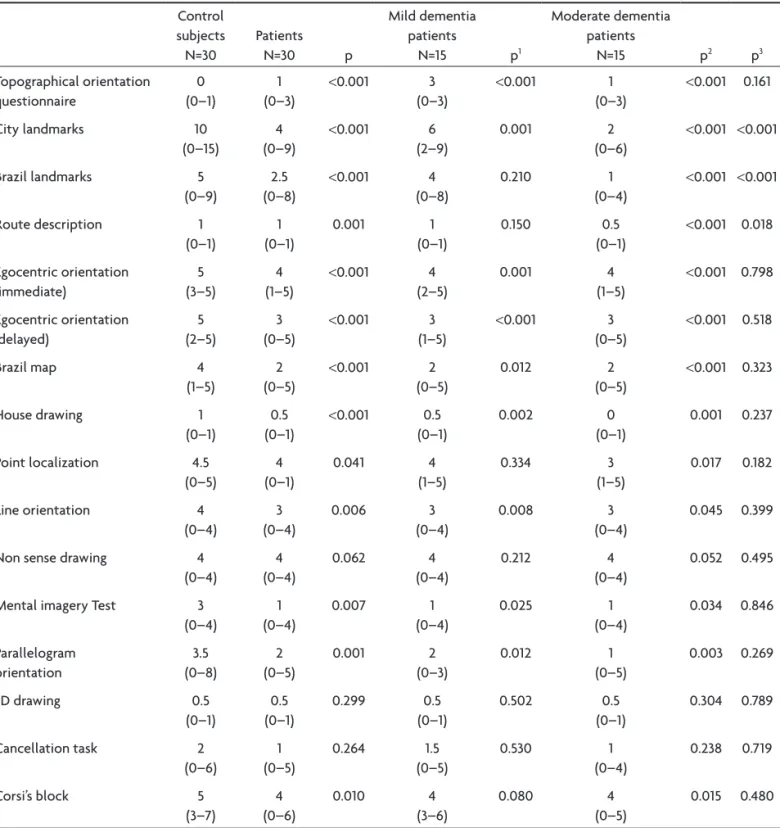
es between patients and control subjects in all tests ex-cept non sense drawing, cancellation test and three di-mension igure copy.
When patients with mild dementia were compared with control subjects there was no difference in the rec-ognition of Brazil landmarks, route description, point lo-calization, Corsi’s block tapping test as well as in non sense drawing, cancellation test and three dimension igure copy. The comparison between mild and moderate
demen-tia patients showed that there was no difference between them in the majority of the tests, except for são Paulo city landmarks, Brazil landmarks and route description.
DISCuSSION
The results of the questionnaire showed that TD was present even in mild dementia of AD. other authors that have applied questionnaires for topographical orientation
in AD had reported similar indings2,3,23.
Table 2. Performance in the questionnaire and in tests related to topographical orientation (median, minimum and maximum values).
Control subjects N=30 Patients N=30 p Mild dementia patients
N=15 p1
Moderate dementia patients
N=15 p2 p3
Topographical orientation questionnaire 0 (0–1) 1 (0–3)
<0.001 3 (0–3)
<0.001 1 (0–3)
<0.001 0.161
City landmarks 10
(0–15)
4 (0–9)
<0.001 6 (2–9)
0.001 2
(0–6)
<0.001 <0.001
Brazil landmarks 5 (0–9)
2.5 (0–8)
<0.001 4 (0–8)
0.210 1
(0–4)
<0.001 <0.001
route description 1 (0–1) 1 (0–1) 0.001 1 (0–1) 0.150 0.5 (0–1)
<0.001 0.018
egocentric orientation (immediate) 5 (3–5) 4 (1–5)
<0.001 4 (2–5)
0.001 4
(1–5)
<0.001 0.798
egocentric orientation (delayed) 5 (2–5) 3 (0–5)
<0.001 3 (1–5)
<0.001 3 (0–5)
<0.001 0.518
Brazil map 4
(1–5)
2 (0–5)
<0.001 2 (0–5)
0.012 2
(0–5)
<0.001 0.323
house drawing 1
(0–1)
0.5 (0–1)
<0.001 0.5 (0–1)
0.002 0
(0–1)
0.001 0.237
Point localization 4.5 (0–5) 4 (0–1) 0.041 4 (1–5) 0.334 3 (1–5) 0.017 0.182
line orientation 4 (0–4) 3 (0–4) 0.006 3 (0–4) 0.008 3 (0–4) 0.045 0.399
Non sense drawing 4 (0–4) 4 (0–4) 0.062 4 (0–4) 0.212 4 (0–4) 0.052 0.495
Mental imagery Test 3 (0–4) 1 (0–4) 0.007 1 (0–4) 0.025 1 (0–4) 0.034 0.846 Parallelogram orientation 3.5 (0–8) 2 (0–5) 0.001 2 (0–3) 0.012 1 (0–5) 0.003 0.269
3D drawing 0.5
(0–1) 0.5 (0–1) 0.299 0.5 (0–1) 0.502 0.5 (0–1) 0.304 0.789
Cancellation task 2 (0–6) 1 (0–5) 0.264 1.5 (0–5) 0.530 1 (0–4) 0.238 0.719
Corsi’s block 5
(3–7) 4 (0–6) 0.010 4 (3–6) 0.080 4 (0–5) 0.015 0.480
p: comparison between patients and controls; p1: comparison between mild dementia patients and controls; p2: comparison between moderate dementia patients and
Although our patients with mild dementia have TD, they were not impaired in point localization, non sense drawing and three dimension igure copy when compared to control subjects. other studies have also reported that spatial and visual perception may not be involved in early
stages of AD23,24 . These indings suggest that TD in AD is
not directly caused by disturbances of less complex spa-tial and visual functions.
spatial memory tasks results showed that spatial work-ing memory disturbances are present in patients with de-mentia compared to controls. however, spatial working memory was not different in patients with mild demen-tia and controls in this study. These indings are different from another study which veriied that were statistical difference in Corsi’s block tapping test between mild
de-mentia patients and control subjects25. It is possible that
impairment of spatial working memory did not contribute to TD in the patients with mild dementia in this study.
Conversely, egocentric and alocentric orientation, landmark recognition, route description, geographic ori-entation, house drawing, line orientation and mental ro-tation are impaired in patients with mild dementia in this study.
egocentric orientation was signiicantly impaired in AD patients in this study, similarly to the indings of
Mo-nacelli et al.5. There was no difference between mild and
moderate AD patients, showing that patients have ego-centric disorientation in early stages of AD.
landmark recognition was also different between con-trol subjects and AD patients, even in mild dementia of
AD, in accordance with other studies. Monacelli et al.5
evaluated landmark recognition in the hospital hall while Uc et al. tested landmark identiication during actual
driv-ing26. Both studies showed that failure in landmark
recog-nition was present in mild AD.
honda et al. evaluated route description by normal adults verifying that the intelligibility of the route de-scription depends on their knowledge of the environment
relationships27. In this sense, the failure of route
descrip-tion by our patients suggests that they have alocentric dis-orientation, and that this disturbance occurs in the ear-ly stages of AD.
landmark recognition and route description were more severe in moderate than in mild dementia in our study. Although this study did not follow AD patients longitudinally, these indings suggest that landmark rec-ognition and route description failures are responsible for the worsening of TD in moderate compared to mild dementia.
Geographic orientation in a Brazil map showed differ-ence between the groups. This task identiied dificulty in mild dementia patients suggesting that the impairment
also occurs since the early stages of AD9.
house drawing results showed difference between mild dementia patients and control subjects. liu et al. studied drawings of mild dementia patients’ houses and
the results were similar to this study28. The results of this
test may be inluenced by impairment of constructional praxis in AD, but dificulty in drawing the relationship be-tween rooms in the house can be also interpreted as an alocentric disorientation.
Although route description and house drawing are probably based on different mental faculties, both may be interpreted as alocentric tests. In our study AD pa-tients with mild dementia were impaired in these tasks, suggesting that alocentric disorientation contributed to TD in these cases.
line orientation test showed difference between pa-tients with mild dementia and controls. These results are different from Uc et al., who also compared line ori-entation test between mild dementia patients (mean MMse=26) and aged-matched control subjects and he did
not ind difference26. Although elementary visual and
per-ception tests apparently were not responsible for TD in AD, more complexes spatial procedures as mental imag-ery and mental rotation seems to contribute to TD, as it was seen on the results of the tests of parallelogram ro-tation and mental imagery tests (counting hidden blocks). Kurylo et al. found similar results in mental rotation test
between control subjects and AD patients24.
These results may be explained by the course of the spread of the pathological process in AD. once the dis-ease starts, the hippoccampal cells area affected and the spreading of the pathological lesions to parahipocampal
gyri may cause landmark recognition6. The involvement of
the retrosplenic cortex may lead to heading impairment3
and as the pathological process affects the parietal
cor-tex, the optic low may be disrupted4.
This study has several limitations. The diagnosis of TD was based only on a questionnaire, which revealed that almost all patients have TD. An environmental test prob-ably would be able to diagnose TD and to classify its se-verity. Another limitation was due to the small number of individuals when the patients were divided into mild and moderate dementia.
REFERENCES
1. Henderson VW, Mack W, Williams BW. Spatial disorientation in Al-zheimer disease. Arch Neurol 1989;46:391-394.
2. Cherrier MM, Mendez M, Perryman K. Route learning performance in Alzheimer disease patients. Neuropsychiatry Neuropsychol Behav Neurol 2001;14:159-168.
3. Takahashi N, Kawamura M, Shiota J, Kasahata N, Hirayama K. Pure topographic disorientation due to right retrosplenial lesion. Neurolo-gy 1997;49:464-469.
4. Tetewsky SJ, Duffy CJ. Visual loss and getting lost in Alzheimer dis-ease. Neurology 1999;52:958-965.
5. Monacelli AM, Cushmagn LA, Kavcic V, Duffy CJ. Spatial disorienta-tion in Alzheimer’s disease. Neurology 2003;61:1491-1497.
6. Aguirre GK, D’Esposito M. Topographical disorientation: a synthesis and taxonomy. Brain 1999;122:1613-1628.
7. Burgess N, Maguire EA, O’Keefe J. The human hippocampus and spa-tial and episodic memory. Neuron 2002;35:625-641.
8. Benton AL, Levin HS, Van Allen MW. Geographic orientation in patients with unilateral cerebral disease. Neuropsychologia 1973;12:183-191.
9. Kandel ER, Schwartz JH, Jessel TM. Principles of neural sciences. 4th
Ed. New York: McGraw-Hill, 2000:1247-1279.
10. Maguire EA, Gadian DG, Johnsrude IS, et al. Navigation-related struc-tural change in the hippocampi of taxi drivers. Proc Natl Acad Sci USA 2000;97:4398-4403.
11. McKhan G, Drachman D, Folstein M, Katzman R, Price D, Stadlan EM. Clinical diagnosis of Alzheimer’s disease: report of the NINCDS-ADRDA Work Group under the auspices of Department of Health and Human Services Task Force on Alzheimer’s Disease. Neurology 1984;34:939-944.
12. Folstein MF, Folstein SE, McHugh PR. Mini-Mental State: a practical method for grading the cognitive state of patients for the clinician. J Psychiatr Res 1975;12:189-198.
13. Brucki SM, Nitrini R, Caramelli P, Bertolucci PH, Okamoto IH. Sugges-tions for utilization of the mini-mental state examination in Brazil. Arq Neuropsiquiatr 2003;61:777-781.
14. Nitrini R, Lefevre BH, Mathias SC, ET al. Neuropsychological tests
of simple application for diagnosing dementia. Arq Neuropsiquiatr 1994;52:457-465.
15. Nitrini R, Caramelli P, Herrera Junior E, et al. Performance of illiter-ate and literilliter-ate nondemented elderly subjects in two tests of long-term memory. J Int Neuropsychol Soc 2004;10:634-638.
16. Nitrini R, Caramelli P, Porto CS, et al. Brief cognitive battery in the di-agnosis of mild Alzheimer’s disease in subjects with medium and high levels of education. Dement Neuropsychol 2007;1:32-36.
17. Benton AL, Varney NR, Hamsher KS. Visuospatial judgment: a clinical test. Arch Neurol 1978;35:364-367.
18. Banich MT. Disruptions in basic spatial processing in humans. In Ban-ich MT (Eds). Neuropsychology: neural basis of mental function. Bos-ton: Houghton Muflin Company, 1997:203-233.
19. Christensen AL. Luria’s neuropsychological investigation manual. Co-penhagen: Munksgaard, 1979.
20. Milner B. Interhemispheric differences in the localization of psycholog-ical processes in man. Br Med Bull 1971;27:272-277.
21. Guariglia CC. Spatial working memory in Alzheimer’s disease. Dement Neuropsychol 2007;1:392-395.
22. Mesulam MM. Principles of behavioral and cognitive neurology. 2.Ed. New York: Oxford University Press, 2000:174-256.
23. Teri L, Borson S, Kiyak A, Yamagishi M. Behavioral disturbance, cog-nitive dysfunction, and functional skill: prevalence and relationship in Alzheimer’s disease. J Am Geriatr Soc 1989;37:109-116.
24. Kurylo DD, Corkin S, Growdon JH, Rizzo JF. Greater relative impair-ment of object recognition than visuospatial abilities in Alzheimer’s disease. Neuropsychology 1996;10:74-81.
25. Grossi D, Becker JT, Smith C, Trojano L. Memory for visuospatial pat-terns in Alzheimer’s disease. Psychol Med 1993;23:65-70.
26. Uc EY, Rizzo M, Anderson SW, Shi Q, Dawson JD. Driver landmark and trafic sign identiication in early Alzheimer’s disease. J Neurol Neuro -surg Psychiatry 2005;76:764-768.
27. Honda A, Nihei Y. Empathy, spatial and verbal abilities characterize one who can best describe a route. Percept Mot Skills 2003;96:861-866 28. Liu L, Gauthier L, Gauthier S. Spatial disorientation in persons with early