326 326
Almeida et al
Postoperative ascending aorta pseudoaneurysm
Arq Bras Cardiol 2001; 76: 326-8.
Instituto de Moléstias Cardiovasculares de Cascavel and Universidade Estadual do Oeste do Paraná
Mailing address: Rui Manuel Sequeira de Almeida - Instituto de Moléstias Cardiovasculares de Cascavel Policlínica de Cascavel Rua Maranhão, 945 -85806-050 - Cascavel, PR, Brazil
English version by Stela Maris C. e Gandour
Rui Manuel Sequeira de Almeida, José Dantas Lima Jr, Thomas Kahrbek, Maurício Tanomaru
Cascavel, PR - Brazil
Surgical Repair of a Pseudoaneurysm of the Ascending Aorta
after Aortic Valve Replacement
Case Report
We report the case of a patient with a pseudoaneurysm of the ascending aortic clinically diagnosed 5 months after surgical replacement of the aortic valve. Diagnosis was confirmed with the aid of two-dimensional echocardiogra-phy and helicoidal angiotomograechocardiogra-phy. The corrective surgery, which consisted of a reinforced suture of the com-munication with the ascending aorta after opening and aspiration of the cavity of the pseudoaneurysm, was suc-cessfully performed through a complete sternotomy using extracorporeal circulation, femorofemoral cannulation, and moderate hypothermia, with no aortic clamping.
Aneurysms of the ascending aorta are rare entities that may occur after cardiac surgeries and that have a significant rate of complication. Surgical repair of ascending aorta pseu-do aneurysms remains a challenge to the ability of surgeons. We report the case of a patient with an ascending aorta pseudoaneurysm that was successfully surgically repaired.
Case Report
An 18-year-old male with characteristics of Marfan’s syndrome underwent surgical replacement of the aortic valve due to a severe aortic insufficiency and ascending aorta ectasia 5 months ago. An ATS Medical Open PivotTM (ATS Medical Inc., Minneapolis, MN, USA) prosthesis, nº 25, was implanted, and in the postoperative period the pa-tient was reoperated upon because of blood dyscrasia and bleeding of the suture of the ascending aorta cannulation. Routine tests were normal on hospital discharge. After this, the patient was referred for clinical follow-up and medicated only with dicumarol. Clinical controls at 30 and 60 days of
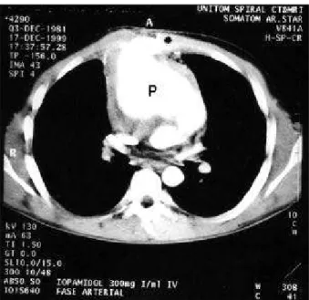
the postoperative period showed a normal evolution. On the 5th postoperative month, the patient sought medical assis-tance because of a mass in the right hemithorax. On physi-cal examination, the mass was pulsatile, measured approxi-mately 4cm, and was located between the 3rd and 4th right in-tercostal spaces, 3cm from the right parasternal line. The chest X-ray was not specific. Two-dimensional Doppler echocardiography showed an aortic prosthesis functioning normally and a huge mass in the anterior portion of the as-cending aorta measuring 10.5cm in its larger axis. The neck of the pseudoaneurysm was small (1cm), located 5cm above the level of the metallic valve, with double leaflet, and had blood flow and intraluminal clots. The patient underwent helicoidal angiotomography with dynamic intravenous infu-sion of nonionic contrast medium through a high-flow digi-tal infusion pump. This helicoidal angiotomography confirmed the diagnosis and showed that the saccular formation occupied the anterior mediastinum in the retros-ternal portion posterior to the manubrium and sretros-ternal body, and caused sternal erosion. This saccular formation measured 11 x 9.8 x 7.8cm in the craniocaudal, laterolateral, and anteroposterior axes, respectively (fig. 1), had a volume of 437cm3, which contained blood. It also showed a semila-minar layer compatible with mural thrombus in the periphe-ral area leading to compression and posterior displacement of the ascending aorta, aortic arch, and pulmonary trunk (fig. 2). Because of these findings, the patient was referred for urgent surgery.
After initial preparation, the common femoral artery and vein were cannulated (femoral cannula kit, Medtronic-DLP, Minneapolis, MN, USA), and extracorporeal circula-tion was installed with moderate hypothermia (24 °C). After a large volume of blood was drained and the extracorporeal circulation was interrupted, but with the heart beating, a sternotomy was performed and the ascending aorta pseu-doaneurysm was directly approached. The pseudoaneu-rysm was aspirated and its neck was identified. The neck could be partially occluded with a finger, and then with 2 stitches of Prolene 3-0 (Ethicon). Extracorporeal circulation was restarted with rewarming. The suture of the
Arq Bras Cardiol 2001; 76: 326-8.
Almeida et al Postoperative ascending aorta pseudoaneurysm
327 327 aneurysm neck was strengthened with straps of bovine
pe-ricardium (Braile Biomédica, São José do Rio Preto, SP, Bra-zil) and 2 stitches of Prolene 3-0. All clots were cleared from the cavity, and surgery was completed in the conventional manner. The postoperative period was uneventful, and the patient stayed in the hospital for 7 days.
In the early postoperative period, a control echocar-diography showed the complete occlusion of the neck of the pseudoaneurysm, and no paraaortic leak was noted.
Discussion
Pseudoaneurysm of the ascending aorta is a rare entity (<1%) and results from complications of cardiac surgery, in
which the ascending aorta is cannulated or incised 1. In our case series of 1,049 patients with extracorporeal circulation, this was the first with this complication (0.09%). Most as-cending aorta pseudoaneurysms occur after surgeries on the aortic valve, coronary revascularizations, usually at the site of the proximal anastomoses of the grafts, aortotomies, or in the cannulation sites. Despite the evidence of some cases of ascending aorta pseudoaneurysm occurring be-cause of infection or in aortas with weak points (Marfan’s syndrome), a great percentage, results from mechanical rup-ture of aortic surup-tures. Most ascending aortic pseudo aneu-rysms are asymptomatic, like the one we presently report, unless they compress important structures, such as corona-ry arteries, venous or arterial grafts, pulmonacorona-ry arteries, or the superior vena cava, causing acute clinical manifestati-ons. The risk of rupture of a pseudoaneurysm should be ta-ken into consideration as an indication for emergency surgery, especially in the case of large masses.
From the surgical point of view, the treatment of ascen-ding aorta pseudoaneurysms remains a challenge. Uninten-tional rupture during the redo sternotomy or mediastinal dissection due to the reduction in pressure around the for-mation when opening and sliding the sternum, are causes of surgical catastrophe. Mortality reported by several authors ranges from 29% to 46% 2,3, and most of the time it is a conse-quence of a fatal hemorrhage due to rupture of the pseudoa-neurysm during surgical maneuvers for its repair.
In our patient, to avoid this situation and due to ero-sion of part of the sternum, femorofemoral extracorporeal cir-culation 4 was started with hypothermia. Hypothermia was required for drainage of a large amount of blood to the extra-corporeal circulation system and arrest of the pump, during the new sternotomy. These maneuvers were required be-cause of the location of the saccular formation and its neck in the ascending aorta, which was depicted in both the two-dimensional echocardiography and the helicoidal angioto-mography. Contrary to findings in some reports 5, echocar-diography was extremely useful in our case. Helicoidal angiotomography is a more accurate imaging technique and provided all details for surgical planning.
In conclusion, a successful complete repair of ascen-ding aorta pseudoaneurysms, avoiascen-ding the risk of fatal in-traoperative bleeding, is due to an objective and accurate planning of surgical tactics and technique 6, which should be based on a careful assessment of the results of examina-tions, such as two-dimensional echocardiography and heli-coidal angiotomography.
Acknowledgements
We thank Mrs. Jussara Planck for editing the text and Mrs. Bernardete Rocha and Mr. Emerson Mendes for their technical support.
Fig. 1 – Axial plane of helical tomography depicting a pseudoaneurysm of the aorta (P) and sternal erosion (*).
328 328
Almeida et al
Postoperative ascending aorta pseudoaneurysm
Arq Bras Cardiol 2001; 76: 326-8.
1. Sabri MN, Henry D, Wechsler AS, Di Sciascio G, Vetrovec GW. Late complications involving the ascending aorta after cardiac surgery. Am Heart J 1991; 121: 1779-83. 2. Sullivan KL, Steiner RM, Smullens SN, Griska L, Meister SG. Pseudo-aneurysm of the ascending aorta following cardiac surgery. Chest 1988; 93: 138-43.
3. Razzouk A, Gundry S, Wang N. Pseudoaneurysms of the aorta after cardiac sur-gery or chest trauma. Am Surg 1993; 59: 818-23.
References
4. Gaudino M, Alessandrini F, Canosa C, Possati G. Repair of an ascending aorta pseudoaneurysm by way of superior ministernotomy. Ann Thorac Surg 1999; 67: 1798-800.
5. Milas BL, Savino JS. Pseudoaneurysm of the ascending aorta after aortic valve replacement. J Am Soc Echocardiogr 1998; 11: 303-6.