online | memorias.ioc.fiocruz.br
Oocyst wall formation and composition in coccidian parasites
Kelly Mai, Philippa A Sharman, Robert A Walker, Marilyn Katrib, David De Souza, Malcolm J McConville, Michael G Wallach, Sabina I Belli, David JP Ferguson, Nicholas C Smith/+
Institute for the Biotechnology of Infectious Diseases, University of Technology, PO Box 123 Sydney, Australia
The oocyst wall of coccidian parasites is a robust structure that is resistant to a variety of environmental and chemical insults. This resilience allows oocysts to survive for long periods, facilitating transmission from host to host. The wall is bilayered and is formed by the sequential release of the contents of two specialized organelles - wall forming body 1 and wall forming body 2 - found in the macrogametocyte stage of Coccidia. The oocyst wall is over 90% protein but few of these proteins have been studied. One group is cysteine-rich and may be presumed to cross-link via disulphide bridges, though this is yet to be investigated. Another group of wall proteins is rich in tyrosine. These proteins, which range in size from 8-31 kDa, are derived from larger precursors of 56 and 82 kDa found in the wall forming bodies. Proteases may catalyze processing of the precursors into tyrosine-rich peptides, which are then oxidatively crosslinked in a reaction catalyzed by peroxidases. In support of this hypothesis, the oocyst wall has high levels of dityrosine bonds. These dityrosine crosslinked proteins may provide a structural matrix for assembly of the oocyst wall and contribute to its resilience.
Key words: Coccidia - oocyst - dityrosine crosslinking - Toxoplasma - Eimeria
Coccidian parasites, which include the genera Toxo-plasma, Neospora, Hammondia, Isospora, Sarcocystis
and Eimeria, amongst others, share many features. For example, like all Apicomplexa, their lifecycle includes asexual stages (sporozoites and merozoites) that pos-sess a phylum-defining feature, the apical complex of organelles, associated with cell invasion. An additional defining feature of the Coccidia is the oocyst; coccidian parasites are transmitted from host to host by acciden-tal ingestion of oocysts that contaminate food or water. Oocysts are remarkably hardy and able to persist in the environment for prolonged periods of time. The “soft-bodied” parasites are safely encapsulated inside a unique structure, the oocyst wall.
The oocyst wall is extremely robust. It is resistant to mechanical and chemical damage; breaking oocysts for laboratory studies requires prolonged, high-speed vor-texing with glass beads and oocysts are routinely cleaned with bleach and stored in the harsh oxidant, potassium dichromate, or the mineral acid, sulphuric acid (Dubey et al.1970, Shirley 1995, Schares et al.2005). The wall is also resistant to proteolysis and impermeable to water- soluble substances, including many detergents and
dis-infectants (Monné & Hőnig 1954, Ryley 1973). This
contributes to the difficulty experienced in attempting to exclude oocysts from, for example, poultry houses.
Financial support: Australian Research Council’s Discovery and Link -age Projects funding schemes (DP0664013, LP0454145), Abic Ltd (Is -rael), Rural Industries Research and Development Corporation’s Post -graduate Scholarship Scheme, ARC/NHMRC research Network for parasitology (RN0460368)
+ Corresponding author: [email protected] Received 16 October 2008
Accepted 4 December 2009
The oocyst wall is, however, permeable to some small molecules and lipid+soluble substances; ammonia and methyl bromide being two of the best known (Monné &
Hőnig 1954, Ryley 1973, Kuticic & Wikerhauser 1996).
The oocyst wall is essentially consistent in structure across different species of coccidian parasites (Belli et al. 2006), but it is the oocyst wall of Eimeria that has been best studied, largely because of the relative ease of acquiring large numbers of oocysts of the parasites of this genera.
Oocyst wall biogenesis
The oocyst wall is formed from the contents of two
specific organelles, wall forming bodies Type 1 (WFB1) and 2 (WFB2), found exclusively in the sexual,
macro-gamete stage of coccidian parasites (Scholtyseck et al.
1969, Scholtyseck 1973, Ferguson et al. 1975, 2003, Pit
-tilo & Ball 1980, Belli et al. 2006). In the early stages of
macrogamete biogenesis, the macrogametocyte contains a centrally-located nucleus and numerous spherical de-posits of electron dense material within swollen regions of the rough endoplasmic reticulum; these represent the
early WFB2. Shortly after this, in the mid-stage of mac -rogamete development, spherical electron dense granules appear in the cytoplasm. These secretory-like granules arise from material that traffics from the Golgi bodies
and represent the early WFB1 (Ferguson et al. 2003). Also present in the early/mid and mid-stage of development are
an additional group of membrane bound vacuoles termed
the veil forming bodies (Ferguson et al. 2000, 2003).
The veil forming bodies vary in appearance between species, being electron dense in Toxoplasma gondii
(Ferguson et al. 2000) and electron lucent in Eimeria maxima (Ferguson et al. 2003). They are secreted dur -ing macrogamete maturation and are responsible for the formation of a veil on the outer surface of the maturing
The outer veil of E. maxima stains positively with anti-bodies that are specific for the apple domains of T. gondii microneme protein 4, a protein associated with oocyst
wall formation in this parasite. This suggests that the
veil contains MIC4-like proteins and that the structure
of the veil is conserved across the Coccidia (Ferguson et
al. 2000, 2003). The veil is lost when oocysts are excreted from the host (Ferguson et al. 1975, Pittilo & Ball 1980);
therefore it is thought that the outer veil plays no role in protection of parasites during passage out of the host or in the external environment (Belli et al. 2006). Its precise role is still unknown, though it seems possible that it pro-vides a temporary “frame” under which the oocyst wall is assembled. This idea is supported by the fact that the veil remains in place throughout the entire intracellular
development phase of the oocyst (Pittilo & Ball 1980). WFB1 in mature macrogametocytes are large, spheri -cal, membrane bound structures with electron dense
con-tents (Pittilo & Ball 1979, Ferguson et al. 2003, Belli et al.
2006). They are located in the periphery of the parasites and have long been thought to contain mucoproteins,
mucopolysaccharides and glycoproteins (McLaren 1969, Scholtyseck 1973). The size of WFBs is species-specific. For example, WFB1 are larger than WFB2 in E. maxima,
Eimeria tenella, Eimeria stieda and Eimeria perforans,
whereas in Eimeria bovis, Eimeria falciformis and T. gondii, the reverse is true (Scholtyseck 1973). WFB2 are less electron dense than WFB1, have a less distinct structure (Ryley 1973, Scholtyseck 1973) and appear as
spherical dilations enclosed within the rough
endoplas-mic reticulum (Ferguson et al. 1975, 2003, Pittilo & Ball 1979). In the mature macrogametocyte, WFB1s locate in the periphery of the parasite inter-mixed with WFB2.
A large number of polysaccharide granules, containing amylopectin and some lipid droplets are also seen in the cytoplasm of the mature macrogametocyte (Ferguson et
al. 1975, Pittilo & Ball 1980).
Once fertilized by a microgamete, a macrogamete develops into a zygote. The oocyst wall starts to form
shortly afterwards (Pittilo & Ball 1980), with WFB1s
aligning beneath the limiting membrane of the zygote
cytoplasm. A recent study (Ferguson et al. 2003) of
oocyst wall formation in E. maxima, using
immuno-cytochemistry with antibodies specific for WFBs, adds
compelling evidence to support the mechanism of oo-cyst wall formation originally proposed by Pittilo and
Ball in 1980. Thus, Ferguson et al. (2003) showed that
oocyst wall formation is a sequential release of the
con-tents of: (i) the veil forming bodies, (ii) WFB1 and (iii) WFB2. It is thought that this mechanism may be con
-trolled at the level of the rough endoplasmic reticulum/
Golgi apparatus. Thus, in the mature macrogametocyte
or zygote, after the veil has been laid down, WFB1 re
-locate to the periphery of the parasite. WFB1s quickly
disaggregate and appear to fuse together at the surface of the parasite, ultimately forming the outer layer of the
oocyst wall. Shortly after WFB1 form the outer layer, the WFB2s, located in the rough endoplasmic reticu -lum are transferred via the Golgi body and secretory granules to the surface where they fuse to create the inner layer of the oocyst wall.
The mature oocyst wall is bilayered. Initially, the outer layer may be as thick as 500-600 nm but, as the development of the wall progresses, the outer layer is compacted to 200 nm or less. An inner zone of
approxi-mately 40 nm separates the outer and inner layers, the latter layer also ultimately being ~40 nm thick (Fergu
-son et al. 2003). The electron dense outer layer is vari -able in size across different genera; for example, it can be ~200 nm thick in E. maxima (Ferguson et al. 2003) but is only 20-40 nm thick in T. gondii (Ferguson et al. 1975, 2000). The electron lucent inner layer, formed
from the contents of WFB2, is more consistent, being around 40 nm thick in most genera studied (Belli et al.
2006). The two layers do not seem to be tightly fused
and can be separated relatively easily (Monné & Hőnig 1954); bleach treatment, for instance, usually strips the
outer layer away from the inner layer.
Biochemical composition of the oocyst wall
The first serious microscopic and chemical examina-tion of the oocyst wall (of E. maxima) was conducted by
Monné and Hőnig (1954), who used a number of destruc -tive treatments that led them to conclude that the outer layer of the oocyst wall contained mainly quinone-tanned proteins without lipids, since the outer layer reacted with ammoniacal silver nitrate solution (an indication of qui-nones). They also noted that the outer layer was stripped off upon exposure to sodium hypochlorite, whereas the structure of the inner layer remained unchanged, leading them to conclude that the inner layer consisted of a lip-id-protein matrix; they believed that lipids were bound firmly to proteins, thus protecting the inner layer against disintegration by sodium hypochlorite.
The first true biochemical examination of the oocyst
wall was carried out by Ryley in 1973 using E. tenella. Ryley (1973) also noted that the outer layer was removed
by sodium hypochlorite and found that it contained carbohydrates and proteins, with high proline content, whereas the inner layer consisted of 1.5% carbohydrates,
30% lipids and 70% proteins. The lipid in the inner layer
was extractable in chloroform and appeared to be a mix-ture of “waxes” containing very small amounts of nitro-gen and phosphorus. However, there are some limitations in this report: first, it did not show detailed analyses of the experimental work and, second, it did not document the metabolites detected in the inner wall.
The most recent biochemical analysis of the oocyst
wall was conducted by Stotish et al., in 1978, using un -sporulated oocysts isolated from the caeca of chickens infected with E. tenella. These researchers proposed that lipids were concentrated in the outer layer whereas the inner layer was composed of mainly glycoproteins. In addition, compositional analysis of the oocyst wall by gas-liquid chromatography and SDS-PAGE indicated
that it was 19% carbohydrates, 14% lipids and 67% protein (Stotish et al. 1978). Lipids identified from the
- mannose, galactose, glucose and hexosamine - were detected. However, some doubts surround the validity of
these conclusions because Stotish et al. (1978) noted that sodium hypochlorite treatment did not affect the struc-ture of the oocyst walls from E. tenella unsporulated oocysts and this was a major basis for their conclusion that lipids are concentrated in the outer layer of the wall (since lipids would be expected to give some protection against stripping by sodium hypochlorite). In direct
con-tradiction to the observations of Stotish et al. (1978), it has been shown that sodium hypochlorite treatment does have an effect on the structure of the oocyst wall in both
E. maxima and E. tenella - the outer layer is stripped off
(Monné & Hőnig 1954, Nyberg et al. 1968, Nyberg & Knapp 1970, Ryley 1973, Belli et al. 2006). Therefore,
the biochemical compositions assigned to the two layers of the oocyst wall of E. tenella by Stotish et al. (1978) could be erroneous. We have, therefore, reinvestigated
the biochemical composition of the oocyst wall using more reliable sensitive modern methods.
We isolated oocyst walls from intact E. maxima and
E. tenella oocysts as published previously by Stotish et
al. (1978). Oocyst walls (equivalent to the wall prepara -tions isolated from 2 x 10⁶ oocysts) were then resuspend-ed in 100 µL of distillresuspend-ed water and treatresuspend-ed with
alpha-amylase to eliminate polysaccharide granules according
to the protocol from Ryley (1973), with slight modifica -tion. Briefly, alpha-amylase was added to a final
con-centration of 0.5 mg/mL and 1 mg/mL for comparison
and samples were incubated at 20ºC overnight. On the next day, the samples were centrifuged at 10,000 g for
10 min at 4ºC, the supernatant was discarded and the
pellet was washed in distilled water. The washing step was repeated three times. The effect of alpha-amylase on elimination of polysaccharide granules was investigated by microscopic examination and transmission electron microscopy (TEM) as published previously by Ferguson
et al. (2003), with a minor modification. Briefly, each
sample of oocyst wall fragments was centrifuged and the
pellets were fixed in 4% glutaraldehyde in 0.1 M phos -phate buffer. The pellets were post-fixed in 1% osmium tetroxide in phosphate buffer, dehydrated in absolute ethanol, treated with propylene oxide and embedded in
Spurr’s epoxy resin. Thin sections of oocyst walls were
then mounted on copper grids and stained with uranyl acetate and lead citrate for routine electron microscopy. In addition, some sections were mounted on formvar-coated gold grids and stained with silver methenamine. The sections were floated on drops of a freshly prepared and filtered mixture containing silver nitrate and hex-amethylene tetramine in a borax-based buffer (pH 9.2).
Sections were stained in the dark at 40°C for 60 min and
washed in water prior to examination. In both cases, sec-tions were examined in a Jeol 1200EX TEM.
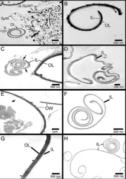
In the absence of treatment with alpha-amylase, the purified oocyst walls appeared to be heavily
contami-nated with polysaccharide granules (Fig. 1A, B). When
alpha-amylase was added to the oocyst walls, the num-ber of polysaccharide granules was reduced markedly, as
shown in the sample treated with 0.5 mg/mL alpha-amy -lase (Fig. 1C) and, more especially, in the sample treated
with 1 mg/mL alpha-amylase (Fig. 1D). Examination
by light and UV microscopy (Fig. 1E, F, respectively) confirmed the characteristic blue autofluorescence of the purified oocyst walls after alpha-amylase treatment, indicating that the basic chemical structure of the walls was not altered significantly by the treatment.
The oocyst walls prepared from E. tenella and E. maxima sporulated and unsporulated oocysts were ex-amined by TEM to confirm the presence of the inner
and/or outer layers. The outer layer of the oocyst walls
(both E. tenella and E. maxima), appeared electron-dense with a roughened appearance on the outer surface, and the inner layer was electron-lucent (Fig. 2A-C, E, G), observations that are consistent with those of
Fer-guson et al. (2003). The oocyst walls from unbleached
samples remained intact (retaining a bi-layered struc-ture) whereas in bleached samples, generally only the inner layer was seen (Fig. 2D, F, H), confirming the observations that bleaching does strip the outer layer of
oocyst walls (Monné & Hőnig 1954, Nyberg et al. 1968, Nyberg & Knapp 1970, Ryley 1973, Belli et al. 2006).
There was a single exception to this pattern; walls pre-pared from a sample of bleached E. tenella sporulated oocysts remained intact as a bi-layered structure (Fig. 2B). This was not always observed and the reason for this exception is not known. However, this exceptional sample proved very useful in subsequent comparisons of wall composition.
The compositional analysis of bleached oocyst walls was carried out as published previously by McConville et al. (1990), with a minor modification (unbleached samples were not analysed due to the presence of various contaminants, including residual amylopectin, which would distort the analyses). Briefly, the oocyst walls
Fig. 1: micrographs of alpha-amylase treated oocyst walls prepared from Eimeria maxima bleached unsporulated oocysts. Each sample contained oocyst walls equivalent to 2 x 10⁶ E. maxima unsporulated
were resuspended in 50 µL of 1:1 chloroform:methanol
and transferred into a clean glass capillary tube with one end flame sealed. Heptadecanoic acid (C17:0; 10 nm) was added to the capillary tube as an internal stand- ard for subsequent quantification of metabolites. The sample was then dried in vacuo at 50ºC. This step was repeated again in the presence of methanol to enable the complete removal of H2O. Methanolic HCl (0.5 M HCl
in methanol; 50 µL) was added to each capillary tube
and sealed under vacuum. The sample was then
incu-bated at 80ºC overnight. Following methanolysis, the
sample was cut open and the acid was neutralized by
the addition of 10 µL pyridine. The neutralized solution
was then transferred to a GC-MS vial insert and dried in vacuo. Trimethylsilyl (N-methyl-N-trifluroacetamide + 1% trimethylchlorosilane; 50 µL) reagent was added to
the tube, which was gently flicked to ensure the sample was well mixed. The sample was then transferred into an insert vial of autosampler vials for compositional analy-ses of carbohydrates and lipids by gas chromatography
(GC; Agilent 6890N GC) and mass spectrometry (MS, Agilent 5973 Mass Selective Detector).
Oocyst wall samples were also hydrolyzed in 6N HCl
as published previously (McConville et al. 1990), with a minor modification. Briefly, the oocyst walls were
resuspended in 100 µL of 1:1 chloroform:methanol and
transferred into GC-MS vial inserts containing 1 nm of scyllo-inositol as an internal control for subsequent
quantification of metabolites. The sample was dried in vacuo at 55º. The vial inserts were then transferred into
reaction vials containing 200 µL 6N HCl, followed by
evacuation under nitrogen. The samples were capped immediately to prevent oxidation by air and incubated at 110ºC over night. The vial inserts were then placed into GC-MS vials and the residual HCl was evaporated under nitrogen. Vial contents were then incubated for 2 h
at rt in 20 µL pyridine to allow resuspension and neu
-tralization of HCl contaminants. N,O-bis[trimethylsilyl] trifluoroacetamide + 1% trimethylchlorosilane (20 µL)
was added to enable trimethylsilyl derivatisation and the
reactions were carried out at 80ºC for 30 min. The amino
acid composition of the oocyst wall proteins was ana-lyzed by GC-MS.
Finally, oocyst wall proteins were extracted using the
method described by Meyers et al. (1998). Oocyst walls (from 2 x 10⁶ oocysts) were resuspended in 100 µL of 1 M NaOH, followed by boiling for 10 min to solubilize
the wall proteins. The lysate was then cooled down at rt for 10 min. Twenty microliters of 5M HCl was added to
neutralize the lysate, which was then brought up to 1 mL by the addition of 880 µLPBS. Absorbance at 230 nm
and 260 nm was measured using a spectrophotometer (GeneQuant, Amersham Biosciences) and protein con-centration was calculated using the following equation:
{[Protein] = (183 x A230) - (75.8 x A280)}.
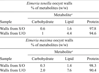
Our analyses revealed remarkable similarities be-tween all the oocyst wall samples (Table I). Thus, the
relative levels, on a percentage w/w basis of carbo -hydrate, lipid and protein were contained in relatively
narrow ranges of 0.6-2.0% carbohydrate, 1.4-7.6% lipid and 90.4-98.3% protein, for unsporulated or sporulated
oocysts, from E. tenella or E. maxima. It is perhaps note-worthy, however, that the lower values for carbohydrates and lipids were seen in the sporulated oocyst samples from both E. tenella and E. maxima. This indicates that the single bilayered oocyst wall sample (the sporulated oocysts of E. tenella) did not exhibit a distinctly differ-ent composition than the single layered sporulated oo-cyst wall sample from E. maxima. In contrast to these re-sults, the percentages of carbohydrate, lipid and protein reported by Stotish et al. (1978) for unsporulated oocysts of E. tenella are quite different, with only 67% protein
and 19% carbohydrate and 14% lipid being recorded. Analysis of 6N HCl hydrolysates of the oocyst walls
of E. tenella and E. maxima revealed that essentially all twenty amino acids are present in the walls of sporulated and unsporulated oocysts. A limited qualitative compar-ison of five of the more abundant amino acids, isoleucine (Ile), aspartic acid (Asp), valine (Val), proline (Pro) and arginine (Arg) was possible by assigning Ile a standard-ized value of 1.0 and presenting the other four amino acids as a ratio of this. It is important to not over interpret this data; it provides a limited snapshot of the relative or-der of abundance of these five amino acids but the ratio values probably do not very precisely reflect their
abun-dance. When examined in this way, the relative abun -dance of the amino acids is similar in all the samples, whether E. tenella or E. maxima, unsporulated or sporu-lated oocysts, and is in the order Ala>Pro>Val>Asp>Ile. The only slight exception to this is the sporulated oocyst Fig. 2: electron micrographs showing the effect of bleach on oocyst
wall (OW) preparations from Eimeria tenella and Eimeria maxima. A, B: E. tenella unbleached and bleached sporulated OW, respectively;
sample of E. maxima, where Asp and Val are reversed, but marginally. The results of Stotish et al. (1978) are in accord with these observations.
Sporulated oocysts of both species contained a number of fatty acids (saponifiable lipids) including palmitic acid
(C16:0), stearic acid (C18:0), oleic acid (C18:1), linoleic acid (C18:2), behenic acid (C22:0), lignoceric acid (C24:0) (Table II). Non-saponifiable lipids such as cholestadiene,
cholestane and cholesterol were also detected. Most of
these were present in a similar w/w percentage in the two
species except that the bilayered E. tenella sample was
comprised of 44.7% oleic acid whereas the E. maxima
sample was only 19.5% oleic acid. The E. tenella sample
also had higher quantities of palmitic acid (13.2% versus
5.9% in E. maxima). In apparent compensation of these differences, walls from E. tenella sporulated oocysts had lower relative percentages of linoleic acid (2.1% in E. tenella versus 8.2% in E. maxima), lignoceric acid (1.8% versus 5.4%) and, especially, cholestane (3.5% versus 11%) and cholesterol (12.1% versus 24.2%) than the E. maxima sporulated oocyst walls.
The levels of lipids in unsporulated oocysts of E. tenella and E. maxima were quite similar to each other but differed from the sporulated wall samples in that they totally lacked linoleic acid. The relative levels of oleic acid in the unsporulated oocyst samples resembled that seen in the E. maxima sporulated oocyst sample and, so, were also different from the levels apparent in the bilayered E. tenella sporulated oocyst sample. The
data from Stotish et al. (1978) for lipid composition of
unsporulated oocyst walls of E. tenella did not resemble the observations made here using GC-MS. First, the samples analysed by Stotish et al. (1978) contained no behenic acid, lignoceric acid, cholestadiene or
choles-tane. Second, Stotish et al. (1978) reported the presence
of myristate, as well as several fatty alcohols (docosanol, tetracosanol, hexacosanol, octacosanol and triacosanol)
not detected here; Stotish et al.(1978) reported hexaco -sanol and phospholipids as being especially abundant.
The carbohydrate content of E. tenella sporulated oocyst walls was very similar to that of sporulated oo-cyst walls of E. maxima, the carbohydrate content being
made up of 4.3-5% mannose, 33.7-37.4% galactose and 58.3-61.3% glucose (the latter possibly reflecting residual contamination with amylopectin). Likewise, the carbohy -drate profile of unsporulated oocyst walls of E. tenella
was similar to that of unsporulated oocyst walls of E. maxima but quite different to that of the sporulated walls,
being dominated by galactose (62.3%-67.6%), followed by glucose (26-27.9%) and mannose (6.4-9.8%). The carbo -hydrate composition of unsporulated oocysts of E. tenella
reported by Stotish et al.(1978) was, however, very differ -ent; they reported that the carbohydrate profile of the wall was 79.6% glucose and 11% hexosamine (which was not detected in the GC-MS analysis reported here) and only 7.2% galactose and 2.2% mannose.
Thus, to summarise, GC-MS analyses of oocyst walls of E. tenella and E. maxima indicates that they are primarily made up of protein (greater than 90%) with relatively small amounts of lipid and carbohydrate. The composition of the oocyst walls of both species was very
TABLE I
The biochemical composition of the oocyst wall of Eimeria
Eimeria tenella oocyst walls
% of metabolites (w/w)
Metabolitea
Sample Carbohydrate Lipid Protein
Walls from S/O 0.6 1.6 97.8
Walls from U/O 1 4.4 94.6
Eimeria maxima oocyst walls
% of metabolites (w/w)
Metabolitea
Sample Carbohydrate Lipid Protein
Walls from S/O 0.3 1.4 98.3
Walls from U/O 2.0 7.6 90.4
a:relative levels of carbohydrate, lipid and protein in oocyst walls prepared from 106 sporulated (S/O) or unsporulated
oocysts (U/O).
TABLE II
The lipid composition of the oocyst wall of Eimeria
Eimeria tenella oocyst walls % of metabolitesa (w/w)
Sample
Lipid Walls from S/O Walls from U/O
Palmitic acid (C16:0) 13.2 13.3
Stearic acid (C18:0) 13.7 22.2
Oleic acid (C18:1) 44.7 16.1
Linoleic acid (C18:2) 2.1 0
Behenic acid (C22:0) 4.5 9.8
Lignoceric acid (C24:0) 1.8 2.9
Cholestadiene 4.4 7.2
Cholestane 3.5 16.7
Cholesterol 12.1 11.8
Eimeria maxima oocyst walls % of metabolitesa (w/w)
Sample
Lipid Walls from S/O Walls from U/O
Palmitic acid (C16:0) 5.9 11.7
Stearic acid (C18:0) 14.4 21.4
Oleic cid (C18:1) 19.5 18.4
Linoleic acid (C18:2) 8.2 0
Behenic acid (C22:0) 6.4 9.6
Lignoceric acid (C24:0) 5.4 10.9
Cholestadiene 5.0 6.4
Cholestane 11.0 7.0
Cholesterol 24.2 14.6
a: relative levels of lipids prepared from 106 sporulated (S/O)
similar, whether sporulated or unsporulated, though there was perhaps slightly more protein (and, result-ingly, proportionally less carbohydrate and lipid) in the sporulated oocyst walls. This may reflect the presence of sporocyst walls, which were occasionally seen in the sporulated oocyst wall samples (Fig. 2A) and, if so, indi-cates that the composition of the sporocyst wall is slight-ly different to that of the oocyst wall. Surprisingslight-ly, the presence of both the outer and inner layer of the oocyst wall (as in the E. tenella sporulated oocyst wall sample) did not greatly affect the overall composition of the wall, indicating that both layers are chemically similar, even though their relative electron densities are different, as indicated by the TEM images.
All of the above results are at odds with those of Sto-tish et al. (1978), who reported much lower protein content and much higher quantities of carbohydrate and lipid in the unsporulated oocyst wall of E. tenella. These discrep-ancies can almost certainly be ascribed to contamination of the walls with the internal contents of the oocyst, as the TEM images presented here document the presence of large numbers of amylopectin granules in the samples prepared following the method described by Stotish et al.
(1978). The high levels of glucose reported by Stotish et al.
(1978)- much higher than detected by the GC-MS analy-ses reported here - support this contention as
amylopec-tin is a polymer of glucose monomers joined by alpha-1,4 linkages (Ryley et al. 1969, 1973).
The lipid compositions of the oocyst wall samples analysed in this study are also different to those reported by Stotish et al. (1978), who failed to detect several of the lipids detected by the sensitive GC-MS methodology used here. Ironically, Stotish et al.also detected several fatty alcohols and phospholipids, as well as hexosamine, none of which were detected in this study. This could reflect the higher stringency of the analyses conducted in our study - GC-MS rules out false positives very ef-fectively - but also probably betrays the fact that the wall
samples prepared by Stotish et al. (1978) were contami-nated with internal contents of the oocysts.
Proteins of the oocyst wall
The observation that the oocyst wall is dominated by protein is an important one as it implies that understand-ing the structure and characteristics of oocyst wall pro-teins - the major building blocks of the oocyst wall - will provide an insight into how the oocyst wall is formed and, potentially, identify vulnerabilities for attack by chemical or immunological agents either within the host and prior to oocyst wall formation or in the environment (e.g., floor litter of poultry houses).
There are only a small number of oocyst wall proteins that have been identified and characterized, mostly from
Eimeria species. Stotish et al. (1978) believed that the protein content of the oocyst wall was predominantly, if not solely, a repeating subunit of ~10 kDa, based on their detection of only a single band on SDS-PAGE gels. Ka-rim et al. (1996) made similar conclusions when describ-ing a monoclonal antibody to a sdescrib-ingle 12 kDa protein band of the oocyst wall of E. tenella; the antibody react-ed with macrogametocytes, as well as the inner wall of
oocysts, and crossreacted with E. maxima. Eschenbacher
et al. (1996) discovered 14 kDa oocyst wall proteins
in E. tenella and Eimeria acervulina, noting that these proteins were characterized by an abundance of amino acids that contain hydroxyl groups in their side-chains, especially serine, tyrosine and threonine. Based on this, they resurrected the idea, first proposed by Monné and
Hőnig (1954), and reitereated by Pittilo and Ball (1980),
that the oocyst wall contains quinone-tanned protein. Studies on the macrogametocyte stage of Eimeria
have yielded valuable information about oocyst wall pro-teins. Mouafo et al. (2002) reported that a monoclonal antibody raised against macrogametocytes of E. tenella, reacted with three proteins of 23, 25 and 30 kDa in the
inner layer of the oocyst wall, in addition to an antigen of 51 kDa in the macrogametocytes. In parallel, detailed
studies on the 56 and 82 kDa proteins from the
macroga-metocytes of E. maxima (EmGam56 and EmGam82) demonstrated (by N-terminal amino acid sequencing and Western blotting) that a series of oocyst wall pro
-teins of 8, 10, 12 and 31 kDa were all derived from these
two precursor proteins (Belli et al. 2003). Like the 14 kDa proteins described by Eschenbacher et al. (1996), these proteins are tyrosine-rich (it seems likely that the
10, 12 and 14 kDa proteins described by Stotish et al. 1978, Eschenbacher et al. 1996, Mouafa et al. 2002, are
essentially the same as the 8, 10 and 12 kDa proteins un -covered by Belli et al.2003 using methods that enabled separation of an apparent single band on an SDS-PAGE gel into three bands). An additional protein of ~29 kDa was also noted but its composition was not defined.
Antibodies to EmGam56 and EmGam82 and to af -finity purified gametocyte antigens (APGA) were sub-sequently used to definitively map the relocation of
pro-teins from WFB1 to the outer layer of the oocyst wall and proteins from WFB2 to the inner layer of the oocyst wall
of E. maxima (Ferguson et al. 2003). We have recently
obtained similar results with both E. tenella and E. acer-vulina, demonstrating the conservation of these proteins and the process of wall formation (SI Belli et al.,
unpub-lished observations). Recently, Krücken et al. (2008) have
confirmed the presence of two E. tenella homologues of EmGam56 and, additionally, reported the discovery of a 22 kDa antigen in the macrogametocytes of E. tenella.
Like the other proteins so far characterized, antibodies to this protein react with WFB2 and the inner layer of the
oocyst wall. Although this 22 kDa protein does contain some tyrosine residues, it is dominated by histidine and proline residues. It is notable for another reason, namely that its gene is present in extremely high copy number (in contrast to the single copies of the genes for, for example,
EmGam56 and EmGam82), indicating that it may be im -portant in oocyst wall formation via a mechanism distinct from that of the tyrosine-rich proteins. As yet, no infor-mation is available about whether this 22 kDa protein is processed into smaller polypeptides nor how it is incorpo-rated into the oocyst wall, though its involvement in sta-bilizing the oocyst wall via cross-links between histidine and catechols, as seen in insect cuticles (Christensen et al. 1991, Xu et al. 1997, Kerwin et al. 1999), is a distinct
The only other oocyst wall proteins to be document-ed thus far are the members of the multigene Cryptospo-ridium oocyst wall protein (COWP), a family of large (174-190 kDa), cysteine-rich proteins that localise to WFB1 of macrogametocytes and the inner wall of the Cryptosporidium oocyst (Spano et al. 1997, Templeton
et al. 2004). Furthermore, the protein also localizes to a
single large vesicle within immature macrogametocytes, indicating that it is a potential early indicator of sexual stage development.
It is thought that COWP forms extensive disulphide
bridges and matrices within the oocyst wall (Spano et al. 1997). This would make it quite distinct from the other oocyst wall proteins described for Eimeria but this fits with the fact that the Cryptosporidium oocyst wall does not autofluoresce blue under UV light, in contrast to those of Eimeria, Toxoplasma, Neospora, Sarco-cystis, Isospora and Cyclospora, which all display the blue autofluorescence characteristic of dityrosine bonds (Belli et al.2006).
An exhaustive analysis of the Cryptosporidium par-vum genome sequence revealed eight putative
paralo-gues of COWP1, all displaying varying degrees of con -servation of the Type I and Type II repeats (Templeton
et al. 2004). While all nine members of this gene family
appeared to contain multiple copies of the cysteine-rich,
Type I repeats, only COWP1, 2 and 3 were observed to
contain the Type II repeats. Furthermore, characteristic histidine-rich domains were observed in all C. parvum COWP sequences with the exception of COWP4 and COWP9. Semi-quantitative RT-PCR analyses revealed
up-regulation in the transcription of all nine genes in parasite stages associated with sexual stage develop-ment and oocyst formation in C. parvum, supporting the
hypothesis that these COWP proteins are involved in the
formation of the oocyst wall.
The study by Templeton et al. (2004) also uncov
-ered a homologue of COWP6 in the T. gondii genome sequence (referred to as TgOWP1), potentially extend-ing this wall-formextend-ing hypothesis to other coccidians. This hypothesis was also validated to some degree by
the observation that no homologue to any of the COWP
proteins could be found in the non-coccidian, apicom-plexan, Plasmodium falciparum. Very recently, we have found two apparent homologues in Eimeria (Walker et
al., unpublished observations). The role of these proteins in oocyst wall formation in Toxoplasma and Eimeria has yet to be investigated.
Proteases, peroxidases, dityrosine bonds and oocyst wall formation
A model for how the tyrosine-rich wall proteins are incorporated into the oocyst wall of Eimeria has been proposed by Belli et al. (2006), based on two well
charac-terised gametocyte proteins, EmGam56 and EmGam82.
Briefly, the model proposes that EmGam56 and
EmGam82 are precursors that are processed into small
tyrosine-rich wall proteins, perhaps by macrogamete-specific proteases. The model goes on to propose that the tyrosine-rich proteins are oxidized, by peroxidase(s), and crosslinked via their tyrosine residues to form a matrix
that subsequently becomes dehydrated (“tanned”) and leads to hardening of the oocyst wall, with its accom-panying, notorious resilience. This model generates sev-eral testable hypotheses: first, precursor proteins found
in WFBs will be processed by gametocyte-specific pro -teases into smaller, tyrosine-rich peptides that are found in the oocyst wall, second, peroxidases will be
specifi-cally located in WFBs and will catalyse cross-linking of
proteins via their tyrosine residues and, third, dityrosine bonds will be present in the oocyst wall. There is some evidence that all these hypotheses are correct.
As mentioned above, it has been shown that
EmGam56 and EmGam82 are processed into small ty
-rosine-rich wall proteins of 8, 10, 12 and 31 kDa (Belli et al. 2003). Thus, antibodies to EmGam56 and EmGam82
also react with these smaller oocyst wall proteins and
N-terminal sequencing maps the location of these pro
-teins within EmGam56 and EmGam82. It is easy to en -visage that this processing is the result of proteolysis by an, as yet, unidentified protease or proteases. If proven, and the proteases that are responsible for the processing of these two gametocyte proteins are identified, then a totally new class of anticoccidial drug - a specific pro-tease inhibitor (or inhibitors) - is not hard to imagine. Thus, future work directed at identifying proteases from the sexual stages of Eimeria is well warranted. Indeed, it has already commenced and our recent work has de-tected several gametocyte-specific proteases (Katrib et al., unpublished observations). Included amongst these are several subtilisins, which are particularly interesting with regard dityrosine bond formation because of their known role in the formation of the cuticle of nematodes. Thus, the assembly of cuticlins and collagens to form the cuticle involves a number of catalytic pathways: (i) col-lagens are synthesised as proproteins that are cleaved at
the N-terminus by a subtilisin-like protease prior to cuti -cle formation (Thacker et al. 1995, 2000, 2006, Thacker
& Rose 2000b), (ii) the collagens (and cuticlins) are held together by di- and tri-tyrosine crosslinks (Page & Win
-ter 2003) and (iii) dual oxidase is the oxidative enzyme responsible for the generation of the tyrosine crosslinks
(Page & Winter 2003). Mutations at any one of these
steps, results in a deficiency of di and tri-tyrosine in the cuticle, the formation of a structurally defective cuticle
and parasite death (Page & Winter2003). Our model of
oocyst wall formation predicts a very similar picture. It has also been shown that the macrogametocytes of
E. maxima have high levels of activity of peroxidases and
that this activity is exquisitely focused within the WFBs
and developing oocyst wall of the parasite (Belli et al.
2003, 2006). An endogenous peroxidase has not yet been isolated from the WFBs of coccidian parasites but we
have been able to demonstrate that exogenous peroxidases from horseradish or Arthromyces can induce crosslinking of EmGam56 in vitro, with accompanying formation of
dityrosine bonds, as detected by HPLC with UV and vis -ible detectors (Mai et al., unpublished observations).
Dityrosine bonds are readily demonstrable in the oocyst wall. Thus, oocysts of all coccidians are well known to autofluoresce blue under UV light in the
of dityrosine bonds (Belli et al. 2006). Furthermore, the oocyst wall of E. maxima contains remarkable levels of
dityrosine and, to a lesser extent, 3,4-dihydroxypheny -lalanine (DOPA), another derivative of tyrosine (Belli et
al. 2003). These levels are much higher than many nor -mal physiological situations and imply that the genera-tion of dityrosine bonds in the oocyst wall is the result of a deliberate enzymatic process engineered by the para-site (Belli et al. 2006).
The hypothesis that dityrosine crosslinking of pro-teins is a key feature of oocyst wall formation and structure helps to explain the incredible environmental resilience of coccidian oocysts - quinone tanning and dityrosine crosslinking, leading to sclerotization, are widespread in the animal kingdom, and beyond, in as-sociation with the construction of structural matrices. Examples include such structures as insect cuticular
resilin (Andersen 1964), exoskeletons of the nematodes, Haemonchus contortus, Caenorhabditis elegans and
Ascaris suum (Fujimoto 1975, Fetterer & Rhoads 1990, Lassandro et al. 1994), ascospore walls from the yeast, Saccharomyces cerevisiae (Briza et al. 1986), cell walls
of Candida albicans (Smail et al. 1995), and the fertiliza-tion membrane of sea urchin eggs, Stronglycocentrotus purpuratus (Foerder & Shapiro 1977). The presence of dityrosine in vertebrate animal proteins is also seen in chicken aorta elastin, collagen and connective tissues from bovines, rats and cats, and in tissues of normal
and diseased humans (LaBella et al. 1968, Amado et al. 1984, Davies et al. 1999). DOPA-containing proteins are
also widely distributed in nature and are involved in the synthesis of extraorganismic structural materials such as helminth worm and mosquito eggshells, egg capsules, cocoons, mussel byssal threads and various biological
glues and cements (Waite 1990, Huggins & Waite 1993).
In addition, phenol oxidase activity (catalyzing oxida-tion of DOPA to dopachrome) is detected only in homo-genates of female Trichuris suis (a parasite inhabiting the intestines of pigs), not males, suggesting that the enzyme is likely to be associated with hardening of eggshell and that the eggshell proteins might consist of
DOPA-con-taining proteins (Fetterer & Hill 1993). Understanding
this process in oocysts may well lead to novel strategies to limit the transmission of coccidian parasites. In fact, there is probably already an outstanding example of this, in the form of a subunit vaccine against coccidiosis in chickens, CoxAbic®.
CoxAbic® is the only commercially available subunit
vaccine against a protozoan parasite. It has been used around the world in trials involving 177 million broil-er chickens and the results from all trials clearly dem-onstrate that, in all aspects of parasite control, broiler growth, flock health and mortality, the performance of CoxAbic® broilers was similar to that of medicated or
live vaccine control groups. Indeed, in most instances, CoxAbic® broilers performed slightly better than flocks reared on prophylactic drugs or live vaccines (M Wallach
et al., unpublished observations). CoxAbic® is composed
of an APGA preparation from E. maxima, dominated by
EmGam56 and EmGam82 (Wallach 2002). Immuniza -tion of broiler breeder hens with the vaccine stimulates
the production of IgG (sometimes referred to as IgY) antibodies that are transferred to the developing broiler chick via the egg yolk. The results presented in this pa-per, together with previous studies on oocyst wall for-mation (Belli et al.2003, Ferguson et al.2003), are con -sistent with the hypothesis that the protective antibodies prevent formation of oocysts by either: (i) protecting the
full length versions of EmGam56 and EmGam82 from degradation/proteolysis into smaller tyrosine-rich poly -peptides destined for incorporation into the oocyst wall or (ii) interfering with the formation of dityrosine
cross-links between the tyrosine-rich polypeptides. Whether
or not, this knowledge can be applied to control of other parasites remains to be seen.
REFERENCES
Amado R, Aeschbach R, Neukom H 1984. Dityrosine: in vitro pro-duction and characterization. Methods Enzymol107: 377-388.
Andersen SO 1964. The crosslinks in resilin identified as dityrosine and trityrosine. Biochim Biophys Acta93: 213-215.
Belli SI, Smith NC, Ferguson DJP 2006. The coccidian oocyst: a tough nut to crack! Trends Parasitol22: 416-423.
Belli SI, Wallach MG, Luxford C, Davies MJ, Smith NC 2003. Role of tyrosine-rich precursor glycoproteins and dityrosine and 3,4-dihydroxyphenylalanine-mediated protein cross-linking in development of the oocyst wall in the coccidian parasite Eimeria maxima. Eukaryot Cell2: 456-464.
Briza P, Winkler G, Kalchhauser H, Breitenbach M 1986. Dityrosine is a prominent component of the yeast ascospore wall: a proof of its structure. J Biol Chem261: 4288-4294.
Christensen AM, Schaefer J, Kramer KJ, Morgan TD, Hopkins TL 1991. Detection of cross-links in insect cuticle by REDOR NMR spectroscopy. J Am Chem Soc113: 6799–6802.
Davies MJ, Fu S, Wang H, Dean RT 1999. Stable markers of oxidant damage to proteins and their application in the study of human disease. Free Radic Biol Med27: 1151-1163.
Dubey JP, Miller NL, Frenkel JK 1970. The Toxoplasma gondii oo-cyst from cat faeces. J Exp Med 132: 636-662.
Eschenbacher KH, Eggli P, Wallach M, Braun R 1996. Characteriza -tion of a 14 kDa oocyt wall protein of Eimeria tenella and E. acervulina. Parasitology112: 169-176.
Ferguson DJP, Belli SI, Smith NC, Wallach MG 2003. The development of the macrogamete and oocyst wall in Eimeria maxima: immuno-light and electron microscopy. Int J Parasitol 33: 1329-1340.
Ferguson DJP, Brecht S, Soldati D 2000. The microneme protein MIC4, or an MIC4-like protein, is expressed within the macro-gamete and associated with oocyst wall formation in Toxoplasma gondii. Int J Parasitol 30: 1203-1209.
Ferguson DJP, Hutchison WM, Siim JC 1975. The ultrastructural de -velopment of the macrogamete and formation of the oocyst wall of Toxoplasma gondii. Acta Pathol Microbiol Scand 83: 491-505.
Fetterer RH, Hill DE 1993. The occurrence of phenol oxidase activity in female Trichuris suis. J Parasitol 79: 155-159.
Fetterer RH, Rhoads ML 1990. Tyrosine-derived cross-linking amino acids in the sheath of Haemonchus contortus infective larvae.
J Parasitol76: 619-624.
Fujimoto D 1975. Occurrence of dityrosine in cuticlin, a structure protein from Ascaris cuticle. Comp Biochem Physiol B Biochem Mol Biol 51: 205-207.
Huggins LG, Waite JH 1993. Eggshell formation in Bdelloura can-dida, an ectoparasitic turbellarian of the horseshoe crab Limulus polyphemus. J Exp Zool265: 549-557.
Karim MJ, Basak SC, Trees AJ 1996. Characterization and immu-noprotective properties of a monoclonal antibody against the major oocyst wall protein of Eimeria tenella. Infect Immun64:
1227-1232.
Kerwin LJ, Turecek F, Xu R, Kramer KJ, Hopkins TL, Gatlin CL, Yates JR 1999. Mass spectrometric analysis of catechol-histidine adducts from insect cuticle. Anal Biochem268: 229-237.
Krücken J, Hosse RJ, Mouafo AN, Entzeroth R, Bierbaum S, Ma -rinovski P, Hain K, Greif G, Wunderlich F 2008. Excystation of
Eimeria tenella sporozoites impaired by antibody recognizing gametocyte/oocyst antigens GAM22 and GAM56. Eukaryot Cell 7: 202-211.
Kuticic V, Wikerhauser T 1996. Studies on the effect of various treat -ments on the viability of Toxoplasma gondii tissue cysts and oo-cysts. Curr Top Microbiol Immunol 219: 261-265.
LaBella F, Waykole P, Queen G 1968. Formation of insoluble gels and dityrosine by the action of peroxidase on soluble collagens. Bio-chem Biophys Res Commun30: 333-338.
Lassandro F, Sebastiano M, Zei F, Bazzicalupo P 1994. The role of dityrosine formation in the crosslinking of CUT-2, the product of a second cuticlin gene of Caenorhabditis elegans. Mol Biochem Parasitol65: 147-159.
McConville MJ, Thomas-Oates JE, Ferguson MA, Homans SW 1990. Structure of the lipophosphoglycan from Leishmania major.
J Biol Chem265: 19611-19623.
McLaren DJ 1969. Observations on the fine structural changes as -sociated with schizogony and gametogony in Eimeria tenella.
Parasitology59: 563-574.
Meyers PR, Bourn WR, Steyn LM, Helden P, Beyers AD, Brown GD 1998. Novel method for rapid measurement of growth of mycobac -teria in detergent-free media. J Clin Microbiol36: 2752-2754.
Monné L, Hönig G 1954. On the properties of the shells of coccidian oocysts. Arkiv för Zoologi7: 251-256.
Mouafo AN, Weck-Heimann A, Dubremetz J-F, Entzeroth R 2002. Monoclonal antibodies specific for the two types of wall-forming bodies of Eimeria tenella macrogametes (Coccidia, Apicomplexa).
Parasitol Res88: 217-224.
Nyberg PA, Bauer DH, Knapp SE 1968. Carbon dioxide as the initial stimulus for excystation of Eimeria tenella oocysts. J Protozool. 15:
144-148.
Nyberg PA, Knapp SE 1970. Effect of sodium hypochlorite on the oo -cyst wall of Eimeria tenella as shown by electron microscopy. Proc Helminthol Soc Wash37: 32-36.
Page AP, Winter AD 2003. Enzymes involved in the biogenesis of the nematode cuticle. Adv Parasitol53: 85-148.
Pittilo RM, Ball SJ 1979. The fine structure of the developing ma-crogamte of Eimeria maxima. Parasitology79: 259-265. Pittilo RM, Ball SJ 1980. The ultrastructural development of the oo
-cyst wall of Eimeria maxima. Parasitology81: 115-122.
Ryley JF 1973. Cytochemistry, physiology, and biochemistry. In DH Hammond, PL Long, The Coccidia: Eimeria, Isospora, Toxo-plasma, and Related Genera, University Park Press, Baltimore, London, p. 145-181.
Ryley JF, Bentley M, Manner DJ, Stark JR 1969. Amylopectin, the storage polysaccharide of the coccidia Eimeria brunetti and E. tenella. J Parasitol 55: 839-845.
Schares G, Pantchev N, Barutzki D, Heydorn AO, Bauer C, Conraths FJ 2005. Oocysts of Neospora caninum, Hammondia heydorni, Toxoplasma gondii and Hammondia hammondi in faeces collect-ed from dogs in Germany. Int J Parasitol 35: 1525-1537.
Scholtyseck E 1973. Ultrastructure. In DH Hammond, PL Long, The Coccidia: Eimeria, Isospora, Toxoplasma, and Related Genera, University Park Press, Baltimore, London, p. 81-144.
Scholtyseck E, Rommel A, Heller G 1969. Light and electron mi -croscopic studies of the formation of the oocyst wall in Eimeria
(Eimeria perforans, E. stiedae and E. tenella) Z Parasitenkd 31:
289-298.
Shirley MW 1995. Eimeria species and strains of chickens. In J Eck-ert, R Braun, MW Shirley, P CoudEck-ert, Biotechnology - Guide-lines on techniques in Coccidiosis research, European Commis-sion, Luxemburg, p. 1-51.
Smail EH, Briza P, Panagos A, Berenfeld L 1995. Candida albicans
cell walls contain the fluorescent cross-linking amino acid dity-rosine. Infect Immun 63: 4078-4083.
Spano F, Puri C, Ranucci L, Putignani L, Crisanti A 1997. Cloning of the entire COWP gene of Cryptosporidium parvum and un-trastructural localization of the protein during sexual parasite development. Parasitology 114: 427-437.
Stotish RL, Wang CC, Meyenhofer M 1978. Structure and composition of the oocyst wall of Eimeria tenella. J Parasitol64: 1074-1081.
Templeton TJ, Lancto CA, Vigdorovich V, Liu C, London NR, Had -sall KZ, Abrahamsen MS 2004. The Cryptosporidium oocyst wall protein is a member of a multigene family and has a homolog in Toxoplasma. Infect Immun 72: 980-987.
Thacker C, Peters K, Srayko M, Rose AM 1995. The bli-4 locus of
Caenorhabditis elegans encodes structurally distinct kex2/sub -tilisin-like endoproteases essential for early development and adult morphology. Genes Dev 9: 956-971.
Thacker C, Rose AM 2000. A look at the Caenorhabditis elegans
Kex2/Subtilisin-like proprotein convertase family. Bioessays22:
545-553.
Thacker C, Sheps JA, Rose AM 2006. Caenorhabditis elegans dpy-5 is a cuticle procollagen processed by a proprotein convertase.
Cell Mol Life Sci63: 1193-1204.
Thacker C, Srayko M, Rose AM 2000. Mutational analysis of bli-4/ kpc-4 reveals critical residues required for proprotein convertase function in C. elegans.Gene 252: 15-25.
Waite JH 1990. The phylogeny and chemical diversity of quinine-tanned glue and varnishes. Comp Biochem Physiol B Biochem Mol Biol 97: 19-29.
Wallach M 2002. The development of CoxAbic® a novel vaccine
against coccidiosis. World Poultry18: 24-26.