New Tissue-engineered Matrix for Periodontal Regeneration
Based on a Biodegradable Material Combined with Canine
Adipose-derived Stem Cells
PhD Dissertation in Veterinary Sciences
João Filipe Martins Freire Requicha
Supervisors:
Professor Carlos Alberto Antunes Viegas
Professor Manuela Estima Gomes
The research described in this Thesis was financially supported by the Portuguese Foundation for Science and Technology (FCT) under the João Filipe Requicha’s PhD scholarship (SFRH/BD/44143/2008) and the project MIT/ECE/0047/2009, and by the
To my Family and Friends
“The universe cannot be read until we have learned the language and become familiar with the characters in which it is written”
A clear sign that a cycle is being fulfilled is when we look behind and remember the people who contributed to it. This work is not the result of only one person’s efforts, but the product of many contributions in different circumstances.
I will start by thanking the University of Trás-os-Montes e Alto Douro (UTAD), in the person of Rector Fontaínhas Fernandes, for authorizing and giving conditions to execute part of this work.
I acknowledge my supervisor, Professor Carlos Alberto Viegas, the great mentor of this journey, for never letting me forget that the practice of Veterinary Medicine and the Research are not opposites, but allies in the search of better animal care. To him, a fraternal and friendly compliment.
To my co-supervisor Professor Manuela Estima Gomes, all my appreciation for the constant words of encouragement which have contributed to my scientific maturity within a multidisciplinary group. Thus, science does not end here.
I thank Professor Rui Reis for hosting me in the 3B's Research Group of the University of Minho where I was able to extend my background into a broader spectrum of knowledge.
To Doctor Fernando Muñoz Guzón I thank him for all the support in the Laboratory of Bone Research of the Veterinary Faculty of Lugo and for his advice on how to conduct preclinical studies. A special word to Doctor Monica Peña for her help with the histology.
I am grateful to have shared one of Doctor Isabel Leonor’s lines of research and for her confidence in the success of this work.
To Professor Fidel San Román, I thank him for his kindness and knowledge transmitted during the Posgraduation in Veterinary Dentistry in Madrid.
To Carlos Albuquerque for the great friendship and collaboration in this work. To my 2001’s friends, Açoriano, Ana Cristina, Filipe, Lixa, Mário, Raq and Sara, and to the newest batch, Alexandra, Ana Jacinto, Cris, Juliana, Marta, Raquel and Sónia, a word of great affection.
shares the taste for Veterinary Dentistry. It was also a good experience to have worked with Francisco Morinha, Teresa Teigão and Tiago Moura.
I acknowledge Professor Isabel Dias for her friendship and constant willingness to help me. To Professor Jorge Azevedo for the statistical analysis of the experimental results. I thank Professor Maria dos Anjos Pires for have received me at the UTAD`s Laboratory of Histology and Anatomical Pathology and to D. Lígia Lourenço for her technical contribution.
Colleagues and friends from the Veterinary Teaching Hospital of the UTAD and from the 3B’s Research Group, an embrace for your camaraderie and for the moments of healthy entertainment.
A word to Doctor Tommaso Rada for welcoming me in the daily routine of cell cultures. I thank Shantesh Hede for his dedication to this work and for his kindness. To Márcia Rodrigues and Albino Martins for the knowledgeable opinions. A special word to Ana Dias, and Ana Catarina, and to Ana Rodrigues, Maria Susano, Luis Mendes, Silvia Mihaila, Pedro Babo, Dianinha, Ana Gonçalves, Praveen, Wojtech, and to the Management Team and the Support and Administration Staff.
To my Celeirós’ housemate Pedro Carvalho, thank you for the moments of nonsense philosophy and for understanding how I idealize Science. A transatlantic kiss to Alessandra and a hug to Helena and Zé.
To the Brites family, a very kind and grateful word for making Tourinhas my second home for more than a decade.
To my Family, my parents, brother, grandmother and uncles, I merely say that this was the result of you being my Family.
To all friends and colleagues who, in one way or another, have been with me, I express my sincere gratitude.
New tissue-engineered matrix for periodontal regeneration based on a biodegradable material combined with canine adipose-derived stem cells
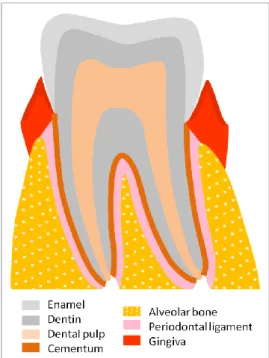
Periodontium is the organ which involves and sustains the tooth and it is constituted by the alveolar bone which forms the dental alveolus, the cementum which surrounds the tooth root, the periodontal ligament, which connects them and form a joint, and the gingiva.
Periodontitis is an inflammatory pathology highly prevalent in both dogs and humans that, when not treated, can lead to tooth exfoliation and also to life threatening systemic implications due to the blood dispersion of inflammatory mediators and pathogenic microorganisms.
The routine clinical therapies currently used to treat periodontal defects are often ineffective. Tissue Engineering has emerged as a valuable alternative approach aiming to regenerate ad
integrum the architecture and the biological function of the damaged tissue by providing the
repair site with a suitable supportive biomaterial seeded with stem cells.
Mesenchymal stem cells from oral and dental origin have the ability to regenerate the periodontium; however, harvesting cells from these sites implies significant local tissue morbidity and low cell yield, as compared to non-oral sources, such as the adipose tissue. Adipose-derived stem cells (ASCs) hold a great potential in cell based therapies for their easiness of harvesting with low morbidity, generating high yields of cells with capacity to differentiate into several lineages.
The main goal of this Thesis was to develop a new Tissue Engineering approach for the treatment of periodontal defects, based on an innovative biodegradable supportive matrix combined with ASCs. As the dog is the most relevant animal model to study human periodontal disease and, simultaneously, an important subject in Veterinary Medicine, we envisioned to validate this Tissue Engineering strategy in a canine model prior to the translation of this strategy for human application. With this in mind, the work aimed to achieve the following distinct objectives:
model;
Develop and characterize a double layer biodegradable scaffold meeting the requirements to achieve different functionalities, targeting regeneration of periodontal defects;
Evaluate the potential of the developed double layer scaffold for Tissue Engineering by culturing it with cASCs,
Assess the in vivo functionality of the scaffold for bone regeneration, using a rat mandibular defect model.
In order to achieve the proposed objectives, in a first stage, canine adipose tissue samples were harvested from two different anatomical sites, namely subcutaneous and omental adipose tissue, and the isolation of cASCs was optimized. The stemness and the osteogenic differentiation potential of these cells were analyzed along passages using real time RT-PCR and staining procedures. Subsequently, the same stem cells were xenotransplanted (into a mouse model) in order to evaluate the response of the xenogenic host against the cASCs.
Concomitantly, a double layer scaffold made of a blend of starch and poly (ε-caprolactone) was designed, comprising a 3D fiber mesh functionalized with silanol groups to promote osteogenesis and a solvent casting membrane aiming to act as a barrier against the migration of gingival epithelium into the periodontal defect. The obtained scaffolds were extensively characterized regarding their mechanical properties, degradation behaviour and effectiveness of the functionalization. Ultimately, the scaffolds were seeded/cultured with cASCs to assess the in vitro functionality of the obtained constructs.
Finally, to assess the ability of the developed scaffold to promote bone regeneration, these were implanted in a circular critical size defected induced in rat mandible, and the formation of new bone was quantified after 8 weeks of implantation.
In general, the stemness and osteogenic differentiation potential of the cASCs was found to decrease along increasing passages. Additionally, it was shown that the anatomical origin of the adipose tissue has a significant effect in the osteogenic differentiation ability of these cells. After the injection of cASCs in a xenogenic host (mouse), it was proved that no significant local immunogenic response was detected against them.
The physicochemical characterization of the newly developed double layer scaffold showed that this matrix presents a morphology, surface composition, degradation behaviour and
and also to promote the osteogenic differentiation on the functionalized fiber mesh.
In the induced mandibular defect, the functionalized scaffold with silanol groups was shown to enhance higher new bone formation in comparison to a collagen commercial membrane and to empty defects. Moreover, the membrane layer was observed to avoid the epithelial and connective tissue ingrowth, which is one of the main requirements for guided tissue regeneration.
In summary, this work demonstrated that the canine adipose tissue provides an alternative source of stem cells with the ability to differentiate into different cellular lineages and which do not induce an in vivo inflammatory response in the xenogeneic host. These findings together with the observed potential of the developed scaffold to accommodate the proliferation and differentiation of the cultured cASCs and to enhance in vivo bone regeneration allows to propose this tissue-engineered matrix for periodontal regeneration as an alternative to the techniques currently used in the dentistry field, both in human and in veterinary medical practice.
Keywords: Periodontium, Periodontal regeneration, Tissue Engineering, Biomaterials,
Nova matriz de engenharia de tecidos para regeneração periodontal baseada num material biodegradável combinado com células estaminais derivadas do tecido adiposo canino
O periodonto é o órgão que envolve e sustenta o dente, sendo constituído pelo osso alveolar que forma o alvéolo dentário, o cemento que envolve a raiz do dente, o ligamento periodontal que estabelece a ligação entre os dois tecidos anteriores formando uma articulação e a gengiva.
A periodontite é uma doença inflamatória de elevada prevalência em cães e seres humanos e que, quando não tratada, pode conduzir à esfoliação dentária e colocar em risco a vida do doente devido às manifestações sistémicas decorrentes pela dispersão sanguínea de mediadores inflamatórios e microrganismos patogénicos.
Os tratamentos que atualmente se usam para tratar esta patologia revelam-se muitas vezes ineficazes. A Engenharia de Tecidos emergiu, recentemente, como uma potencial terapia alternativa. Esta abordagem consiste no fornecimento de um biomaterial de suporte semeado com células estaminais que visa a regeneração ad integrum da arquitetura e da função biológica do tecido afetado.
As células estaminais mesenquimatosas de origem oral e dentária têm a capacidade de regenerar os tecidos periodontais. No entanto, as células provenientes destes locais pressupõem uma morbilidade significativa associada ao local da colheita e possuem um baixo rendimento celular em comparação com fontes não-orais, como por exemplo o tecido adiposo.
As células estaminais derivadas do tecido adiposo (ASCs) possuem um grande potencial de aplicação em terapias celulares devido à sua facilidade de colheita e baixa morbidade associada, ao alto rendimento de células isoladas e à capacidade demonstrada para se diferenciarem em inúmeras linhagens celulares.
O principal objetivo da presente Tese consistiu no desenvolvimento de uma nova abordagem de Engenharia de Tecidos para o tratamento de defeitos periodontais com base numa matriz biodegradável combinada com ASCs.
doença periodontal em humanos, pretende validar-se esta estratégia de Engenharia de Tecidos em modelo canino antes da aplicação translacional para a Medicina Humana, sem descuidar nunca que esta doença assume também grande importância na Medicina Veterinária. Tendo isto em consideração, o trabalho desenvolvido nesta Tese teve como principais objetivos:
Otimizar a colheita e o isolamento de ASCs caninas (cASCs) provenientes de diferentes regiões anatómicas e caracterizar a sua estaminalidade e o seu potencial osteogénico em diferentes passagens;
Avaliar a resposta à implantação de cASCs em modelo subcutâneo de murganho saudável;
Desenvolver e caracterizar um material de suporte (scaffold) biodegradável de dupla camada que vise a regeneração de defeitos periodontais;
Avaliar o potencial in vitro do scaffold de dupla camada desenvolvido para uso em Engenharia de Tecidos através da sua cultura com cASCs;
Avaliar a funcionalidade in vivo do scaffold para a regeneração óssea, utilizando um modelo de defeito mandibular em rato.
A fim de alcançar os objetivos propostos, numa primeira etapa, as amostras de tecido adiposo canino foram colhidas a partir de duas localizações anatómicas diferentes, a saber, tecido subcutâneo e tecido omental, e o isolamento das cASCs foi posteriormente otimizado. A estaminalidade destas células e o potencial de diferenciação osteogénica foram analisadas ao longo das diferentes passagens através de RT-PCR em tempo real e histologia. Posteriormente, as mesmas cASCs foram xenotransplantadas em murganho, a fim de avaliar a resposta xenogénica do hospedeiro contra as mesmas.
Concomitantemente, desenvolveu-se um scaffold composto por uma mistura de amido e poli-ε-caprolactona e compreendendo duas camadas: uma malha tridimensional de fibras (“fiber mesh”) funcionalizadas com grupos silanol para promover a osteogénese ao nível do osso alveolar, e uma membrana obtida por evaporação de solvente com o intuito de atuar como barreira contra a migração de epitélio gengival para o interior do defeito periodontal.
Os scaffolds obtidos foram extensivamente caracterizados quanto às suas propriedades mecânicas, ao comportamento de degradação e à eficácia da técnica de funcionalização. De seguida, os scaffolds foram semeados com cASCs a fim de avaliar a funcionalidade in vitro das construções obtidas.
induzido na mandíbula do rato, e a formação de novo osso foi determinada após oito semanas de implantação.
Em geral, a estaminalidade e o potencial de diferenciação osteogénica das cASCs decresceu ao longo de passagens. Além disso, demonstrou-se também que a origem anatómica do tecido adiposo tem um efeito significativo nas duas características analisadas. Após a injeção de cASCs num hospedeiro xenogénico (murganho), não foi observada nenhuma resposta inflamatória local exuberante contra as mesmas.
A caracterização físico-química do scaffold desenvolvido demostrou que esta matriz apresenta uma morfologia, composição da superfície, comportamento de degradação e propriedades mecânicas adequadas para auxiliar a regeneração periodontal. Os estudos biológicos com cASCs revelaram o potencial que ambas as camadas têm de permitir a adesão e proliferação celular, bem como de promover a diferenciação osteogénica na malha funcionalizada.
Nos defeitos mandibulares experimentais, o scaffold funcionalizado com grupos silanol induziu uma maior formação de osso novo em comparação com a membrana comercial de colagénio e com os defeitos vazios. Além disso, a face composta pela membrana evitou o crescimento do tecido epitelial e conjuntivo para o interior do defeito, o qual é um dos requisitos para o sucesso da regeneração guiada de tecidos.
Em resumo, este trabalho demonstrou que o tecido adiposo canino fornece uma fonte alternativa de células estaminais com capacidade de diferenciação osteogénica e que estas não induzem uma resposta inflamatória exuberante no hospedeiro xenogénico. Estes resultados, juntamente com o potencial observado do scaffold desenvolvido para acomodar a proliferação e diferenciação das cASCs cultivadas e para promover a regeneração de osso
in vivo, permite propor esta construção como uma alternativa às técnicas atualmente
utilizadas na regeneração periodontal, tanto no homem como no cão.
Palavras-chave: Periodonto, Regeneração periodontal, Engenharia de Tecidos, Biomateriais, Células estaminais derivadas do tecido adiposo, Cão, Modelos animais
Acknowledgements ... vii
Abstract ... ix
Resumo ...xiii
Table of Contents ... xvii
Index of Tables ... xxvi
Index of Figures... xxvii
Abbreviations List ... xxxi
Short Curriculum vitae ... xxxv
List of Publications ... xxxvii
Section I - General Introduction ... 1
Chapter I - Periodontal tissue engineering strategies based on non-oral stem cells ... 3
Abstract ... 3
1. Introduction ... 5
2. Perspective on the current periodontal therapies ... 6
3. Tissue Engineering... 7
4. Bone marrow stem cells ... 9
5. Adipose-derived stem cells ... 13
6. Embryonic stem cells ... 14
7. Induced pluripotent stem (iPS) cells ... 14
8. Periosteal progenitor cells ... 15
Section II - Detailed description of experimental Materials and Methodologies ... 25
Chapter II - Materials and Methods ... 27
1. Canine adipose-derived stem cells (cASCs): harvesting of source tissue, isolation, expansion and differentiation ... 27
1.1. Harvesting of the canine adipose tissue... 28
1.1.1. Animal enrolment ... 28
1.1.2. Anaesthesia ... 28
1.1.3. Surgical procedure ... 28
1.1.4. Adipose tissue storing ... 29
1.2.Isolation of the canine ASCs ... 29
1.3.Expansion of canine ASCs ... 30
1.4.Differentiation of the canine ASCs ... 30
1.4.1. Osteogenic differentiation of the canine ASCs ... 30
1.4.2. Chondrogenic differentiation of canine ASCs ... 31
1.5.Characterization of the canine ASCs ... 31
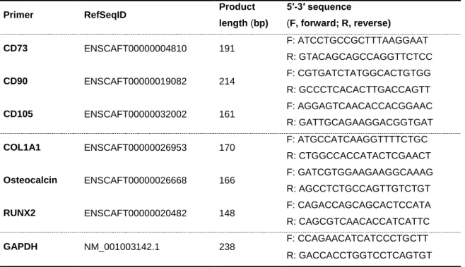
1.5.1. Gene expression analysis ... 31
1.5.2. Cytology ... 35
2. Subcutaneous implantation of the canine ASCs in healthy mice... 36
2.1.Preparation of the canine ASCs suspension ... 36
2.2.Study animals ... 36
2.3.Subcutaneous injection of the canine ASCs ... 37
2.4.Euthanasia and explants collection ... 37
2.7.Immunohistochemistry ... 37
3. Development of a double layer scaffold for periodontal regeneration ... 39
3.1.Description of the raw material – Blend of starch and poly (ɛ-caprolactone) ... 39
3.2.Production of the SPCL solvent casting membranes ... 40
3.3.Production of the SPCL wet-spun fiber meshes ... 40
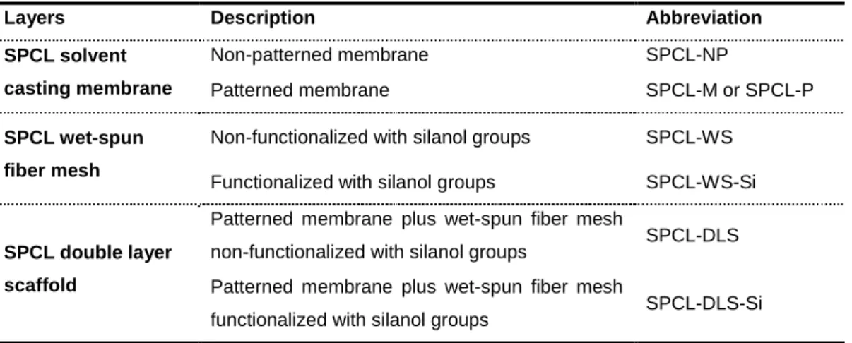
3.4.Production of the SPCL double layer scaffolds ... 41
3.5.Characterization of the morphology and structure of the developed scaffolds ... 42
3.5.1. Scanning electron microscopy ... 42
3.5.2. Micro-computed tomography ... 42
3.6.Characterization of the surface chemical composition ... 42
3.7.Characterization of the degradation behaviour... 43
3.7.1. Water uptake and weight loss ... 43
3.7.2. Assessment of the morphology of samples after degradation ... 43
3.7.3. α-Amylase activity ... 43
3.7.4. Calcium and silicon concentration of the degradation solutions ... 44
3.8.Characterization of the mechanical behaviour ... 44
4. Seeding/culturing of the canine ASCs onto the materials ... 44
4.1.Seeding/culturing onto solvent casting membranes ... 45
4.2.Seeding/culturing onto wet-spun fiber meshes... 45
4.3.Characterization of the canine ASCs-material constructs ... 45
4.3.1. Scanning electron microscopy ... 45
4.3.5. Gene expression analysis of the osteoblastic markers ... 47
4.3.6. Histology – Donath technique ... 47
4.3.7. Lévai Laczkó staining ... 48
5. In vivo evaluation of the double layer scaffold in a rat mandibular defect ... 49
5.1.Study animals ... 49
5.2.Experimental design ... 49
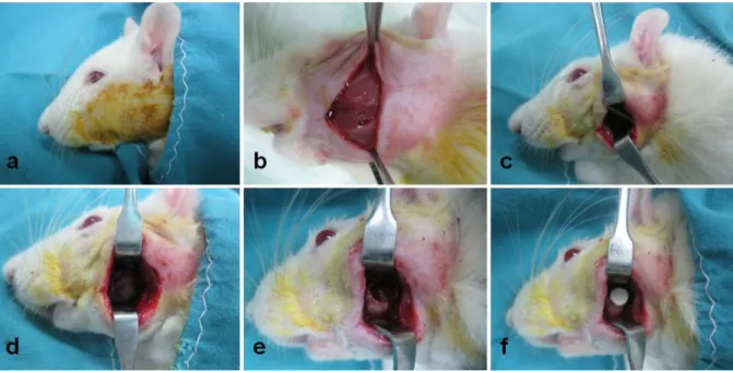
5.3.Surgical procedure ... 50
5.4.Postoperative care ... 51
5.5.Euthanasia and explants collection ... 52
5.6.Histology ... 52
5.7.Histomorphometric analysis ... 52
6. Statistical analysis ... 53
7. References ... 54
Section III - New tissue-engineered matrix for periodontal regeneration based on a biodegradable material combined with canine adipose-derived stem cells ... 59
Chapter III - Effect of anatomical origin and cell passage number on the stemness and osteogenic differentiation potential of canine adipose-derived stem cells ... 61
Abstract ... 61
1. Introduction ... 63
2. Materials and Methods ... 64
2.1.Harvesting of canine adipose tissue ... 64
2.2.Isolation and expansion of canine ASCs ... 65
2.3.2. Chondrogenic differentiation... 65
2.4.Canine ASCs characterization ... 66
2.4.1. Gene expression analysis of MSCs markers ... 66
2.4.2. Canine ASCs osteogenic potential assessment ... 67
2.4.3. Canine ASCs chondrogenic potential assessment ... 68
2.5.Statistical analysis ... 68
3. Results ... 69
3.1.Effect of the passage number in the undifferentiated canine ASCs ... 69
3.2.Effect of the anatomical site in the undifferentiated canine ASCs ... 70
3.3.Osteogenic differentiation assessment ... 71
3.4.Chondrogenic differentiation assessment ... 73
4. Discussion ... 74
5. Acknowledgments ... 79
6. References ... 80
Chapter IV - Evaluation of the response to the implantation of canine adipose-derived stem cells in a healthy mice subcutaneous model ... 87
Abstract ... 87
1. Introduction ... 89
2. Materials and Methods ... 90
2.1.Surgical harvesting of the canine adipose tissue ... 90
2.2.Isolation and expansion of the canine ASCs ... 90
2.6.Euthanasia and explants collection ... 91
2.7.Preparation of the canine ASCs’ cytoblocks ... 91
2.8.Hematoxylin and eosin ... 92
2.9.Immunohistochemistry ... 92
3. Results ... 93
3.1.Histology of the explants ... 93
3.2.Immunohistochemistry of the explants ... 94
4. Discussion ... 95
5. Conclusion ... 97
6. Acknowledgments ... 97
7. References ... 98
Chapter V - Design and characterization of a biodegradable double layer scaffold aimed at periodontal tissue engineering applications ... 103
Abstract ... 103
1. Introduction ... 105
2. Materials and methods ... 106
2.1.Production of the materials ... 106
2.2.Morphology characterization ... 108
2.2.1. Scanning electron microscopy ... 108
2.2.2. Micro-computed tomography ... 108
2.3.Fourier transform attenuated total reflectance infrared spectroscopy ... 108
2.4.Degradation behaviour ... 109
2.4.3. α-Amylase activity ... 110
2.4.4. Calcium and silicon concentration of the degradation solutions ... 110
2.5.Mechanical behaviour ... 110
2.6.Culturing of canine adipose-derived stem cells ... 110
2.6.1. Scanning electron microscopy ... 111
2.6.2. dsDNA quantification ... 111
2.6.3. Real time RT-PCR analysis ... 111
2.7.Statistical analysis ... 112
3. Results ... 112
3.1.Morphology characterization ... 112
3.2.Fourier transform attenuated total reflectance infrared spectroscopy ... 113
3.3.Degradation behaviour ... 114
3.3.1. Water uptake and weight loss ... 114
3.3.2. Morphology after degradation ... 114
3.3.3. α-Amylase activity ... 116
3.3.4. Calcium and silicon concentration of the degradation solutions ... 117
3.4.Mechanical behaviour ... 117
3.5.Culturing of canine ASCs ... 118
4. Discussion ... 119
5. Conclusion ... 123
6. Acknowledgements ... 123
biodegradable double layer scaffold and adipose-derived stem cells ... 131
Abstract ... 131
1. Introduction ... 133
2. Materials and Methods ... 134
2.1.Preparation of the materials ... 134
2.2.Seeding/culturing of the canine ASCs onto materials... 135
2.3.Characterization of the membranes/fiber meshes cultured with canine ASCs ... 136
2.3.1. Cellular morphology ... 136
Scanning electron microscopy ... 136
Histology – Donath technique ... 137
2.3.2. Cellular metabolic activity ... 137
2.3.3. Cellular proliferation ... 137
2.3.4. Osteogenic differentiation of the canine ASCs on the wet-spun fiber meshes138
Alkaline phosphatase activity ... 138
Gene expression of specific osteogenic markers ... 138
2.4.Statistical analysis ... 139
3. Results ... 140
3.1.Cellular morphology ... 140
3.2.Cellular metabolic activity – MTS assay ... 142
3.3.Cellular proliferation – dsDNA assay ... 142
3.4.Osteogenic differentiation assessment ... 143
4. Discussion ... 145
5. Conclusion ... 148
Chapter VII - Evaluation of a starch-based double layer scaffold for bone regeneration
in a rat model ... 153
Abstract ... 153
1. Introduction ... 155
2. Materials and Methods ... 156
2.1.Production of the materials ... 156
2.2.Animals... 156 2.3.Surgical procedure ... 157 2.4.Histology ... 157 2.5.Statistical analysis ... 158 3. Results ... 158 3.1.Histology ... 158 3.2.Bone histomorphometry ... 159 4. Discussion ... 160 5. Conclusion ... 162 6. Acknowledgments ... 162 7. References ... 164
Section III - Final Conclusions ... 167
Chapter I – Periodontal tissue engineering strategies based on non-oral stem cells Table 1. 1 – Summary table describing tissue engineered approaches for periodontal
regeneration based on the use of BMSCs ...12
Table 1. 2 – Summary table describing tissue engineered approaches for periodontal
regeneration based on the use of ASCs ...14
Chapter II – Materials and Methods
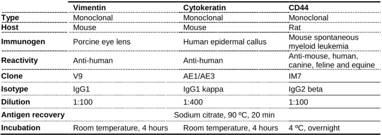
Table 2. 1 – Primer sequences for targeted cDNAs ...34 Table 2. 2 – Characteristics of the antibodies used in this study ...38 Table 2. 3 – Summary of the components of the double layer scaffold which were developed
and characterized ...41
Chapter III – Effect of anatomical origin and cell passage number on the stemness and osteogenic differentiation potential of canine adipose-derived stem cells
Table 3. 1 – Primer sequences for targeted cDNAs ...68 Chapter IV – Evaluation of the response to the implantation of canine adipose-derived stem cells in a healthy mice subcutaneous model
Table 4. 1 – Characteristics of the antibodies used in this study ...92 Chapter V – Design and characterization of a biodegradable double layer scaffold aimed at periodontal tissue engineering applications
Table 5. 1 – Mechanical properties of the SPCL-DLS and SPCL-DLS-Si ... 118 Chapter VI – A tissue engineering approach for periodontal regeneration based on a biodegradable double layer scaffold and adipose-derived stem cells
Chapter I – Periodontal tissue engineering strategies based on non-oral stem cells Figure 1. 1 – Schematic representation of the dental and periodontal anatomy ... 5 Figure 1. 2 – Periodontal disease aspect in humans. A: healthy gingiva; B: gingivitis; C:
periodontitis ... 5
Figure 1. 3 – Schematic image of the several stem cells sources for application in
periodontal regeneration ... 8
Chapter II – Materials and Methods
Figure 2. 1 – Schematic representation of the rat mandible ...50 Figure 2. 2 – Surgical procedure to induce the defect and implant the material ...51 Chapter III – Effect of anatomical origin and cell passage number on the stemness and osteogenic differentiation potential of canine adipose-derived stem cells
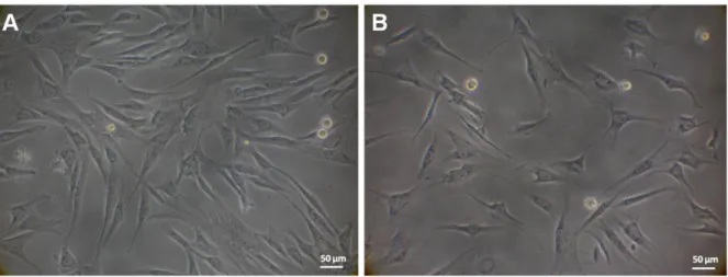
Figure 3. 1 – Representative light microscopy images of cASCs obtained by enzymatic
digestion from different anatomical sites, namely subcutaneous and omental, cultured in basal medium ...69
Figure 3. 2 – Real time RT-PCR analysis of various MSCs genes, namely CD73, CD90 and
CD105, in canine ASCs cultured in basal medium ...70
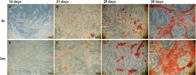
Figure 3. 3 – Light microscopy images of cASCs cultured in osteogenic conditions, stained
with Alizarin Red...71
Figure 3. 4 – Real time RT-PCR analysis results of the various osteoblastic genes, namely
COLIA1, RUNX2 and Osteocalcin, in cASCs, obtained cultured in osteogenic conditions ....72
Figure 3. 5 – Representative light microscopy images of subcutaneous cASCs cultured in
chondrogenic conditions stained with Toluidine Blue, Safranin O, Alcian Blue and H&E ...74
Chapter IV – Evaluation of the response to the implantation of canine adipose-derived stem cells in a healthy mice subcutaneous model
Figure 4. 1 – Histological images of the observed clusters on the dermis and the cASCs’
aimed at periodontal tissue engineering applications
Figure 5. 1 – Schematic representation of the developed double layer scaffold comprising
the membrane and the fiber mesh. Schematic picture of the implantation of the double layer scaffold in a periodontal defect. Table summarizing the components of the double layer scaffold that were developed and characterized ... 107
Figure 5. 2 – SEM micrographs of the SPCL-M, SPCL-WS and SPCL-WS-Si. At the right,
the micro-CT image of the double layer scaffold and the FTIR-ATR spectrum of the SPCL-WS and SPCL-SPCL-WS-Si ... 113
Figure 5. 3 – SEM micrographs and degradation profile in terms of water uptake and weight
loss of the SPCL-DLS and SPCL-DLS-Si ... 115
Figure 5. 4 – Cumulative release of reducing sugars as function of degradation time of
SPCL-DLS and SPCL-DLS-Si ... 116
Figure 5. 5 – Cumulative release of calcium and silicon as function of degradation time of
SPCL-DLS-Si ... 117
Figure 5. 6 – Canine ASCs proliferation in the SPCL-M, SPCL-WS and SPCL-WS-Si
cultured in basal medium ... 118
Figure 5. 7 – Osteocalcin gene expression in cASCs cultured onto WS and
SPCL-WS-Si in basal and osteogenic medium ... 119
Chapter VI – A tissue engineering approach for periodontal regeneration based on a biodegradable double layer scaffold and adipose-derived stem cells
Figure 6. 1 – Schematic representation of the envisioned tissue engineering strategy based
on the use of a double layer scaffold with two different target functionalities to culture the harvested/isolated canine ASCs. Description of the different components ... 135
Figure 6. 2 – SEM micrographs of SPCL-P and SPCL-NP cultured with cASCs in basal
medium, and SPCL-WS and SPCL-WS-Si cultured with cASCs in both basal medium and osteogenic medium ... 141
Figure 6. 3 – Optical microscopy images of cASCs cultured onto the WS and
SPCL-WS-Si in basal and in osteogenic medium (Lévai Laczkó staining) ... 141
Figure 6. 4 – cASCs metabolic activity and proliferation upon culturing onto the SPCL-NP
and SPCL-P membranes in basal medium, and culturing onto the SPCL-WS and SPCL-WS-Si in basal and osteogenic medium ... 142
Figure 6. 6 – Real time RT-PCR analysis of osteoblastic genes, namely COLIA1, RUNX2
and Osteocalcin, in cASCs cultured onto the SPCL-WS and SPCL-WS-Si in basal and osteogenic medium ... 144
Chapter VII – Evaluation of a starch-based double layer scaffold for bone regeneration in a rat model
Figure 7. 1 – Representative images of the mandibular defect without scaffold (empty) and
filled with collagen, SPCL and SPCL-Si scaffolds (Lévai Laczkó staining) ... 159
Figure 7. 2 – Box plot of the percentage of new bone formation in empty defects, collagen,
A
ALP – Alkaline phosphatase
ANOVA – Analysis of variance
ASCs – Adipose-derived stem cells
αMEM – Alpha Minimum Essential Medium Eagle
B
BMSCs – Bone marrow stem cells
BM – Basal medium
BMP – Bone morphogenetic protein
C
cASCs – Canine adipose-derived stem cells
CD – Cluster of differentiation
cDNA – Complementary DNA
CO2 – Carbon dioxide
COLIA1 – Collagen type I alpha 1
CT – Computed tomography
D
DGAV – Direção-Geral de Alimentação e Veterinária
DLS – Double layer scaffold
DMEM – Dulbecco’s Minimum Essential Medium Eagle
DNA – Deoxyribonucleic acid
DPSCs – Dental pulp stem cells
dsDNA – Double strain DNA
DNS – Dinitrosalicyclic acid
E
ESCs – Embryonic stem cells
F
FBS – Fetal bovine serum
FCT – Portuguese Foundation for Science and Technology
FELASA – Federation for Laboratory Animal Science Associations
spectroscopy with attenuated total reflectance
G
GAPDH – Glyceraldehyde-3-phosphate dehydrogenase
GTR – Guided tissue regeneration
H
HA – Hydroxyapatite
H&E – Hematoxylin and Eosin
I
IGF – Insulin growth factor
IHC – Immunohistochemistry
ITS – Insulin-Transferrin-Selenium
M
mCT – Micro-computed tomography
mRNA – Messenger RNA
MSCs – Mesenchymal stem cells
MTS – 3- (4,5-dimethylthiazol-2-yl)-5- (3-carboxymethoxyphenyl)-2- (4-sulfophenyl)-2H-tetrazolium OD – Optical density OM – Osteogenic medium Om – Omental P P – Passage
PBS – Phosphate buffer saline
PCL – Polycaprolactone
PD – Periodontal disease
PDGF – Platelet derived growth factor
PDL – Periodontal ligament
PDLSCs – Periodontal ligament stem cells
PLA – Polylactic acid
PGLA – Poly (lactic-co-glycolic acid)
PRP – Platelet rich plasma
PTFE – Polytetrafluoroethylene
R
RNA – Ribonucleic acid
RT-PCR – Reserve transcription combined with polymerase chain reaction
RUNX2 – Runt-related transcription factor 2
S
Sc – Subcutaneous
SEM – Scanning electron microscopy
SPCL – Blend of starch and poly (ɛ-caprolactone)
SPCL-M – SPCL membrane
SPCL-NP – SPCL non-patterned membrane
SPCL-P – SPCL patterned membrane
SPCL-WS – SPCL wet-spun fiber mesh non-functionalized with silanol groups
SPCL-WS-Si – SPCL wet-spun fiber mesh functionalized with silanol groups
SPCL-DLS – SPCL double layer scaffold comprising a membrane and a wet-spun fiber mesh non-functionalized with silanol groups
SPCL-DLS-Si – SPCL double layer scaffold comprising a membrane and a wet-spun fiber mesh functionalized with silanol groups
TCP – Tricalcium phosphate
TE – Tissue Engineering
TGF – Transforming growth factor
W
João Filipe Requicha was born on the 6th October 1983, in Viseu, Portugal and he obtained the degree in Veterinary Medicine in 2007 by the University of Trás-os-Montes e Alto Douro (UTAD; Vila Real, Portugal).
After graduation, he worked as a clinical practitioner in small animal medicine in a private veterinary clinic and in the Veterinary Teaching Hospital of the UTAD, and collaborated in the lecturing of subjects, such as Veterinary Anatomy and Veterinary Dentistry and Small Animal Internal Medicine. During that period, he developed his interest, knowledge and skills in Veterinary Dentistry.
In 2008 he was awarded with a Portuguese Science Foundation (FCT) PhD scholarship and started his PhD program in Veterinary Sciences at the University of Trás-os-Montes e Alto Douro, under the supervision of Carlos A. Viegas, in collaboration with the 3B’s Research Group of the University of Minho (Guimarães, Portugal), under the supervision of Manuela E. Gomes. He spent some periods during his PhD in the Doctor Fernando Muñoz’s Laboratory of Bone Research of the Veterinary Faculty of the University of Santiago de Compostela (Lugo, Spain).
Meanwhile, in 2011 he also completed a Master’s degree in Veterinary Medicine by the University of Trás-os-Montes e Alto Douro with a thesis focused on the canine oral cavity neoplasia, and the Post graduation in Veterinary Dentistry and Maxillofacial Surgery of the University Complutense of Madrid (Spain).
In 2013 obtained the accreditation as Researcher-Coordinator on Animal Experimentation, issued by the Direção-Geral de Alimentação e Veterinária (DGAV, the Portuguese Veterinary Authority), after attending the Laboratory Animal Science course by Federation for Laboratory Animal Science Associations (FELASA).
João Requicha is an active member of several scientific organizations such as the Tissue Engineering and Regenerative Medicine International Society – European Chapter (TERMIS-EU), the Portuguese Society for Stem Cells and Cell Therapies (SPCE-TC) and the European Veterinary Dentistry Society (EVDS). He is also a founder member of the Portuguese Society of Veterinary and Experimental Dentistry being, at the present moment, a board member.
submitted to national (FCT) and European entities (EC 7 Framework Program), as well as in the organization of national and international scientific meetings, such as the 1st Iberian Congress of Veterinary Dentistry in 2010 (Vila Real), the XXXVIII Congress of the European Society for Artificial Organs in 2011 (Porto, Portugal), the 21st European Congress of Veterinary Dentistry in 2012 (Lisbon, Portugal) and the TermStem Congress in 2012 (Guimarães, Portugal). He has reviewed manuscripts for international scientific journals in the field of Tissue Engineering and Regenerative Medicine.
As a result of his research work, João F. Requicha has attended to some of the most relevant national and international conferences in his research field, presenting 6 oral communications and 4 poster presentations. He is the first author of 3 accepted papers in international refereed journals and 3 submitted papers for publication.
The work performed under the scope of this PhD Thesis resulted in the publications listed below.
International Refereed Journals
Requicha JF, Moura T, Muñoz F, Leonor IB, Martins T, Gomes ME, Reis RL, Viegas CA.
Evaluation of a starch-based double layer scaffold for bone regeneration in a rat model.
Submitted
Requicha JF, Carvalho PP, Pires MA, Dias I, Gomes ME, Reis RL, Viegas CA.
Evaluation of the response to the implantation of canine adipose-derived stem cells in a healthy mice subcutaneous model. Submitted
Requicha JF, Viegas CA, Leonor IB, Reis RL, Gomes ME. A tissue engineering
approach for periodontal regeneration based on a biodegradable double layer scaffold and adipose-derived stem cells. Submitted
Requicha JF, Viegas CA, Hede S, Leonor IB, Reis RL, Gomes ME. Design and
characterization of a biodegradable double layer scaffold aimed at periodontal tissue engineering applications. Journal of Tissue Engineering and Regenerative Medicine. doi:
10.1002/term.1816
Requicha JF, Viegas CA, Muñoz F, Reis RL, Gomes ME. Non-oral stem cells on
periodontal regeneration strategies. The Anatomical Record. doi: 10.1002/ar.22797
Requicha JF, Viegas CA, Albuquerque CM, Azevedo JM, Reis RL, Gomes ME. 2012.
Effect of anatomical origin and cell passage number on the stemness and osteogenic differentiation potential of canine adipose-derived stem cells. Stem Cell Reviews and
Reports, 8 (4): 1211-1222
Non-indexed International Refereed Journal
JF Requicha, ME Gomes, IR Dias, RL Reis, CA Viegas. 2013. Ingeniería de tejidos
Oral communications
Requicha JF, Leonor IB, Muñoz F, Moura T, Carvalho P, Anjos M, Azevedo J, Reis RL, Gomes ME, Viegas CA. Development of a new approach to the periodontal regeneration. 101st FDI Annual World Dental Congress, Istanbul, Turkey; 28-31 August 2013.
Requicha JF, Moura T, Leonor IB, Muñoz F, Gomes ME, Reis RL, Viegas CA.
Assessment of a scaffold for periodontal regeneration in a rodent model. XXII European
Congress of Veterinary Dentistry and XII World Veterinary Dental Congress. Prague, Czech Republic; 23-26 May 2013
Requicha JF, Viegas CA, Albuquerque CM, Azevedo JM, Reis RL, Gomes ME. Canine
adipose stem cells: the influence of the anatomy and passaging on the stemness and osteogenic differentiation potential. Term Stem 2012. Guimarães, Portugal; 9-12 October
2012
Requicha JF, Leonor IB, Viegas CA, Reis RL, Gomes ME. Tissue engineered constructs
for periodontal regeneration based on adipose stem cells and a newly designed polymeric scaffold. XXXVIII Congress of the European Society for Artificial Organs
(ESAO 2011) and IV Biennial Congress of the International Federation on Artificial Organs (IFAO 2011). Oporto, Portugal; 9-12 October 2011
Requicha JF, Leonor IB, Albuquerque C, Gomes ME, Reis RL, Viegas CA. New
biodegradable membrane for periodontal tissue engineering. XX European Congress of
Veterinary Dentistry. Chalkidiki, Greece; 1-3 September 2011
Requicha JF, Leonor IB, Viegas CA, Reis RL, Gomes ME. Biodegradable double layer
scaffold for periodontal engineering. European Chapter of the Tissue Engineering and
Regenerative Medicine International Society (TERMIS) 2011 Annual Meeting. Granada, Spain; 7-10 June 2011
Poster presentations
Requicha JF, Leonor IB, Muñoz F, Moura T, Azevedo J, Viegas CA, Reis RL Gomes ME. A novel tissue engineering concept targeting the regeneration of periodontal defects. European Chapter of the Tissue Engineering and Regenerative Medicine International Society (TERMIS) 2013 Annual Meeting, Istanbul, Turkey, June 2013.
CA. Tissue engineered scaffold for periodontal regeneration: laboratorial and preclinical characterization. I Simpósio Inter-Universitário de Investigação em Medicina Dentária. Coimbra, Portugal, March 2013
Requicha JF, Leonor IB, Muñoz F, Moura T, Azevedo JM, Gomes ME, Reis RL, Viegas CA. In vitro and in vivo assessment of an innovative double layer scaffold for periodontal tissue engineering. II 3B’s-ICVS Associate Laboratory Meeting. Braga, Portugal; May 2012
Requicha JF, Hede S, Leonor IB, Viegas CA, Reis RL, Gomes ME. Characterization of canine adipose derived stem cells and its potential in periodontal tissue engineering applications. The 6th Annual International Meeting of the SPCE-TC, Cantanhede, Portugal, April 2011
Section I
General Introduction
Chapter I
Periodontal tissue engineering strategies based on non-oral stem cells
Abstract
Periodontal Disease is an inflammatory disease which constitutes an important health problem in humans, due to its enormous prevalence and life threatening implications on systemic health. Routine standard periodontal treatments include gingival flaps, root planning, application of growth/differentiation factors or filler materials and guided tissue regeneration. However, these treatments have come short on achieving regeneration ad
integrum of the periodontium, mainly due to the presence of tissues from different embryonic
origins and their complex interactions along the regenerative process.
Tissue Engineering (TE) aims to regenerate damaged tissue by providing the repair site with a suitable scaffold seeded with sufficient undifferentiated cells and thus constitutes a valuable alternative to current therapies for the treatment of periodontal defects. Stem cells from oral and dental origin are known to have potential to regenerate these tissues. Nevertheless, harvesting cells from these sites implies a significant local tissue morbidity and low cell yield, as compared to other anatomical sources of adult multipotent stem cells. This manuscript reviews studies describing the use of non-oral stem cells in Tissue Engineering strategies, highlighting the importance and potential of these alternative stem cells sources in the development of advanced therapies for periodontal regeneration.
______________________________________________________
*This Chapter is based on the following publication:
1. Introduction
Periodontium is an organ constituted by various tissues namely the alveolar bone, which forms the dental alveolus, the cementum which surrounds the root, the periodontal ligament (PDL) which is sustained by the bone and the cementum, bonding them, and finally the gingiva, which involves all the tissues above (1, 2) (Fig. 1.1).
Figure 1. 1 – Schematic representation of the dental and periodontal anatomy.
The periodontium is often affected by the Periodontal Disease (PD), an inflammatory disease which includes two different inflammatory stages: an initial form called gingivitis and an advanced form called periodontitis (Fig. 1.2), which usually progresses with bone resorption, cementum necrosis and gingival recession or hyperplasia. Ultimately, when left untreated, it leads to the formation of a periodontal pocket with permanent loss of tooth support, and consequently increasing mobility and teeth loss (3-5).
Periodontal disease has a multifactorial aetiology involving microbial, behavioural (absence of oral hygiene and diet), local (malocclusions and overcrowding), systemic (immunodeficiency, infections, metabolic disease and nutritional disturbances) and genetic factors (6-8). The PD is an important health problem in Medicine, due to its enormous prevalence and life threatening implications on systemic health. It has been reported a correlation between PD and preterm birth, low weight at birth and neonatal morbidity, diabetes, respiratory, osteoarticular and cardiovascular diseases (4). Chronic periodontitis affects about 30% of the adult population and 7-13% of them have the severe form of the disease (9).
Periodontal regeneration consists of a complex and orchestrated sequence of biological events at a molecular and cellular level, accomplishing success when the following goals are met: formation of new cementum in the radicular surface, restoration of the alveolar crest till the cementum-enamel junction, reestablishment of the junctional epithelium and formation of a dense set of periodontal ligament fibers obliquely oriented assuring its functionality (1, 10, 11). This phenomenon involves the pool of progenitor cells present in the periodontium, clustered near the periodontal ligament blood vessels, which form the fibroblasts, osteoblasts, and cementoblasts (2) helped by progenitor cells existent in the bone endosteal spaces which migrate to the PDL (12).
2. Perspective on the current periodontal therapies
The main aims of the current periodontal therapies include the infection control and the re-establishment of a periodontal tissue apparatus. As the dental plaque is the primary cause of PD, their removal from the tooth crown, gingival sulcus and root surfaces is essential for the prevention and/or control of periodontal disease. Plaque removal can be accomplished by a combination of home care procedures that include mechanical and chemical plaque reduction techniques. In order to control the microbial population, clindamycin hydrochloride, amoxicillin/clavulanate and metronidazole seem to be particularly effective systemic antimicrobials. Locally delivered antimicrobials (perioceutics), such as doxycycline gel, can be applied to teeth that have been cleaned and polished (6, 7, 13).
However, when the disease progresses, advanced periodontal surgery becomes necessary. To treat patients with deep pockets and bone loss, mucogingival surgery (flap exposure), open curettage (5, 10) and the root conditioning with demineralizing agents (5, 10, 11, 14) are required. In order to stimulate the periodontal regeneration, the clinicians sometimes inject into the defects cocktails of growth factors, such as enamel matrix derivatives (EMD) or
platelet-rich plasma (PRP) (15, 16) or specific growth/differentiation factors (e.g. PDGF, IGF-1, BMP-2 and BMP-7) (5, 11). In some cases, depending on the dimensions of the defect and the degree of osteolysis and/or tooth root exposition, it is advised the application of filler materials such as autografts, allografts and alloplastic materials (most frequently, hydroxyapatite-HA and tricalcium phosphate-TCP) (5, 10, 14) and/or guided tissue regeneration (GTR) membranes.
GTR techniques were firstly proposed in the 80s (11, 14) and have the objective to guide selectively the cell proliferation in different compartments, namely the alveolar bone, cementum and PDL, using ePTFE, PLA, PGLA or collagen membranes as a physical barrier (10, 13, 17). In fact, this was one of the first techniques which considered the physiopathology of the periodontal regeneration, avoiding some of the major drawbacks reported for the other existing therapies, namely, the gingival epithelium and connective tissue expansion, ankylosis and radicular resorption phenomenon and the difficulty to avoid the collapse of the periodontal defect (1, 5, 11, 17). However, none of these procedures, even when used in combination (for example, simultaneous application of filler materials with GTR membranes) have revealed enough efficacy to achieve full periodontal regeneration.
3. Tissue Engineering
Recently, Tissue Engineering (TE) and other cell based therapies have emerged as an alternative approach for the regeneration of several tissues damaged by disease or trauma, including the periodontium. TE involves the use of a support material – membrane or scaffold – where cells are seeded and cultured in order to obtain hybrid materials which can induce the regeneration of the target tissue.
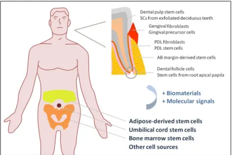
Several materials have been proposed to be used in TE, mostly biodegradable polymers of synthetic and natural origin (17-19) aimed at acting as a support for cells and new tissue ingrowth until complete regeneration of the tissue defect is accomplished (11, 18, 20, 21). Tissue engineering approaches usually rely on the use of adult mesenchymal stem cells (MSCs) (Fig. 1.3), that have the capacity to differentiate in various types of cells, as for example, osteoblasts, chondroblasts, adipocytes, cardiomyocytes, neuronal cells or periodontal ligament cells (10, 22, 23).
Figure 1. 3 – Schematic image of the several stem cells sources for application in periodontal
regeneration.
Periodontium is a highly specialized and complex organ and is derived from the dental follicle and the neural crest cells. These cells constitute a multipotent cell population in vertebrates and participate in the embryonic development of most dental tissues including the gingiva, the dental follicle, the periodontal ligament and the alveolar bone (24, 25).
From the embryonary dental structures, Gronthos and colleagues identified for the first time, in 2000, the dental pulp stem cells (DPSCs) (26). Stem cells have also been isolated from human exfoliated deciduous teeth (SHEDs) (27). In the periodontal ligament have been described the PDL fibroblasts (28) and the PDL stem cells (PDLSCs) (29), which are the most studied cell source from periodontal origin. From the gingiva, it is possible to obtain distinct cell populations, such as the gingival fibroblasts and gingival precursor cells (30, 31). Other undifferentiated dental stem cells have been referred in the literature along the years, namely, the dental follicle cells (DFCs) (32), the stem cells from the apical papilla (SCAP) (33) and the cementum-derived cells (CDCs) (34). Regarding the osseous component of the periodontium, McCulloch et al. proposed, in 1987, that paravascular cells from the endosteal spaces of alveolar bone communicate with the PDL and may contribute to its cell populations (12). Recently, undifferentiated stem cells were isolated from the alveolar bone margin (35).
However, the development of new strategies for periodontal regeneration requires significant amounts of cells with regenerative potential to have therapeutic efficacy, combined or not with a biodegradable supportive matrices. Furthermore, autologous approaches are usually preferred for safety reasons and, for this purpose, it is very important to collect these cells
without sacrifice of the tissues that are intended to regenerate, namely, the periodontium or even the teeth. Thus, it is of utmost relevance to consider the use of adult MSCs from other origins. Among those cells, the bone marrow stem cells and the adipose-derived stem cells assume an important role in the present and future research in this field. Therefore, this review will focus on the application of undifferentiated stem cells from dental and non-periodontal origin in Tissue Engineering approaches targeting the regeneration of non-periodontal defects.
4. Bone marrow stem cells
Bone marrow stem cells (BMSCs) were firstly identified in 1968 (36) and since then they have been extensively studied, being considered the gold standard in cell-base therapies and other regenerative medicine approaches. In 2009, Kramer et al. after inject male DiI-labeled BMSCs into a female rat periodontal pocket, observed for the first time that these cells obtain PDL morphology and PDL protein markers in vivo over a 6-week period (37).
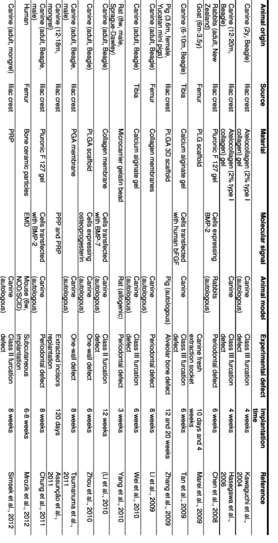
Along the years, as described below, different Tissue Engineering approaches combining distinct types of supportive matrices (gels or scaffolds) with undifferentiated BMSCs were developed and assessed in pre-clinical models, as summarized in the Table 1.1.
One of the first reports on the use of BMSC in periodontal regeneration therapies refers to a study by Kawaguchi and colleagues (2004) that proposed the auto-transplantation of canine BMSCs on an atelocollagen gel into dog furcation defects. It was observed that cementum with extrinsic fibers was formed along almost denuded root surfaces (38), providing one of the first data on periodontal regeneration in a superior animal model with resemblances to human. In a subsequent study, the same researchers transplanted BMSCs labeled with green fluorescence proteins (GFP), using the same gel and experimental model, and identified cementoblasts, osteoblasts, osteocytes and fibroblast derived from those cells, concluding that they participate effectively in the periodontal regeneration (39). This data was corroborated by Wei et al. who used BMSCs labeled with bromodeoxyuridine (BrdU) mixed with alginate gel in the same model (40) and observed new bone, PDL, cementum and blood vessels formation and expression of typical surface markers of osteoblasts and fibroblasts.
Another approach (41) described the implantation of BMSCs engineered to express BMP-2 in a pluronic F-127 hydrogel into rabbit periodontal defects where it was demonstrated that this growth/differentiation factor enhances the regeneration of periodontal tissues comparing
A distinct study reported the use of goat BMSCs seeded onto a PLG scaffold that was fitted around a titanium fixture immediately after tooth removal. The results showed the formation of new bone and periodontal tissue-like bundles of fibers extending from the periphery of the wound toward the surface of implant, suggesting that BMSCs-PLG construct promoted osteointegration (42). In the same year, Zhang and his team implanted a tooth/bone construct seeded with BMSCs into mandible defects in swines and observed the formation of small tooth-like structures consisting of organized dentin, enamel, pulp, cementum, periodontal ligament resembling Sharpey’s fibers and alveolar bone (43).
Rat BMSCs (GFP+) have also been loaded into gelatin microbeads and transplanted into a surgically created rat periodontal defect (44). After three weeks of implantation, it was found higher evidence of bone, cementum and PDL regeneration in defects filled with microbeads-BMSCs than in empty defects or defects filled with the beads alone.
Recently, a study performed by Tsumanuma and colleagues compared the performance of BMSCs with PDLSCs and alveolar periosteal cells (APCs), when applied as cell sheets in a biodegradable polymer membrane for periodontal regeneration. The obtained histological data showed that the alveolar bone regeneration ratio was higher when using the PDLSCs, as well as the cementum thickness and the quality of PDL fibers orientation (45). Moreover, Wei and his team (2012) reported that the formation of human PDLSCs sheets was promoted by vitamin C induction and extrapolated this finding to both human BMSCs and UCSCs by culturing them with 20 mg/ml of vitamin C (46). Recently, several molecular factors have been proposed to promote the regenerative mechanism of the periodontium, for example, the platelet-rich plasma (15), the enamel matrix derivatives (EMD) (47, 48) or other specific growth/differentiation factors (e.g. PDGF, IGF-1, BMP-2 and BMP-7) (5, 49-51). For examples, Assunção et al. evaluated the effect of platelet-poor plasma (PPP), calcium chloride-activated platelet-rich plasma (PRP/Ca), calcium chloride- and thrombin-activated PRP (PRP/Thr/Ca), as well as BMSCs with PRP/Ca (BMSCs/PRP/Ca) on the healing of replanted dog teeth. The obtained outcomes suggest that thrombin activation is more effective in the healing, and that the use PRP/Ca with BMSCs show advantages in comparison to PRP/Ca alone (52). A study comparing the use BMSCs plus PRP and PRP or autogenous cortical bone alone concluded that no differences were observed between the treatments (53).
Zhou et al. described a study in which canine BMSCs modified by osteoprogesterin gene were seeded onto a PLGA scaffold and implanted in an autologous periodontal defect, leading to enhanced formation of new alveolar bone and cementum as well as new connective tissue, as compared to the controls (without cells and material). This study shows
that gene therapy utilizing osteoprogesterin may be used as an adjuvant in periodontal TE therapies/strategies (54).
In the same year, it was reported the use of a collagen membrane as a vehicle for canine BMSCs transfected with BMP-7, revealing higher bone formation and new cementum length upon implantation in a canine furcation defect, comparing to the material with non-transfected cells (49). Chung et al. engineered these cells with BMP-2 and obtained promising results in terms of periodontal regeneration (51). Bone marrow stem cells transfected with human β-FGF have also shown to accelerate significantly the periodontal regeneration in dog class III furcation defects (55).
A work focused on using either human BMSCs or PDLFs seeded onto bone ceramic particles with EMD in a subcutaneous mice model, revealed no differences between the two cell sources in terms of ectopic bone formation. In contrast, a large amount of bone/cementum-like tissue and PDL-like fibrous tissue was found when used implants containing PDLFs, as compared to those based on BMSCs (48).
Another comparative study demonstrated that co-cultured human PDLSCs and BMSCs subsequently combined with an osteoinduced ceramic bovine bone led to the formation of a neovascularized ectopic cementum/PDL-like complex resembling the physiologic PDL Sharpey’s fibers (56) in a subcutaneous mice model.
Apart of the effect of molecular stimuli on the cells behaviour, other stimuli has been studied, as recently referred by Cmielova et al. who concluded that BMSCs, as well as PDLSCs, respond to ionizing radiation by induction of senescence without affect viability (57).
Bone marrow stem cells have also been proposed in several strategies for regenerate the alveolar bone, a particular component of the periodontium, combined with different materials, namely β-TCP (58), HA/TCP (59) and fluorohydroxyapatite (60), nano-hydroxyapatite block scaffolds (61), chitosan-gelatin scaffold (62) and fibrin glue (63), or even combined with PRP for improve the osteointegration of hydroxyapatite-coated dental implants (64).
Table 1. 1 – Summary table describing tissue engineered approaches for periodontal
5. Adipose-derived stem cells
Adipose-derived stem cells (ASCs) were firstly identified in 2001 by Zuk (65) and since then they have been the focus of many studies on regenerative medicine (66, 67). In fact, the adipose tissue exhibits several attractive advantages over other stem cells sources such as the bone marrow, mainly due to its wide availability (68), the relative simplicity of the harvesting procedures (69, 70) and their great differentiation potential (22, 71). In fact, it is possible to obtain a large quantity of cells without causing high morbidity in the harvesting site, as for example, from 1 g of adipose tissue it can be isolated about 5,000 stem cells, whereas the yield from BMSCs is 100-1,000 cells/ml of bone marrow (72). Therefore, ASCs are considered a very attractive stem cell type for the development of new Tissue Engineering and other cell-based therapies, including for periodontal regeneration. Table 1.2 summarizes recent studies focusing on the use of ASCs in periodontal TE strategies.
The use of ASCs in periodontal regeneration strategies was reported for the first time in 2008 (15). In the referred study, rat ASCs were mixed with PRP and implanted into periodontal defects induced in rats. After 2 and 4 weeks of implantation, a small amount of regenerated alveolar bone was observed. Moreover, 8 weeks after implantation, it was observed the formation of a PDL-like structure along with alveolar bone (15).
Hung et al. (2011) demonstrated that implants composed of collagen gels with either rabbit ASCs or rabbit DPSCs, were able to promote the growth of self-assembled new teeth in adult rabbit extraction sockets with high success rate. Furthermore, rabbit ASCs demonstrated a higher growth rate and a better senescence resistance in culture, when compared to rabbit DPSCs (73).
An interesting in vitro study by Wen et al. described the potential of ASCs for periodontal regeneration showing that when incubated in dentin non-collagenous proteins and dental follicle cell conditioned medium adopted flat, cuboidal or polygonal shapes, features typical of a cementoblast-like morphology. Additionally, under these conditions, cells showed in vitro mineralization ability, ALP activity and expression of bone sialoprotein, type I collagen, osteonectin and osteocalcin characteristic of cementoblasts (74).
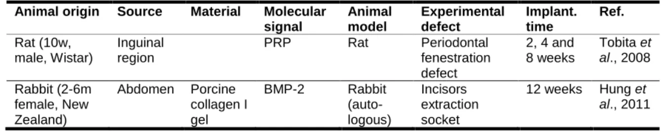
Table 1. 2 – Summary table describing tissue engineered approaches for periodontal regeneration based on the use of ASCs.
Animal origin Source Material Molecular signal Animal model Experimental defect Implant. time Ref. Rat (10w, male, Wistar) Inguinal region PRP Rat Periodontal fenestration defect 2, 4 and 8 weeks Tobita et al., 2008 Rabbit (2-6m female, New Zealand) Abdomen Porcine collagen I gel BMP-2 Rabbit (auto-logous) Incisors extraction socket 12 weeks Hung et al., 2011
6. Embryonic stem cells
Embryonic stem cells (ESCs) are derived from the inner cell mass of blastocysts and are pluripotent stem cells capable of differentiating into almost all kinds of cells of the adult body (75).
Only a few studies have addressed the potential use of ESCs in periodontal regeneration. Inanç and colleagues (2007) characterized the osteogenic differentiation potential of human embryonic stem cells (hESCs) under the inductive influence of human PDL fibroblast monolayers (76). The same authors have also achieved with success the differentiation of hESCs (HUES-9) into periodontal ligament fibroblastic progenitors (76, 77). A study on the differentiation capacity of hESCs toward the periodontal compartment cells and their relationship with tooth root surfaces in vitro, demonstrated that hESCs differentiation is influenced by tooth structures (extracted tooth root slices), PDLFs, and osteogenic medium, resulting in increased propensity toward mesenchymal lineage commitment, and formation of soft-hard tissue relationship in close contact areas (78). These reports suggest that human ESCs may be considered an important cell source for periodontal Tissue Engineering applications.
7. Induced pluripotent stem (iPS) cells
The reprogramming of somatic cells into induced pluripotent stem (iPS) cells by forced expression of a small number of defined factors (e.g., Oct3/4, Sox2, Klf4 and c-Myc) (79, 80) has great potential for tissue-specific regenerative therapies, avoiding ethical issues surrounding the use of ESCs and problems with rejection following implantation of non-autologous cells.
So far, the only study on iPS cells focusing in periodontal regeneration showed that iPS cells combined with EMD were found to provide a valuable tool for periodontal Tissue Engineering